
2.0 J M2 0.9 - Blood Journal
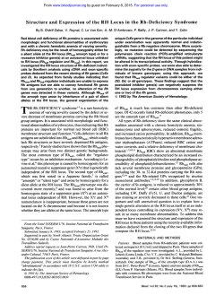
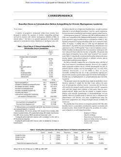
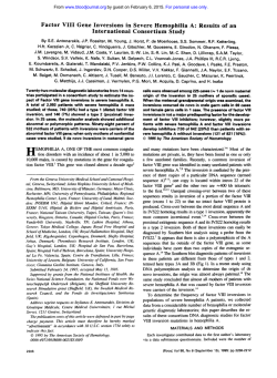
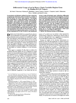
From www.bloodjournal.org by guest on February 6, 2015. For personal use only.
1351
CORRESPONDENCE
Factor Vlll Gene Rearrangementin Hemophilia A Carrier Detection: A Word of Caution
To the Editor:
Carrier detection and prenatal diagnosis is now facilitated by the
discovery that a unique inversion of the tip of chromosome X is
responsible for 50% of cases of severe hemophilia A.'.*We found
a similar frequency (27 of 54) of factor VI11 gene inversions in
severe unrelated hemophilia A patients in Israel. The mechanism
proposed for the inversion involves disruption of the factor VI11
gene and displacement of exons 1-22 to a site about 1 Mb from the
5' end of the normal gene.',' It is the purpose of this letter to show
that, in carriers of the inversion, crossing over between the normal
factor VI11 gene and the inverted gene may complicate diagnosis.
Coagulation studies performed in 1979 in a family with a sporadic
case of severe hemophilia A suggested that the sister and the mother
of the patient had significant posterior probabilities of being carriers,
ie, 0.7 and 0.2. respectively4 (Fig 1). Reevaluation of this family by
DNA analysis of the factor VI11 gene showed that the sister of the
patient inherited from her mother the non-hemophilic allele (6.2 kb)
at the Xba I polymorphic site of intron 22. This finding strongly
suggested that she is not a carrier. In addition, crossing over was
detected between the factor VI11 gene and the extragenic markers
in the intron 22 homologous regions, at DXS52 and at DXS 15 (Fig
1). More recently, Southern blot analysis for the newly discovered
factor VI11 rearrangement showed the inverted factor VI11 gene pattern in the patient, his sister, his mother, and grandmother (Fig 1).
However, the latter findings do not define the sister as a carrier in
viewofthe crossing over found earlier. Depending on the exact
location of the crossover, the sister may be a carrier if the break
occurred inside the normal factor VI11 gene, for which the chances
are very smatl yet not entirely negligible, particularly in view of her
suggestive coagulation tests. If a recombination occurred between
the normal and the inverted factor VI11 gene, the sister's maternal
X chromosome would bear both the normal and the inverted factor
VI11 genes and, consequently, she maynotbe
a carrier. Such a
recombination should occur in about 2% of such cases because this
is the frequency of recombinations between markers in the factor
VI11 gene and markers in intron 22 homologous regions that we
found in our material (1 of 57 informative meioses) and as reported
by others.' Practically, in future pregnancies, fetal blood sampling
will be necessary in the sister of the patient for establishing a final
diagnosis in male fetuses who will bear the inversion.
Thus, the possibility of crossing over between the normal and
inverted FVIII gene has to be taken into account when counselling
hemophilia A families in which the severe hemophilia A is related
to factor VI11 gene inversions.
Hava Peretz
Sali Usher
Uri Martinovitz
Uri Seligsohn
Institute of Thrombosis and Haemostasis
National Haemophilia Center Tel-Hashomer
Chemical Pathology Laboratory
1
I
'P1
M1
I En
I H
2.0
4.5
5.8
UI
J
M2
4.8
0.9
2.0
4.5
5.8
M2
4.8
0.9
2.0
4.5
5.8
1.6
5.3
5.8
M2
P2
PIIW
4.8
C.O.
0.9
2.0
4.5
5.8
Fig 1. A pedigreeof a family with a sporadic case of hemophilia A
showing the results of DNA analyses.
).1 The hemophilic patient;(01
unaffected males;(0)female carriers;(01suspected carrier. The
bars
represent parts of the X chromosome containing the normal I O )
I factor Vlll genes. The numbers adjacent to the
bars representthe lengths (in kilobases) ofthe polymotphicfragments
at the following loci: 0.9/1.2-&/
I site in intron 18; 4.8/6.2-Xbs 1
site in intron 22; 5.4/-extragenic Xb8 I in intron 22 homologous
region;1.6/2.O-Msp
I and4.5/5.3-Taq
I sites at DXS52;and
2.8/5.8--8gl site at DXS15. The family was not informative for the
(CAIn polymorphism in intron 13. Two alleles of the (CAln polymorphism in intron22 were in complete linkage dimquilibtiumwith
the intron 22 Xba I polymorphism. P1, P2 and M1, M2 denote the
paternal and meternal haplotypes, respectively. The arrow points
to
the npparent region on
the X chromosomes wherathe crouing over
(CO) could have occurred
to produce the P1/M2 haplotype.
From www.bloodjournal.org by guest on February 6, 2015. For personal use only.
1352
CORRESPONDENCE
False-Positive Residual Disease Assessment After Bone Marrow Transplant
in Acute Lymphoblastic Leukemia
To the Editor:
Polymerase chain reaction(PCR) of gene rearrangements provides
a sensitive widely applicable technique for the detection of minimal
residual disease (MRD) in childhood acute lymphoblastic leukemia
(ALL).' Generally, remission bone marrow (BM) DNA is amplified.
dot blotted. and hybridized with tumor-specific probes. The signal
obtained is compared with that generated by an equivalent amount
of normal BM mononuclear cell DNA. Two recent reports in Blood
describe the use of such a method.',' In this report. we highlight a
potential pitfall of this approach that may occur at times of rebound
polyclonallymphocytosissuchas
that seenafterBMtransplant
(BMT)."
A retrospective analysis of MRD after BMT was performed in a
2-year-old boy with common ALL. He presented with a white cell
@ P
A
treated accordingtothe
count of 125 X lO"/L andwasinitially
Medical Research Council UKALL x D protocol. Marrowremission
was documented at day 28. A fully matched unmanipulated sibling
therallograft was performed4 months after diagnosis. Conditioning
apy was cyclophosphamide at 120 m g k p and single fraction total
body irradiation. Cyclosporin alone was administered as
graft-versus-hostdisease(GVHD)prophylaxis.Thetransplantcoursewas
uneventfulandhedid
not haveanymajorepisodes
of sepsis or
require treatment for GVHD. He remains well and in remission 3
years after BMT.
28 postinduction.at
BM DNAobtainedatpresentation.atday
BM harvest. and at 3.6. 12, and 18 months post-BMT was analyzed
for MRD by Ig heavy chain (IgH) PCR as previously described.'
Five microliters of each product was dot blotted and probed with a
D28 BMH 3 6 9 12 18-2 -3 -4 -5 N N C
100bp>
82bp >
P
B
D28 BMH 3 6 9 12 18
-2 -3 -4 -S N N C
C
P
D28
BMH 3
6
918 12
-3 -4
-5 N
N
c
Fig 1. PAGE analysis and hybridization. (A) An 846 nondenaturing PAGE gel showing amplified BM DNA samples collected
at presentation, at day 28, at BM
harvest (BMHI and at 3, 6, 12,
and 18 months post-BMT. Logarithmic dilutions of tumor DNA
in normal BM DNA (-2 t o -51.
normal BM DNA (NI, and a no
template DNA control (C) are included. (B) Dot blot analvsis of
duplicate
products
hybridized
with a '*P-labeled 20-base oligonucleotide DNJ regionprobe. (C)
Electroblot analysis. Products
were resolved by 8% nondenaturing PAGE and electrophoretically transferred t o nylon membranes (Hybond N+; Amersham
International, Amenham, UK)
with a Millipore "MilliBlot" apparatus at a currentof 4 mAlcm'
for 30 minutes. Membranes
were fixed in 0.4 N NaOH, neutralized in 2 x SSC, and air-dried
before probe hybridization. The
second band observed in the dilution lanes is artefactual and
does not compromise residual
disease assessment.
From www.bloodjournal.org by guest on February 6, 2015. For personal use only.
1994 84: 1351-1352
Factor VIII gene rearrangement in hemophilia A carrier detection: a
word of caution [letter]
H Peretz, S Usher, U Martinovitz and U Seligsohn
Updated information and services can be found at:
http://www.bloodjournal.org/content/84/4/1351.citation.full.html
Articles on similar topics can be found in the following Blood collections
Information about reproducing this article in parts or in its entirety may be found online at:
http://www.bloodjournal.org/site/misc/rights.xhtml#repub_requests
Information about ordering reprints may be found online at:
http://www.bloodjournal.org/site/misc/rights.xhtml#reprints
Information about subscriptions and ASH membership may be found online at:
http://www.bloodjournal.org/site/subscriptions/index.xhtml
Blood (print ISSN 0006-4971, online ISSN 1528-0020), is published weekly by the American
Society of Hematology, 2021 L St, NW, Suite 900, Washington DC 20036.
Copyright 2011 by The American Society of Hematology; all rights reserved.


© Copyright 2026