Juvenile Genetic Hemochromatosis Is Clinically and
From www.bloodjournal.org by guest on February 6, 2015. For personal use only.
CORRESPONDENCE
2979
ACKNOWLEDGMENT
Supported by grants from the Medical Research Council of Canada
and the National Cancer Institute of Canada.
Trenna Sutcliffe
Loning Fu
Jacinth Abraham
Homayoun Vaziri
Samuel Benchimol
Division of Cellular/Molecular Biology
Ontario Cancer Institute
Princess Margaret Hospital
Department of Medical Biophysics
University of Toronto
Toronto, Ontario, Canada
REFERENCES
1. Lai JL, Preudhomme C, Zandecki M, Flactif M, Vanrumbeke M,
Lepelley P, Wattel E, Fenaux P: Myelodysplastic syndromes and acute
myeloid leukemia with 17p deletion. An entity characterized by specific
dysgranulopoiesis and a high incidence of p53 mutation. Leukemia
9:370, 1995
2. Fenaux P, Jonveaux P, Quiquandon I, Lai JL, Pignon JM, LoucheuxLefebvre MH, Bauters F, Berger R, Kerckaert JP: p53 gene mutations in
acute myeloid leukemia with 17p monosomy. Blood 78:1652, 1991
3. Soenen V, Preudhomme C, Rournier C, Daudignon A, Lai JL,
Fenaux P: 17p deletion in acute myeloid leukemia with myelodysplastic
syndrome. Analysis of breakpoints and deleted segments by fluorescence in situ. Blood 91:1008, 1998
4. Zhu YM, Bradbury D, Russell N: Expression of different conformations of p53 in the blast cells of acute myeloblastic leukaemia is
related to in vitro growth characteristics. Br J Cancer 68:851, 1993
5. Zhang W, Hu G, Estey E, Hester J, Deisseroth A: Altered
conformation of the p53 protein in myeloid leukemia cells and
mitogen-stimulated normal blood cells. Oncogene 7:1645, 1992
6. Bueso-Ramos CE, Yang Y, deLeon E, McCowan P, Stass SA,
Albitar M: The human MDM-2 oncogene is overexpressed in leukemias. Blood 82:2617, 1993
7. Watanabe T, Ichikawa A, Saito H, Hotta T: Overexpression of the
MDM2 oncogene in leukemia and lymphoma. Leuk Lymphoma 21:391,
1996
8. Seliger B, Papadileris S, Vogel D, Hess G, Brendel C, Storkel S, Ortel
J, Kolbe K, Huber C, Huhn D, Neubauer A: Analysis of the p53 and MDM-2
gene in acute myeloid leukemia. Eur J Haematol 57:230, 1996
9. Kuerbitz SJ, Plunkett BS, Walsh WV, Kastan MB: Wild-type p53
is a cell cycle checkpoint determinant following irradiation. Proc Natl
Acad Sci USA 89:7491, 1992
10. Pietenpol JA, Tokino T, Thiagalingam S, El-Deiry WS, Kinzler
KW, Vogelstein B: Sequence-specific transcriptional activation is
essential for growth suppression by p53. Proc Natl Acad Sci USA
91:1998, 1994
11. Crook T, Marston NJ, Sara EA, Vousden KH: Transcriptional
activation by p53 correlates with suppression of growth but not transformation. Cell 79:817, 1994
12. Yang GS, Minden MD, McCulloch EA: Influence of schedule on
regulated sensitivity of AML blasts to cytosine arabinoside. Leukemia
7:1012, 1993
13. Funk WD, Pak DT, Karas RH, Wright WE, Shay JW: A
transcriptionally active DNA-binding site for human p53 protein
complexes. Mol Cell Biol 12:2866, 1992
14. Di Leonardo A, Linke SP, Clarkin K, Wahl GM: DNA damage
triggers a prolonged p53-dependent G1 arrest and long-term induction
of Cip1 in normal human fibroblasts. Genes Dev 8:2540, 1994
15. El-Deiry WS, Harper JW, O’Connor PM, Velculescu VE,
Canman CE, Jackman J, Pietenpol JA, Burrell M, Hill DE, Wang Y,
Wiman KG, Mercer WE, Kastan MB, Kohn KW, Elledge SJ, Kinzler
KW, Vogelstein B: WAF1/CIP1 is induced in p53-mediated G1 arrest
and apoptosis. Cancer Res 54:1169, 1994
16. Dulic V, Kaufmann WK, Wilson SJ, Tlsty TD, Lees E, Harper
JW, Elledge SJ, Reed SI: p53-dependent inhibition of cyclin-dependent
kinase activities in human fibroblasts during radiation-induced G1
arrest. Cell 76:1013, 1994
17. Slebos RJ, Lee MH, Plunkett BS, Kessis TD, Williams BO, Jacks
T, Hedrick L, Kastan MB, Cho KR: p53-dependent G1 arrest involves
pRB-related proteins and is disrupted by the human papillomavirus 16
E7 oncoprotein. Proc Natl Acad Sci USA 91:5320, 1994
18. Fu L, Benchimol S: Participation of the human p53 38UTR in
translational repression and activation following g-irradiation. EMBO J
16:4117, 1997
19. Goldman SC, Chen CY, Lansing TJ, Gilmer TM, Kastan MB:
The p53 signal transduction pathway is intact in human neuroblastoma
despite cytoplasmic localization. Am J Pathol 148:1381, 1996
20. Maestro R, Gloghini A, Doglioni C, Piccinin S, Vukosavljevic T,
Gasparotto D, Carbone A, Boiocchi M: Human non-Hodgkin’s lymphomas
overexpress a wild-type form of p53 which is a functional transcription
activator of the cyclin-dependent kinase inhibitor p21. Blood 89:2523, 1997
21. Butz K, Shahabeddin L, Geisen C, Spitkovsky D, Ullmann A,
Hoppe-Seyler F: Functional p53 protein in human papillomaviruspositive cancer cells. Oncogene 10:927, 1995
Juvenile Genetic Hemochromatosis Is Clinically and Genetically Distinct
From the Classical HLA-Related Disorder
To the Editor:
Genetic hemochromatosis (GH) is a common HLA-linked recessive
disorder characterized by progressive parenchymal iron loading and the
appearance of clinical manifestations in the fifth decade of life, predominantly in males. HFE has been recently identified as the candidate gene, with
most patients being homozygous for a Cys-282 = Tyr (C282Y) mutation
and others being compound heterozygotes for C282Y and a second mutation,
His-63 = Asp (H63D).1 Homozygosity for C282Y is found in more than
90% of North European patients,2 but in only 64% of severely iron-loaded
Italian individuals.3 This finding may suggest that various genetic iron
overload syndromes exist in addition to the HFE-related one.
Fifteen years ago, we described cases of juvenile GH suggesting that
this was a distinct disease entity.4 In the juvenile condition, males and
females appear to be equally affected. Patients present with hypogonadotropic hypogonadism and, unless proper treatment is started, die early
because of cardiac dysfunction. We now provide further evidence that
the juvenile condition is clinically and genetically distinct from the
classical adult disorder.
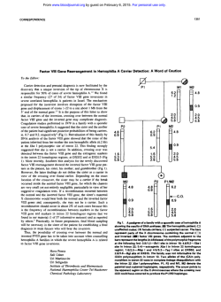
The pedigrees of our two Italian families with juvenile GH are shown
in Fig 1. The clinical features of family 1 were reported in 1983,4
whereas family 2 has never been described. Of the four affected individuals,
three presented with hypogonadotropic hypogonadism at 14 to 21 years of
age. The affected male of family 2 presented with cardiac failure at 20 years
of age and died at 21 years of age of congestive cardiomyopathy.
From www.bloodjournal.org by guest on February 6, 2015. For personal use only.
2980
CORRESPONDENCE
Fig 1. Pedigree of the two
families with juvenile genetic hemochromatosis. Circles denote
female family members and
squares denote male family
members. Solid symbols indicate probands. In family 2, the
parents were consanguineous
(first cousins). Each rectangle indicates the HLA-A locus and the
presence (1) or absence (2) of
the C282Y and H63D mutations.
All individuals but the probands
had normal body iron status.
Some individuals in family 2 were
heterozygous for b-thalassemia
(bNbT), whereas the remaining
ones had normal b-gene (bNbN).
There is no relationship in family 1 between HLA-A antigens and
iron overload. In fact, although the two probands are HLA identical, the
youngest sibling has the same HLA constellation, but a fully normal
body iron status at 30 years of age. Family 2 is not informative with
respect to HLA linkage, but helps to exclude any interaction between
hemochromatosis and b-thalassemia.
All of the family members we examined using the approach of Lynas5
were found to be negative for the C282Y and H63D mutations in the
HFE gene (Fig 1). In a study on 7 Italian patients belonging to 5
unrelated families with juvenile GH, Camaschella et al6 have indepen-
dently excluded the HFE gene as responsible for this condition.
Segregation analysis of 6p markers closely associated with HFE
showed, in fact, that juvenile GH is unlinked to 6p and thus genetically
distinct from HFE.
Both patients of family 1 and the young lady of family 2 underwent
regular phlebotomies. Based on the amount of iron mobilized by
bleedings, we estimated that these subjects had body iron stores ranging
from 220 to 329 mg/kg of body weight at the time of diagnosis at 17 to
21 years of age. Once the three patients became iron deficient at the end
of the intensive phlebotomy program, they underwent regular venesec-
From www.bloodjournal.org by guest on February 6, 2015. For personal use only.
CORRESPONDENCE
2981
tions to maintain serum ferritin levels less than 100 µg/L. At the time of
our writing, they have been observed in this way for 7 to 16 years. Based on
phlebotomy requirements for maintenance of normal iron balance, the rate of
estimated iron accumulation (positive iron balance) ranged from 3.2 to 3.9
mg/d. This was clearly higher than the rate of 0.8 to 1.6 (1.2 6 0.3) mg/d
found in 5 adult males who are homozygous for the C282Y mutation. This
remarkable difference in iron overprocurement (1 to 2 v 3 to 4 mg/d) suggests
completely different pathogenetic mechanisms.
In conclusion, juvenile GH is an autosomal recessive condition
clinically and genetically different from the classical adult disorder.
HLA-typing and HFE-based genotyping are useless, and diagnosis must
rely on conventional evaluation of body iron status. Identification of the
candidate gene through positional cloning could unveil a new crucial
protein of iron metabolism in humans.
ACKNOWLEDGMENT
Supported by grants from IRCCS Policlinico S. Matteo, Pavia, and
MURST, Rome, Italy.
Mario Cazzola
Paola Cerani
Andrea Rovati
Angioletta Iannone
Giovanni Claudiani
Gaetano Bergamaschi
Department of Internal Medicine and Medical Oncology
Department of Immunohaematology and Transfusion
University of Pavia
IRCCS Policlinico S. Matteo
Pavia, Italy
Atri Hospital
Atri, Italy
REFERENCES
1. Feder JN, Gnirke A, Thomas W, Tsuchihashi Z, Ruddy DA,
Basava A, Dormishian F, Domingo R Jr, Ellis MC, Fullan A,
Hinton LM, Jones NL, Kimmel BE, Kronmal GS, Lauer P,
Lee VK, Loeb DB, Mapa FA, McClelland E, Meyer NC,
Mintier GA, Moeller N, Moore T, Morikang E, Prass CE, Quintana L,
Starnes SM, Schatzman RC, Brunke KJ, Drayna DT, Risch NJ,
Bacon BR, Wolff RK: A novel MHC class I-like gene is mutated
in patients with hereditary haemochromatosis. Nat Genet 13:399,
1996
2. Jouanolle AM, Fergelot P, Gandon G, Yaouanq J, Le Gall JY,
David V: A candidate gene for hemochromatosis: Frequency of the
C282Y and H63D mutations. Hum Genet 100:544, 1997
3. Piperno A, Sampietro M, Pietrangelo A, Arosio C,
Lupica L, Montosi G, Vergani A, Fraquelli M, Girelli D, Pasquero P,
Roetto A, Gasparini P, Fargion S, Conte D, Camaschella C:
Heterogeneity of hemochromatosis in Italy. Gastroenterology 114:996,
1998
4. Cazzola M, Ascari E, Barosi G, Claudiani G, Dacco` M,
Kaltwasser JP, Panaiotopoulos N, Schalk KP, Werner E: Juvenile idiopathic hemochromatosis: A life-threatening disorder
presenting as hypogonadotropic hypogonadism. Hum Genet 65:149,
1983
5. Lynas C: A cheaper and more rapid polymerase chain reactionrestriction fragment length polymorphism method for the detection of
the HLA-H gene mutations occurring in hereditary hemochromatosis.
Blood 90:4235, 1997
6. Camaschella C, Roetto A, Cicilano M, Pasquero P, Bosio S,
Gubetta L, Di Vito F, Girelli D, Totaro A, Carella M, Grifa A, Gasparini
P: Juvenile and adult hemochromatosis are distinct genetic disorders.
Eur J Hum Genet 5:371, 1997
Hypermethylation of p15INK4B Gene in a Patient With Acute Myelogenous Leukemia
Evolved From Paroxysmal Nocturnal Hemoglobinuria
To the Editor:
Paroxysmal nocturnal hemoglobinuria (PNH) is an acquired disease
resulting from somatic mutations in the PIG-A gene involving primitive
hematopoietic stem cells. The PNH clones may have growth or survival
advantages relative to normal clones that may promote their expansion, resulting in the development of overt PNH. However, little is
known about how PNH clones gain growth advantage. Recent studies
demonstrated preferential hematopoiesis by PNH clones in vivo.1,2
However, proliferation may be affected similarly in PIG-A–deficient
clones and in normal clones,3 suggesting that PIG-A abnormalities
alone may not be sufficient to confer a growth advantage on PNH
clones.
A proportion of PNH patients terminate in severe pancytopenia with
dysplasia, ie, myelodysplastic syndrome (MDS), and rarely progress to
acute leukemia. We previously reported in BLOOD that specific
p15INK4B gene inactivation by promoter hypermethylation may be
associated with the development of MDS,4 because it may confer a
growth advantage on cells. One overt leukemia patient analyzed in this
study in whom PNH evolved through MDS (PNH/MDS-OL) showed
intense hypermethylation of the p15INK4B gene. Surface marker analysis
of his leukemic blasts showed low levels of expression of
CD59, suggesting that leukemic blasts were derived from the PNH
clone. So, to clarify at what point the p15INK4B gene was densely
methylated and inactivated in this PNH/MDS-OL patient and
whether this p15INK4B gene methylation is related to the expansion of
PNH clones, we analyzed this patient and an additional 17 PNH
patients.
We obtained clinical samples after receiving informed consent from a
total of 18 patients (4 men and 14 women) who were positive for sugar
water test and/or acidified serum (Ham) test and were diagnosed as
PNH based on clinical manifestations. They included 12 female patients
(unique patient no. [UPN] 1 through 12) analyzed in our previous study
in which we demonstrated the existence of monoclonal populations
with PNH phenotype by clonality analysis using X-chromosome
inactivation and assessment of expression of glycophosphatidylinositolanchored proteins by flow cytometry.5 We extracted DNA from
polymorphonuclear cells (PMNCs) and mononuclear cells (MNCs) (or
T lymphocytes), as described previously,5 and analyzed the methylation
status of the p15INK4B gene by the simple and sensitive methylationspecific polymerase chain reaction (MSP) method.6 Because MSP can
detect 1023 methylated alleles among unmethylated alleles,6 changes of
the methylation status in PNH clones could be detected even if they
are present at a very low incidence. After bisulfite modification, we
amplified their DNAs with each primer set specific for unmethylated and methylated DNA in a thermal cycler (TAKARA, Kyoto,
Japan).
From www.bloodjournal.org by guest on February 6, 2015. For personal use only.
1998 92: 2979-2981
Juvenile Genetic Hemochromatosis Is Clinically and Genetically Distinct
From the Classical HLA-Related Disorder
Mario Cazzola, Paola Cerani, Andrea Rovati, Angioletta Iannone, Giovanni Claudiani and Gaetano
Bergamaschi
Updated information and services can be found at:
http://www.bloodjournal.org/content/92/8/2979.full.html
Articles on similar topics can be found in the following Blood collections
Information about reproducing this article in parts or in its entirety may be found online at:
http://www.bloodjournal.org/site/misc/rights.xhtml#repub_requests
Information about ordering reprints may be found online at:
http://www.bloodjournal.org/site/misc/rights.xhtml#reprints
Information about subscriptions and ASH membership may be found online at:
http://www.bloodjournal.org/site/subscriptions/index.xhtml
Blood (print ISSN 0006-4971, online ISSN 1528-0020), is published weekly by the American Society of
Hematology, 2021 L St, NW, Suite 900, Washington DC 20036.
Copyright 2011 by The American Society of Hematology; all rights reserved.
© Copyright 2026

