
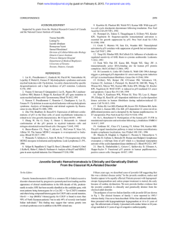
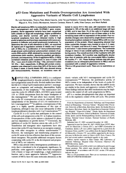
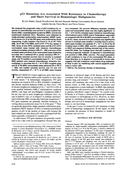
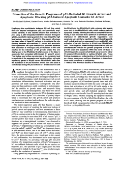
p53 Gene Mutation in B-Cell Chronic Lymphocytic Leukemia
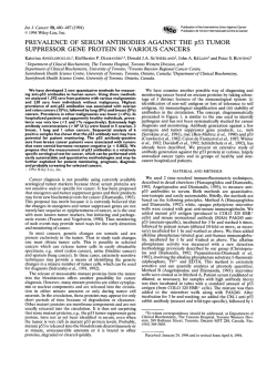
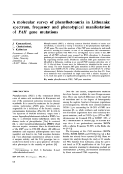
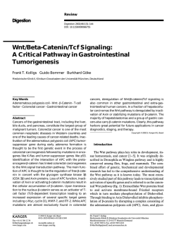
From www.bloodjournal.org by guest on February 6, 2015. For personal use only.
p53 Gene Mutation in B-Cell Chronic Lymphocytic Leukemia Is Associated
With Drug Resistance and Is Independent of M D R l / M D R 3 Gene Expression
By Soumaya El Rouby, Anju Thomas, Dan Costin, Carl R . Rosenberg, Milan Potmesil,
Robert Silber, and Elizabeth W. Newcomb
W e studied 5 3 patients with B-cell chronic lymphocytic
leukemia (B-CLL) and found mutations of the p53 gene in
15%. Patients with p53 gene mutations were found to
have an aggressive form of B-CLL disease characterized by
advanced Rai stage, rapid lymphocytedoublingtime (LDT),
and resistance to chemotherapy. While 27 of 29 treated
patients (93%) without p53 mutations achieved a partial
remission, only one of seven treated patients (14%)with
p53 mutations achieved a partial remission (P = .00009).
Adjusting for prognostic factors (age, sex, race, and Rai
stage), patients with p53 gene mutations had a 13-fold
greater risk of death than patients without p53 mutations
(P = .013). In addition to examining the clinical relevance
of p53 gene mutations in B-CLL, w e investigated the possible role of p53 gene regulation in the expression of the
multidrug resistance genes M D R I and MDR3. W e quantitated MDRI and MDR3 mRNA expression by reverse transcription-polymerase chain reaction (RT-PCR). Expression
of both the MDRI and MDR3 genes was independent of
p53 gene mutation or prior drug treatment, and did not
predict for clinical response. Our findings indicate that p53
gene mutations in B-CLL are associated with a poor clinical
outcome and may be a prognostic indicator for drug resistance.
0 1993 by The American Society of Hematology.
B
mia (ANLL),l6-l9acute lymphoblastic leukemia (ALL),”,”
chronic myelogenous leukemia (CML),22 multiple mye l ~ m a , ’and
~ non-Hodgkin’s lymphoma.24While the product of the MDR3 gene has been c h a r a c t e r i ~ e dthe
, ~ ~function of its product in drug resistance is less well understood
than that of the MDR 1 gene. The expression of both these
genes has been analyzed in B-CLL patient^.^^.^' Although
no link between MDRI expression and aggressive disease
has been observed, a possible association between MDR3
overexpression and advanced Rai stages has been suggested.”
In the present study, we investigated the role of p53 gene
mutations in the regulation of MDR gene expression and its
possible clinical relevance. We correlate the occurrence of
p53 gene mutations with Rai stages, responsiveness to chemotherapy, and overall prognosis. In addition, we show that
MDR gene overexpression in these patients is independent
of p53 gene mutation and clinical response to chemotherapy.
-CELL CHRONIC lymphocytic leukemia (B-CLL) is a
disease characterized by the clonal expansion of CD5’
B cells. While chemotherapy can effectively control lymphocyte proliferation in most patients, there are few complete remissions. In addition, a small percentage of patients
do not respond to any therapy. Prognostic factors that indicate shorter survival duration have been described. These
include advanced clinical stage,’-3a diffuse pattern of bone
marrow in~olvement,~
a lymphocyte doubling time (LDT)
of less than 12 month^,^ and the presence of chromosomal
abn~rmalities.~,~
Attention has recently focused on tumor-suppressor
genes as possible prognostic indicators in human cancer.’-’’
Structural alterations and point mutations of the p53 tumor-suppressor gene have been demonstrated in 10% to
15% of B-CLL patients.”-’3 Wild-type p53 protein has been
shown to repress the activity of the human MDRl gene
promoter in vitro, while the mutant p53 protein hasa stimulatory effect on this a ~ t i v i t y .Increased
’~
expression of the
MDR 1 gene is considered a common mechanism for drug
resistan~e.’~
Overexpression of the MDR 1 gene product,
P- 170, has been associated with drug resistance in hematologic malignancies, including acute nonlymphocytic leuke-
From the Departments of Pathology, Medicine, Radiology, and
Environmental Medicine, New York University School of Medicine
and Kaplan Comprehensive Cancer Center, New York, NY.
Submitted April 26, 1993; accepted August 12, 1993.
Supported in part by National Cancer Institute Grants No. CA535 72, CA-50529, and CA-54484, and by the Marcia Slater Society
for Research in Leukemia and the Harry and Gussie Wallerstein
Foundation. D.C. is a Fellow of the American Cancer Society.
Address reprint requests to Elizabeth W. Newcomb, PhD, Department of Pathology MSB531, New York University Medical Center
and Kaplan Comprehensive Cancer Center, 550 First Ave, New
York, NY 10016.
The publication costs of this article were defrayed in part by page
charge payment. This article must therefore be hereby marked
“advertisement” in accordance with 18 U.S.C.section 1734 solely to
indicate this fact.
0 I993 by The American Society of Hematology.
0006-4971/93/82l0-0028$3.00/0
3452
MATERIALS AND METHODS
Patient population. Fifty-three patients with B-CLL, seen at
New York University Medical Center, were studied. The diagnosis
of B-CLL required the demonstration of at least 10 X 109/Lmonoclonal B lymphocytes positive for CD5, CD19, and CD20. The
disease was staged according to Rai et al.’ Some of the clinical
characteristics of the patient population, including LDT, are listed
in Table 1. Indications for treatment included symptomatic adenopathy, hepatomegaly or splenomegaly, or disease progression to Rai
stage 111 or IV. Most patients were treated either with chlorambucil
or with chlorambucil and prednisone. Nine patients received cyclophosphamide, five patients fludarabine, two patients 2‘-deoxycoformycin, and one patient an Adriamycin-containing regimen (Adria
Labs, Columbus, OH). Several patients were treated with multiple
drug regimens. A complete response to treatment was defined by a
lymphocyte count of less than 4 X 10⁹/L, a granulocyte count
greater than 1.5 X 10⁹/L, a platelet count greater than 100 X IO9,
and a normal bone marrow examination.” A partial response was
defined by a decrease of at least 50%in the diameter of the enlarged
lymph nodes and a decrease of the lymphocyte count by 75%.30,31
Enrichment of B lymphocytes and DNA extraction. After obtaining informed consent, samples of heparinized blood were obtained from B-CLL patients or normal healthy volunteers. MononuBlood, Vol82, No 11 (December 1). 1993: pp 3452-3459
From www.bloodjournal.org by guest on February 6, 2015. For personal use only.
p53 MUTATION AND DRUG RESISTANCE IN B-CLL
3453
Table 1. Summarv of Clinical Parameters of
Age at
Diagnosis
(vr)
LDT
(mo)
Patients with p53 mutations
1
2
3
4
5
6
7
8
75
49
76
55
76
75
67
67
36
3
18
6
7
2
<1
12
Patients without p53 mutations
9
10
11
12
13
14
15
16
17
18
19
20
21
22
23
24
25
26
27
28
29
30
31
32
33
34
35
36
37
38
39
40
41
42
43
44
45
46
47
48
49
50
51
52
53
36
64
59
68
65
69
56
80
56
65
79
58
57
53
60
60
55
36
65
60
55
70
62
47
65
60
60
58
41
49
61
74
73
65
74
70
37
54
76
42
76
68
77
48
54
6
8
20
Patient No
20
34
NA
18
20
30
14
12
NA
NA
24
24
NA
NA
36
36
NA
37
6
NA
15
12
9
NA
24
NA
18
>38
>42
>92
>18
41
>38
>80
>26
>23
48
>28
>85
>79
NA
>92
53 B-CLL Patients
Rai
Stage
Therapy
Response
II
II
111
I1
111
II
IV
II
NT
247
112
103
124
28
NT
109
CP, c
FLU, CHOP, CAE, SPX
CP, C E
CP, FLU
CP
CP
CP, CVP, FLU, M, E, dCF
None
No
No
No
No
No
Yes
No
II
34
8
NT
19
NT
NT
NT
NT
15
NT
66
NT
NT
13
NT
NT
NT
41
11
NT
NT
NT
NT
23
NT
38
NT
NT
NT
23
36
24
NT
66
33
110
46
13
23
34
123
NT
NT
NT
36
CLB, C
CP, CLB
CP
CP
CP, SPX
CP, CLB, FLU
CP
CLB
CLB
CLB, CP
CLB
CLB, C
CLB
CP
CLB
CLB
CP, c P, c
CLB
CLB
CP
CP
FLU
CP
CLB, CP
CP, CLB, C, C E, C
CLB
CL8 dCF P
CLB
c, CVP
None
None
None
None
None
None
None
None
None
None
None
None
None
None
None
None
Yes
Yes
Yes
Yes
Yes
Yes
Yes
Yes
Yes
Yes
Yes
Yes
No
Yes
Yes
Yes
Yes
Yes
Yes
Yes
Yes
Yes
Yes
Yes
Yes
Yes
Yes
Yes
No
111
I
I
II
Ill
I1
II
I
I
I
II
I
I
II
Ill
II
I
II
II
I
I
I
I
IV
II
I
1
I
II
II
0
I
I
0
I
0
I
0
I
II
0
I
0
I
I
+
+
+
+
+
+P
-
Abbreviations: NA. not assessable; IC50(CLB),concentration of CLB in pmol/L that inhibits viability of 50% of the lymphocytes in vitro; C, cytoxan
(Bristol-Myers. Evansville. IN); CAE, Cytoxan Adriamycin etoposide-16; CLB, chlorambucil; CP, chlorambucil + prednisone; CVP, cytoxan
vincristine + prednisone; CHOP, Cytoxan + Adriamycin vincristine + prednisone; FLU, fludarabine; dCF, 2’-deoxycoformycin; M, mitoxantrone; E,
etoposide; C P, Cytoxan + prednisone; P, prednisone; SPX, splenectomy; NT, not tested before initial chemotherapy.
+
+
+
+
+
From www.bloodjournal.org by guest on February 6, 2015. For personal use only.
EL ROUBY ET AL
3454
clear cells were isolated by centrifugation on a Ficoll-Hypaque gradient. B lymphocytes were further enriched by rosetting T cells with
sheep red blood cells (SRBC rosetting) (Crane Labs, NY) as described.” The final preparation from B-CLL patients contained
greater than 90% B lymphocytes. B lymphocytes or monocytes
from normal healthy donors were further purified by sorting using
fluorescein isothiocyanate (F1TC)-labeled monoclonal antibody to
CD19 (clone 89B, IgGlK; Coulter, FL) and phycoerythrin-labeled
mAb to CD14 (clone Cris-6, IgGI; Olympus Corp, NY), respectively. Cells were incubated for 30 minutes with the appropriate
dilution of the mAbs in ice-cold phosphate-buffered saline (PBS)
containing 1% bovine serum and 1% human AB serum (GIBCO,
BRL, NY). Cells were washed, resuspended in PBS, and then applied to an Ortho Cytofluorograph equipped with an argon ion laser
(Ortho Instruments, MA). Sorted cells were collected into ice cold
RPMI with 20% fetal calf serum (FCS). High-molecular weight
DNA was isolated by digestion with proteinase K (Boehringer, Indianapolis, IN), extraction with phenol/chloroform, and ethanol
pre~ipitation.~’
In vitro chemosensitivity assay. B lymphocytes obtained from
patients before initial therapy were cultured on microtiter plates
(2.5 X lo6cells/well) in 180 pL of RPMI 1640 plus 10%FCS in the
presence ofchlorambucil or without the drug for 72 hours followed
by addition of 3-(43 dimethylthiazol-2-yl)-2,5-diphenyltetrazolium bromide (MTT) as previously de~cribed.’~.’~
The IC,, is defined as that concentration ofthe drug (pmol/L) that inhibits viability by 50%.”
Oligonucleotide primers. Primers used to amplify exons 4
through 9 of the p53 gene have been previously de~cribed.’~
Amplimers used for amplification of MDRl and 0-actin-specific sequences have also been rep~rted.”.’~MDR3-specific sequences
were amplified using the following primers: sense strand, GCTTCAGGAATTGTTGA (residues 2602 to 262 I), and antisense strand,
TCGAAAACAACCGGCATAGG (residues 285 1 to 2870). These
primers generate a 268-kb PCR product. All oligonucleotide
primers were either synthesized with an Applied Biosystems Synthesizer (model 380A) or purchased from OPERON (Technologies
Inc, CA).
Detection ofp53 mutations. Mutations of p53 in B-CLL patients were detected by single-strand conformation polymorphism
(SSCP) analysis of PCR products. Primer pairs for exons 4 through
9 were used to amplify p53 coding sequence, using 100 ng of genomic DNA isolated from the lymphocytes of each patient. The PCR
mixture, the amplification conditions, and direct sequencing of
PCR products performed with the sequencing kit (United States
Biochemical) were described p r e v i o ~ s l yWhen
. ~ ~ screening for mutations in the SSCP assay and DNA sequencing, genomic DNA
from samples containing known wild-type and mutant p53 alleles
were amplified by PCR and run in parallel as controls.
Detection of gene expression by reverse transcription and
PCR. Total cellular RNA was prepared by guanidine isothiocyanate e~traction.’~
Poly A mRNA was purified by affinity adsorbtion to oligo-(dT)-cellulose using the Fast Track mRNA isolation
kit (In Vitrogen, CA). Complementary DNA (cDNA) was prepared
by reverse transcription (RT) of mRNA (0.5 pg) from purified B
lymphocytes or cell lines using 50 ng of random hexadeoxynucleotide primer and 200 U of MO MULV reverse transcriptase (Superscript RNase H-Reverse transcriptase; BRL) under conditions recommended by the supplier. cDNA aliquots equivalent to 0.05 pg
@-actin) and 0.25 pg (MDRI and MDR3) RNA were used for enzymatic amplification by PCR using 2 U of Amplitaq polymerase
(Perkin-Elmer/Cetus, Nonvalk, CT). PCR was performed in 50 pL
containing I pmol/L of specific primers. Twenty-five cycles were
+
performed: each cycle included I minute ofdenaturation at 94”C, I
minute of primer annealing (55”C, MDRI; 57”C, MDR3; 60°C,
0-actin) and 2 minutes of extension at 72°C. PCR products (20
pL/reaction) were analyzed by electrophoresis on 1.2% agarose gel
and visualized after ethidium bromide staining. Gels were processed for Southern blotting. The membranes containing the DNA
fragments were hybridized with specific oligonucleotides end-labeled with [y3’P]-ATP,’’ washed, and exposed to x-ray film at
-70°C for 2 hours: MDR I , ACTAGAAGGTGCTGGGAAG;
MDR3, GGACAGTTGTGTCTTTGACC; 0-actin, GGAGTCCTGTGGCATCCAC. The hybridization signals were quantified
by densitometric scanning of the autoradiographs (LKB Ultrascan
XL Laser Densitometer). The human leukemia cell line CCRFCEM and its vinblastine-resistant derivative CEM/VLB 100 served
as negative and positive controls, respectively, for MDRl gene expression.40The human liver cell line HepC2 served as a positive
control for MDR3 gene expression4’ and the CEM/VBL100 cell
line was used as the negative control.
Statistical analysis. Comparison between patients by p53 status was assessed using the Mann-Whitney test (continuous variables) and Fisher’s exact test (nominal variable^).^^ Survival distributions for the two groups of patients were compared using the
Kaplan-Meier method. The difference between the two survival
curves was assessed using Gehan’s generalized Wilcoxon test. To
control for the effects of confounding variables, ie, prognostic factors that differed by p53 status, the data were subjected to the Cox
proportional hazards model. Using this procedure, a time-weighted
relative risk for death was estimated and was adjusted for pertinent
confound er^.^' P values in all cases were two-tailed. All statistical
calculations were performed using SAS procedure^.^^
RESULTS
Identgfication ofp53 mutations. Mutations of p53 were
detected in B lymphocytes from eight of 53 patients ( 1 5%)
by SSCP analysis (Fig I). At the time of p53 gene analysis,
six of these eight patients had been untreated. Mutations
were confirmed by DNA sequencing (Fig 2) and the data are
listed in Table 2. Six of the mutations were missense mutations, resulting in an amino acid substitution, and two ofthe
mutations were nonsense mutations, resulting in premature
termination of protein translation. The mutations were located in the highly conserved domains of the p53 gene.
Three mutations occurred in exon 5 (37%), two in exon 6
(25%), and one each in exons 4, 7, and 8.
Patient characteristics and clinical course. The 45 patients without p53 mutations had a median age at diagnosis
of 62 years (range, 36 to 80) and the eight patients with
mutations had a median age of 67 (range, 49 to 81) ( P =
NS). There was no difference in gender between the two
groups. As shown in Table I , no patient with a p53 gene
mutation was in Rai stage 0 or I, compared with 57% of
patients without p53 mutations ( P = .002). A LDT of less
than 8 months was observed in five of eight patients (63%)
with p53 mutations, compared with only 9% of the assessable patients without mutations ( P = .003).
Of the 45 patients without p53 mutations, 29 (64%) required therapy, and 27 of these patients (93%) had a partial
remission. In contrast, 7 of 8 patients (87%)with p53 mutations required therapy, but only one patient (14%) achieved
a partial remission (P= .00009). A shorter survival duration
From www.bloodjournal.org by guest on February 6, 2015. For personal use only.
p53 MUTATION AND DRUG RESISTANCE IN B-CLL
3455
WILD-TYPE
MUTATED
1
2
3
4
5
6
A C G T
-
m-
G
cC-l;i
-
EXON 4
A C G T
G
C
C
-M
~-
c-
EXON 5: Codon 152
1
(
1
2
3
4
5
6
7
8
1
EXON 6:Codon 209
Fig 2. Representative DNA sequence analyses show p53 gene
mutations detected by SSCP in 6-CLL patients. Mutation in codon
152 results from insertion of a C (GCC to GCCC); mutation in codon
209 (AGA to ACA) results from a single base change.
EXON 5
I
1
2
3
4
5
6
7
8
EXON 8
was also observed in patients with p53 mutations (Fig 3). Six
of eight patients (75%) with p53 mutations have died, compared with eight of45 patients ( 18%) without p53 mutations
(relative risk, 4.66: P = .I5 by Gehan's generalized Wil-
apy, the B lymphocytes from 28 patients (six with p53 mutations) were tested for drug resistance with the MTT chemosensitivity assay (Table I). Because IC,, values are
significantly increased following chlorambucil treatment:'
Patient
No.'
Fig 1. Detection of p53 gene mutations in 6-CLL patients by
PCR-SSCP analysis. DNA from each patient's lymphocytes was
amplified by PCR in the presence of w3*PdCTP for each of the
corresponding exons 4 through 9 of the p53 gene. The radiolabeled
DNA fragments are separated on a nondenaturinggel. Representative SSCP migration patterns of some samples are shown for exons
4, 5, and 8. DNA samples with a p53 mutation show a shift in
electrophoretic mobility as compared with other DNA samples on
the same gel: exon 4 (lane 6, patient no. 8). exon 5 (lane 7, patient
no. 5). and exon 8 (lane 5, patient no. 1).
8
4
5
6
2
3
7
1
Exon
Codon
Mutation
Amino Acid
4
5
5
5
6
6
7
8
47
168
152
177
209
209
241
286
CCG-ICG
CAC-CSC
Insert of C
Pro to Ser
His to Pro
ccc-CIC
Pro to Leu
Arg to Lys
Arg to Thr
Ser to Phe
Glu to stop
AGA-AAA
AGA-ACA
TCC-EC
GAA-IAA
Six of eight patients did not receive chemotherapy before DNA analysis for p53 gene mutations.
From www.bloodjournal.org by guest on February 6, 2015. For personal use only.
EL ROUBY ET AL
3456
loo
L
I
0
I
40
I
80
I
120
I
I
I
160
200
240
Months After Diagnosis
Fig 3. Kaplan-Meier survival curves after diagnosis of 6-CLL
based on the presence or absence of p53 gene mutations.
expression (Fig 4 and Table 3). There was no association
between MDR 1 expression and the following: p53 gene mutations, prior drug treatment, clinical course of the disease,
and Rai stages.
MDR3 gene expression was sevenfold greater in the human liver cell line HepG2 relative to the negative cell line
CEM/VBLIOO. A twofold to threefold increase in MDR3
expression was observed in sorted CDI 9' cells and no expression was observed in CD14' cells from normal healthy
donors (Table 3). Of 16 patients analyzed for MDR3 gene
expression (Fig 4 and Table 3), 12 expressed the MDR3
gene. Eight patients showed intermediate to high levels, four
showed low expression (absorbance value less than sorted
CD 19' cells), and four showed no expression of the MDR3
gene. Intermediate to high expression of the MDR3 gene
was detected in 43% of patients with p53 mutations and
56% of patients without p53 mutations (Table 3). There was
no association between prior drug treatment, Rai stage,
clinical response to treatment, and MDR3 gene expression.
In addition, there was no association between the levels of
expression of MDR3 and MDRl in the same patient.
DISCUSSION
ICs0 data as a predictive indicator are presented only for
those patients who were tested before therapy. The mean
ICs0of chlorambucil was 3.4-fold greater when p5 3-positive
lymphocytes were tested compared with p53-negative lymphocytes (P= .006).
MDRl and MDRS gene expression. RNA samples obtained from B lymphocytes of 16 B-CLL patients were analyzed for coexpression of the MDRI, MDR3, and 0-actin
genes using the RT-PCR method. The results of Southern
blot analysis of gene expression for MDR I , MDR3, and
0-actin are shown in Fig 4. The oligonucleotides used to
probe the Southern blots for either the MDRl or the MDR3
gene were specific and hybridized only with their respective
cDNA products. Seven of the patients had p53 mutations
(lanes a through g). Of the nine patients without p53 mutations (lanes h through p), six had received drug treatment
(lanes h through m), while three other patients were without
drug treatment (lanes n through p). mRNA from human
cell lines specific for MDRl (CEM/VBLlOO) and MDR3
(HepG2) gene expression served as positive controls (lanes
1). mRNA from human cell lines with basal levels of MDR 1
(CCRF-CEM) and MDR3 (CEM/VBL100) expression
served as negative controls (lanes 2). Measurement of P-actin gene expression was used as an internal control for RNA
recovery from each patient. Densitometric quantitation of
MDRl and MDR3 gene expression of B lymphocytes from
B-CLL patients and sorted CD 19' and CD 14' cells from
normal healthy donors are summarized in Table 3.
The drug-resistant cell line CEM/VLB 100 expressed a
12-fold greater level ofthe MDR 1 gene relative to the parental drug-sensitive CCRF-CEM cell line. A twofold increase
in MDRl expression was observed in sorted CD19+ cells
and no expression was observed in sorted CD14+ cells from
normal healthy donors (Table 3). All 16 patients analyzed
for MDRl gene expression showed an intermediate to high
Mutations in the p53 tumor suppressor gene were identified in eight of 53 B-CLL patients. It is less likely that chemotherapy selected out p53 mutant clones of B lymphocytes,
since six of eight patients with p53 mutations were untreated at the time of DNA analysis. In the other two patients, DNA was not available before treatment. Patients
with p53 gene mutations differed from those without mutations in several ways: they were more likely to be in Rai
stage I1 or higher, to have a decreased LDT, to require chemotherapy, to show resistance to chlorambucil in vitro, to
have a poor clinical response to therapy, and to have a
shorter survival duration.
In view ofthe report that p53 mutations may be involved
in regulation ofthe MDRl gene,14 we examined if p53 mutations were associated with modulation of the genes involved in multidrug resistance in B-CLL patients. The B
lymphocytes from all patients expressed high levels of the
MDR I gene. MDR 1 gene overexpression was not dependent on the presence of p53 mutations, suggesting that
MDRI overexpression in B-CLL lymphocytes is not regulated by the p53 gene product. Similarly, it has been recently
suggested that MDR 1 in AML expression is not affected by
the presence of p53 mutations.46
Overexpression of MDR 1 has been associated with resistance to chemotherapy in many m a l i g n a n ~ i e sDrugs
. ~ ~ used
for ANLL therapy, such as the anthracyclines, are susceptible to the action of the P-170 efflux pump. In untreated
ANLL, 67% of patients with high MDRI levels fail to go
into complete remission after one cycle of anthracyclinebased treatment, compared with 33% of ANLL patients
with low MDR 1 expres~ion.'~
In a prospective study of 122
patients with untreated ANLL, 68% of P- 170-positive patients did not achieve complete remission compared with
19% of the P-170-negative patients.'* In our study, the absence of any association between MDRl expression and
From www.bloodjournal.org by guest on February 6, 2015. For personal use only.
.
3457
p53 MUTATION AND DRUG RESISTANCE IN B-CLL
1 2 a b c d e
MDR1
m
-
f g h i j k l m n o p
0
-
Fig 4. Southem blot analysisof gene expression by RT-PCR of RNA from E-CLL patients. The coexpression of MDRl ,MDRB. and &actin
genes was analyzed in 1 6 B-CLL patients (lanes a-g, patients no. 1-7; lanes h-m, patients no. 9-1 4; lanes n-p, patients no. 38-40) whose
clinical parameters are detailed in Table 1. mRNA from human cell lines CEM/VLBlOO and HepG2 sewed as positive controls (lane 1)for the
expression of MDRl and MDR3 genes, respectively. mRNA from human cell lines with basal levels of M D R l (CCRF-CEM) and MDR3
(CEM/VBL100) sewed as negative controls (lane 2) for MDRl and MDR3 gene expression, respectively. PCR-amplified products were
separated on agarose gels, Southern blotted and hybridized to 32P-labeledoligonucleotideprobes specific for each gene product. Films were
exposed for 2 hours at 70°C. The signals on the autoradiographs were scanned by a laser densitometerto quantitate the relative levels of
gene expression for each patient (see Table 3). The expression of @-actinsewed as an internal control for the recovery of RNA from each
patient.
-
clinical response to therapy may be due to the fact that most
of the drugs used to treat B-CLL are not affected by the
MDR 1 gene product, P- 170.
Our results show intermediate to high levels of MDRI
gene expression in B lymphocytes from all 16 B-CLL patients tested. High expression of the MDRl gene in B lymphocytes from 90% to 100%of B-CLL patients has been
shown in two other
In contrast, we and
~ t h e r sfound
~ ~ . low
~ ~expression of MDR I in sorted C D 19'
B lymphocytes from normal healthy donors. Similar results
of low MDRl expression have been reported for enriched
populations of normal B lymphocytes.26All ofthe studies to
date show that MDRl expression is usually low in B lymphocytes obtained from normal donors compared with B
lymphocytes obtained from B-CLL patients. Further studies on MDR I expression in CD5' B lymphocytes from normal donors and B-CLL patients are needed to understand
this difference in the pattern of MDR I gene expression and
its role in the disease.
MDR3 is the second human multidrug resistance gene to
be identified by its homology with the MDRl gene. A physiological role of MDR3 in the phenomenon of multidrug
resistance is not yet established. In this study, we observed a
low expression of MDR3 in normal sorted C D 19' cells and
highly variable MDR3 gene expression in B-CLL patients.
Previous studies have showed high levels of MDR3 expression in untreated patients with prolymphocytic leukemia,"
or in patients in advanced Rai stages 111 and IV?' Our study
showed no association between the level of MDR3 expression and Rai stage, previous drug treatment or the presence
of p53 gene mutations. Additional studies of MDR3 expression in a large group of B-CLL patients will be required to
determine whether there is an association with any of the
clinical parameters.
This report demonstrates an association between p53
gene mutations and an aggressive form of B-CLL characterized by advanced Rai stage, rapid LDT, increased drug resistance as measured by a poor response to chemotherapy, and
shortened survival duration. Mutations of the p53 gene
have been associated with disease progression and decreased
survival in many other human cancers, including colon adenocarcinoma,' breast cancer,' CML,'' multiple m y e l ~ m a , ~ '
transitional cell bladder cancer,52gastric cancer,53and lung
~ a n c e r . No
~ . ~other
~ studies to date have linked the presence
of p53 mutations in B-CLL with decreased response to chemotherapy, in vitro drug resistance, and a shortened survival duration. Our results suggest that p53 mutations may
become a marker for drug resistance in B-CLL. Since the
From www.bloodjournal.org by guest on February 6, 2015. For personal use only.
EL ROUBY ET AL
3458
Table 3. Summary of MDR Gene Expression
MDR Gene
Expression'
Rai Stage
Sample
Controls
CEM/VBL100
CEM
HepG2
CD19+cells
Donor 1
Donor 2
Donor 3
CD14+cells
Donor 1
Donor 2
Patients with p53 mutations
1
2
3
4
5
6
7
Patients without p53 mutations
9
10
11
12
13
14
38
39
40
II
II
111
II
Ill
II
IV
II
Ill
I
I
II
Ill
II
0
I
Clinical
Response
MDRl
MDR3
2.4
0.2
ND
0.2
0.4
0.5
0.1
0.5
0.1
0.1
0.0
0.0
No
No
No
No
No
Yes
No
1.4
1.4
2.0
1.7
1.9
1.8
2.0
0.4
0.0
0.3
0.1
1.4
1.o
1.o
Yes
Yes
Yes
Yes
Yes
Yes
NT
NT
NT
1.7
1.2
1.3
1.6
0.0
0.1
0.5
1.5
1.5
1.3
0.6
2.1
0.8
2.0
2.0
2.0
2.2
2.4
0.5
1.5
0.6
0.0
Abbreviations: NT, not treated; ND, not determined.
The values given are the difference between the minimum and maximum absorbance of each sample.
overall frequency of p53 mutations is relatively low in BCLL, further studies in a larger population of patients will
be needed to establish the reliability of a p53 mutation as a
prognostic indicator.
ACKNOWLEDGMENT
The authors thank Laura Morse and John Drygas for their assistance in separation of B lymphocytes used in this study, Henry
Cohen for graphical and statistical assistance, and Drs Joel Buxbaum and Riccardo Dalla-Favera for their critical reading of the
manuscript. The human cell lines CCRF-CEM and CEM/VBL 100
were generously provided by Dr William T. Beck. We are grateful to
Dr John Hint of the Cell Sorting Unit of the Kaplan Comprehensive Cancer Center for help with cell sorting.
REFERENCES
1. Rai KR, Sawitsky A, Cronkite EP, Chanana AD, Levy RN,
Pasternack BS: Clinical staging of chronic lymphocytic leukemia.
Blood 46919, 1975
2. Binet JL, Lepomer M, Dighiero G, Charron D, Athus D,
Vaughier G, Beral HM, Nataki JC, Araphael M, Nizet B, Follezou
JY: A clinical staging system for chronic lymphocytic leukemia:
Prognostic significance. Cancer 402355, 1977
3. Binet JL, Catovsky D, Chandra P, Dighiero G, Montserrat E,
Rai KR, Sawitsky A: Chronic lymphocytic leukaemia: Proposals
for a revised prognostic staging system. Br J Haematol48:365, 198 1
4. Rozman C, Montserrat E, Rodriguez-Fernandez JM, Ayats
R, Vallespi T, Parody R, Rios A, Prados D, Morey M, Gomis F,
Alcala A, Gutierrez M, Maldonado J, Gonzalez C, Giralt M, Hernandez-Nieto L, Cabrera A, Fernandez-Ranada JM: Bone marrow
histologic pattern-The best single prognostic parameter in chronic
lymphocytic leukemia: A multivariate survival analysis of 329
cases. Blood 64:642, 1984
5. Lee JS, Dixon DO, Kantarjian HM, Keating MJ, Talpaz M:
Prognosis of chronic lymphocytic leukemia: A multivariate regression analysis of 325 untreated patients. Blood 69:929, 1987
6. Han T, Henderson ES, Emrich U, Sandberg AA: Prognostic
significance of karyotypic abnormalities in B cell chronic lymphocytic leukemia: An update. Semin Hematol 24:257, 1987
7. Scott N, Sagar P, Stewart J, Blair GE, Dixon MF, Quirke P
p53 in colorectal cancer: Clinicopathological correlation and prognostic significance. Br J Cancer 63:317, 1991
8. Allred DC, Clark GM, Elledge R, Fuqua SAW, Brown RW,
Chamness GC, Osborne CK, McGuire WL: Association ofp53 protein expression with tumor cell proliferation rate and clinical outcome in node-negative breast cancer. J Natl Cancer Inst 85:200,
1993
9. Quinlan DC, Davidson AG, Summers CL, Warden HE, Doshi HM: Accumulation of p53 protein correlates with a poor prognosis in human lung cancer. Cancer Res 52:4828, 1992
10. Nakai H, Misawa S, Toguchida J, Yandell DW, Ishizaki K
Frequent p53 gene mutations in blast crisis of chronic myelogenous
leukemia, especially in myeloid crisis harboring loss of a chromosome 17p. Cancer Res 52:6588, 1992
1 I. Gaidano G, Ballerini P, Gong JZ, Inghirami G, Neri A, Newcomb EW, Magrath IT, Knowles DM, Dalla-Favera R: p53 mutations in human lymphoid malignancies: Association with Burkitt
lymphoma and chronic lymphocytic leukemia. Proc Natl Acad Sci
USA 885413, 1991
12. El Rouby S, Bayona W, Pisharody SM, Newcomb E W p53
mutations in B-cell chronic lymphocytic leukemia. Cum Top Immunol Microbiol 182:3 13, 1992
13. Fenaux P, Preudhomme C, Lai' JL, Quiquandon I, Jonveaux
P, Vanrumbeke M, Sartiaux C, Morel P, Loucheux-Lefebvre MH,
Bauters F, Berger R, Kerckaert J P Mutations of the p53 gene in
B-cell chronic lymphocytic leukemia: A report on 39 cases with
cytogenetic analysis. Leukemia 6:246, 1992
14. Chin K-V, Ueda K, Pastan I, Gottesman MM: Modulation
of activity of the promoter of the human MDRl gene by ras and
p53. Science 255:459, 1992
15. Chen C, Chin JE, Ueda K, Clark DP, Pastan I, Gottesman
MM, Roninson IB: Internal duplication and homology with bacterial transport proteins in the mdrl (p-glycoprotein) gene from multidrug-resistant human cells. Cell 47:38 l , 1986
16. Zhou D-C, Marie J-P, Suberville A-M, Zittoun R: Relevance
of mdrl gene expression in acute myeloid leukemia and comparison of different diagnostic methods. Leukemia 6379, 1992
17. Sat0 H, Preisler H, Day R, Raza A, Larson R, Browman G,
Goldberg J, Vogler R, Grunwald H, Gottlieb A, Bennett J, Gottesman M, Pastan I: MDRl transcript levels as an indication of resistant disease in acute myelogenous leukaemia. Br J Haematol
75:340, 1990
18. Campos L, Guyotat D, Archimbaud E, Calmard-Ono1 P,
Tsuruo T, Troncy J, Treille D, Fiere D: Clinical significance of
multidrug resistance p-glycoprotein expression in acute nonlymphoblastic leukemia cells at diagnosis. Blood 79:473, 1992
From www.bloodjournal.org by guest on February 6, 2015. For personal use only.
p53 MUTATION AND DRUG RESISTANCE IN 6-CLL
19. Michieli M, Damiani D, Geromin A, Michelutti A, Fanin R,
Raspadon D, Russo D, Visani G, Dinota A, Pileri S, Tsuruo T,
Grandi M, Baccarani M, Tura S: Overexpression of multidrug resistance-associated p 170-glycoprotein in acute non-lymphocytic leukemia. Eur J Haematol48:87, 1992
20. Marie J-P, Zittoun R, Sikic BI: Multidrug resistance (mdrl)
gene expression in adult acute leukemias: Correlations with treatment outcome and in vitro drug sensitivity. Blood 78586, 1991
21. Rothenberg ML, Mickley LA, Cole DE, Balis FM, Tsuruo T,
Poplack DG,Fojo AT: Expression of the mdr- 1/P- 170 gene in patients with acute lymphoblastic leukemia. Blood 741388, 1989
22. Kuwazuru Y, Yoshimura A, Hanada S, Ichikawa M, Saito
T, Uozumi K, Utsunomiya A, Arima T, Akiyama S-I: Expression
of the multidrug transporter, pglycoprotein, in chronic myelogenous leukaemia cells in blast crisis. Br J Haematol 74:24, 1990
23. Epstein J, Xiao H, Oba BK: P-glycoprotein in plasma-cell
myeloma is associated with resistance to VAD. Blood 74:9 13, 1989
24. Dalton WS, Grogan TM, Meltzer PS, Scheper RJ, Dune
BGM, Taylor CW, Miller TP, Salmon SE: Drug-resistance in multiple myeloma and non-Hodgkin’s lymphoma: Detection of pglycoprotein and potential circumvention by addition of verapamil to
chemotherapy. J Clinical Oncol 7:4 15, 1989
25. Van der Bliek AM, Kooiman PM, Schneider C, Borst P
Sequence of mdr3 cDNA encoding a human p-glycoprotein. Gene
71:401, 1988
26. Holmes JA, Jacobs A, Carter G, Whittaker JA, Bentley DP,
Padua RA: Is the mdrl gene relevant in chronic lymphocytic leukemia? Leukemia 4:216, 1990
27. Henveijer H, Sonneveld P, Baas F, Nooter K Expression of
mdrl and mdr3 multidrug-resistance genes in human acute and
chronic leukemias and association with stimulation of drug accumulation by cyclosporine. J Natl Cancer Inst 82:1133, 1990
28. Sonneveld P, Nooter K, Burghouts JThM, Herweijer H,
Adriaansen HJ, van Dongen JJM: High expression of the mdr3
multidrug-resistance gene in advanced stage chronic lymphocytic
leukemia. Blood 79: 1496, 1992
29. International workshop on chronic lymphocytic leukemia:
Chronic lymphocytic leukemia: Recommendations for diagnosis,
staging, and response criteria. Ann Intern Med 110:236, 1989
30. French cooperative group on chronic lymphocytic leukemia:
Long-term results ofthe CHOP regimen in stage C chronic lymphocytic leukemia. Br J Haematol 73:334, 1989
3 1. French Cooperative Group on Chronic Lymphocytic Leukemia: A randomized clinical trail of chlorambucil versus CHOP in
stage B chronic lymphocytic leukemia. Blood 75: 1422, 1990
32. Potmesil M, Hsiang Y-H, Liu LF, Bank B, Grossberg H,
Kirschenbaum S, Penzinger A, Kanganis D, Knowles D, Traganos
F, Silber R: Resistance of human leukemic and normal lymphocytes to drug-induced DNA cleavage and low levels of DNA topoisomerase 11. Cancer Res 48:3537, 1988
33. Maniatis TE, Fritsch EF, Sambrook J: Molecular Cloning: A
Laboratory Manual. Cold Spring Harbor, NY, Cold Spring Harbor
Laboratory, 1989
34. Mosman T Rapid colorimetric assay for cellular growth and
survival: Application to proliferation and cytotoxicity assays. J Immunol Methods 6555, 1983
35. Silber R, Costin D, Newcomb EW, Degar B, Mani M, Rosenberg CR, Morse L, Drygas J, Canellakis ZN, Potmesil M: Resistance of chronic lymphocytic leukemia B lymphocytes to chlorambucil or fludarabine and sensitivity to camptothecin analogues. (in
preparation)
36. Frankel RH, Bayona W, Koslow M, Newcomb E W p53
mutations in human malignant gliomas: Comparison of loss of
heterozygosity with mutation frequency. Cancer Res 52: 1427, 1992
3459
37. Noonan KE, Beck C, Holzmayer TA, Chin JE, Wunder JS,
Andrulis IL, Gazdar AF, Willman CL, Griffith B, Von Hoff DD,
Roninson IB: Quantitative analysis of MDR 1 (multidrug resistance) gene expression in human tumors by polymerase chain reaction. Proc Natl Acad Sci USA 87:7160, 1990
38. Kasaian MT, Ikematsu H, Casali P Identification and analysis of a novel human surface CD5-B lymphocyte subset producing
natural antibodies. J Immunol 148:2690, 1992
39. Chomczynski P, Sacchi N: Single-step method of RNA isolation by acid guanidinium thiocyanate-phenol-chloroform extraction. Anal Biochem 162:156, 1987
40. Beck WT, Mueller TJ, Tanzer L R Altered surface membrane glycoproteins in vinca alkaloid resistant human leukemic
lymphoblasts. Cancer Res 39:2070, 1979
41. Van der Bliek AM, Baas F, Ten Houte de Lange T, Kooiman
PM, Van der Velde-Koerts T, Borst P The human mdr3 gene encodes a novel p-glycoprotein homologue and gives rise to alternatively spliced mRNAs in liver. EMBO J 6:3325, 1987
42. Zar JH: Biostatistical Analysis. Englewood Cliffs, NJ, Prentice-Hall, 1984
43. Lee E T Statistical Methods for Survival Data Analysis. Belmont, CA, Lifetime Learning Publications, 1980
44. SAS Technical Report: Additional SAS/STAT Procedures.
Cary, NC, SAS Institute, 1988
45. Costin D, Potmesil M, Morse L, Mani M, Canellakis ZN,
Silber R: Sensitivity of chronic lymphocytic leukemia B-lymphocytes to chlorambucil, fludarabine, and camptothecin analogs.
Blood 80:175, 1992 (suppl, abstr)
46. Zhao B, Drach D, Hu G, Squires J, Drach J, Deisseroth A,
Andreeff M: Absence of regulation of MDRl by p53 in normal
hematopoiesis and acute myelogenous leukemia. Blood 80:797,
1992 (suppl, abstr)
47. Goldstein U,Galski H, Fojo A, Willingham M, Lai S-L,
Gazdar A, Pirker R, Green A, Crist W, Brodeur GM, Lieber M,
Cossman J, Gottesman MM, Pastan I: Expression of a multidrug
resistance gene in human cancers. J Natl Cancer Inst 81:116, 1989
48. Drach D, Zhao S, Drach J, Mahadevia R, Gattringer C,
Huber H, Andreeff M: Subpopulations of normal peripheral blood
and bone marrow cells express a functional multidrug resistant phenotype. Blood 802729, 1992
49. Chaudhary PM, Mechetner EB, Roninson IB: Expression
and activity of the multidrug resistance p-glycoprotein in human
peripheral blood lymphocytes. Blood 80:2735, 1992
50. Nooter K, Sonneveld P, Janssen A, Oostrum R, Boersma T,
Henveijer H, Valerio D, Hagemeijer A, Baas F Expression of the
mdr3 gene in prolymphocytic leukemia: Association with cyclosporin-A-induced increase in drug accumulation. Int J Cancer
45:626, 1990
5 1. Neri A, Baldini L, Trecca D, Cro L, Polli E, Maiolo T: p53
gene mutations in multiple myeloma are associated with advanced
forms of malignancy. Blood 8 I :128, I993
52. Sarkis AS, Dalbagni G, Cordon-Cardo C, Zhang Z-F, Sheinfeld J, Fair WR, Herr HW, Reuter VE: Nuclear overexpression of
p53 protein in transitional cell bladder carcinoma: A marker for
disease progression. J Natl Cancer Inst 8553, 1993
53. Martin HM, Filiple MI, Moms RW, Lane DP, Silvestre F
p53 expression and prognosis in gastric carcinoma. Int J Cancer
502359, 1992
54. Horio Y, Takahashi T, Kuroishi T, Hibi K, Suyama M,
Niimi T, Shimokata K, Yamakawa K, Nakamura Y, Ueda R, Takahashi T: Prognostic significance of p53 mutations and 3p deletions in primary resected non-small cell lung cancer. Cancer Res
53:1, 1993
From www.bloodjournal.org by guest on February 6, 2015. For personal use only.
1993 82: 3452-3459
p53 gene mutation in B-cell chronic lymphocytic leukemia is
associated with drug resistance and is independent of MDR1/MDR3
gene expression
S el Rouby, A Thomas, D Costin, CR Rosenberg, M Potmesil, R Silber and EW Newcomb
Updated information and services can be found at:
http://www.bloodjournal.org/content/82/11/3452.full.html
Articles on similar topics can be found in the following Blood collections
Information about reproducing this article in parts or in its entirety may be found online at:
http://www.bloodjournal.org/site/misc/rights.xhtml#repub_requests
Information about ordering reprints may be found online at:
http://www.bloodjournal.org/site/misc/rights.xhtml#reprints
Information about subscriptions and ASH membership may be found online at:
http://www.bloodjournal.org/site/subscriptions/index.xhtml
Blood (print ISSN 0006-4971, online ISSN 1528-0020), is published weekly by the American
Society of Hematology, 2021 L St, NW, Suite 900, Washington DC 20036.
Copyright 2011 by The American Society of Hematology; all rights reserved.


© Copyright 2026