
Spring/Summer 2014 - Pediatrics Nationwide
Spring/Summer 2014
Pediatrics
NATIONWIDE
Gene Therapy’s
Road to
Redemption
Advancing the Conversation
on Child Health
16
16
10
4
26
28
In Practice: News from the Field
In Sight: A Brave New World of Data
Second Opinions: Impacting Child Poverty
GENE THERAPY’S
ROAD TO
REDEMPTION
30
Connections: Continuing the
Conversation
Fifteen years ago, gene therapy
suffered a highly visible fatality,
leaving the field in shambles. Now,
one team’s efforts at gene therapy
for muscular dystrophy suggest
the field may finally be on track
to deliver on its initial promise.
Table of Contents
D E PA R T M E N T S
F E AT U R E S
10
14
16
22
Off Target
Aiming for Zero
Gene Therapy’s Road
to Redemption
22
Evolution of an Atlas
The value of …
transparency is
that it holds us
accountable to
the standard of
care that we
believe in.
– Paul Levy, MD, former CEO
of Beth Israel Deaconess
Medical Center (page 15)
The diagnosis of NEC is very subjective, so having an
objective test … could have meaningful impact.
– Karl Sylvester, MD, Lucile Packard Children’s Hospital (page 7)
2
PediatricsNationwide.org | Spring/Summer 2014
Spring/Summer 2014 |
PediatricsNationwide.org
3
In Practice
CHIP at a Crossroads
Funding is running out for the Children’s Health Insurance Program, a state-federal partnership
that prevents low-income children from falling through the cracks.
F
ive years since it was reauthorized, the Children’s
Health Insurance Program is again facing an
uncertain future. Funding for the state-federal
partnership will run out next year, putting in jeopardy
a program that has resulted in dramatic reductions in
uninsured children.
CHIP, as it’s commonly called, covers 8 million children
whose families earn too much to qualify for Medicaid but
not enough to afford high-quality private health insurance.
Since the initial law was approved in 1997, the number of
uninsured children in the United States has dropped by 40
percent to a record low of 7.2 million. In 2009, President
Barack Obama reauthorized CHIP, expanding eligibility,
simplifying enrollment and offering incentives for states to
add more children to the program.
But the program could go away if Congress doesn’t act
soon. While Obama reauthorized CHIP through 2019,
funding will end in October 2015. The Affordable Care
Act, approved 13 months after the CHIP reauthorization,
provided funding for the program but also cast doubt
on its future. Under the ACA, children could be moved
from CHIP to the ACA’s health insurance marketplaces.
That scenario concerns CHIP supporters. “CHIP is a
program that was designed for children,” says Alison
Buist, director of child health at the Children’s Defense
Fund in Washington, D.C. “It really does have
appropriate health benefits and networks for children.”
William Cotton, MD, medical director of the Primary
Care Centers at Nationwide Children’s Hospital, says
CHIP — though not perfect — is a better option than
trying something new. “Pediatricians are pretty scared
about that,” Dr. Cotton says. “We think CHIP has been
very successful and that the Affordable Care Act won’t
cover children as well.”
With states already planning for next year, Congress will
need to act in the coming months to make sure children
in the CHIP program don’t lose their coverage. The idea,
however, could face opposition. Though originally
bipartisan, CHIP has become more controversial in recent
years. President George W. Bush twice vetoed CHIP
reauthorization legislation. Plus, the political fallout from
the ACA makes any kind of health care proposal more
controversial these days. “We have our work cut out for
us,” says Dr. Buist.
— Dave Ghose
By the Book
AHA releases first evidence-based guidelines on anticoagulation in congenital heart disease.
A
nticoagulation is a key element in managing
patients with congenital heart disease. Despite the
therapy’s widespread use, there are no established
guidelines on anticoagulants in this patient population,
which often leads to inconsistency and guesswork in
how and when to use the drugs.
A new document from the American Heart Association
and the American College of Cardiologists is changing
that with the first-ever evidence-based recommendations
on preventing and treating thrombosis in all types of
congenital heart disease. The report, published in
December in Circulation, is the result of an exhaustive
review of published research on anticoagulation in children
and adults. Dozens of cardiologists from around the
country spent nearly three years on the project, part of a
larger initiative by the two groups to develop a series of
tools to help cardiologists better care for their patients.
“When writing guidelines, a group of experts is assembled
and literature is reviewed,” says Craig Sable, MD, chair
of the AHA Council on Cardiovascular Disease in the
Diagnosis Focus
Up to one in 10 blood clots in women occur in teen contraceptive users.
W
hen a teenage girl presents with chest pain,
most doctors likely think first of anxiety,
muscle injury or heartburn. In most cases, this
is entirely appropriate. But if the girl is taking birth
control pills, the problem could be a pulmonary
embolism — and failing to diagnose it could be fatal.
More than half of all sexually active teen girls take
combination hormonal contraceptives and many more
teens take them to treat other health conditions.
Contraceptive users are up to six times more likely
to have a blood clot than non-users and recent data
indicate that as many as one in 10 females who
experience blood clots are younger than 20.
“I tell my patients that if they are otherwise healthy
and walk into an ER with leg or back pain or shortness
of breath, no one is ever going to think of a blood
clot — they have to tell the doctor they’re on a birth
control pill,” says Sarah O’Brien, MD, a hematologist
at Nationwide Children’s Hospital who studies venous
thromboembolism (VTE) among contraceptive users.
Emergency nurses and physicians have the
responsibility to respond appropriately as well,
she says. “If the patient is on combined hormonal
birth control, pulmonary embolism automatically
has to be on the list of things to rule out,” says Dr.
O’Brien.
Young and a cardiologist at Children’s National
Medical Center in Washington, D.C. “In pediatrics,
it is rare to have multicenter randomized controlled
studies, so much of our guidelines are based on smaller
studies or expert consensus.”
The 82-page report addresses the more common questions
about anticoagulant use, such as whether to give postoperative aspirin therapy to patients with single ventricle
defects. In the past, some cardiologists would give their
patients low-dose aspirin following surgery, while others
might have used a higher dose and still others wouldn’t
have given aspirin at all. Under the new guidelines,
low-dose aspirin is recommended.
Dr. O’Brien’s latest research suggests that reducing
the incidence of VTE in teen girls could begin with
thrombophilia screening of patients with a family
history of the problem or who have other risk factors,
such as smoking or obesity. But she cautions against
screening too broadly. As many as 5 percent of U.S.
Caucasians are carriers of the most common type of
inherited thrombophilia, 90 percent of whom will
never experience a complication.
Denying so many women the convenience of
estrogen-containing birth control could actually cause
more VTE-related problems than it avoids, Dr. O’Brien
says, since the risk of blood clot during pregnancy is
far greater than any increase from contraceptive use.
Prescribing an estrogen-free birth control pill or finding
alternative contraceptives are preferable solutions, she
says. These changes could potentially reduce the risk
for VTE in the highest-risk population and
avoid underdiagnosis of VTE in
emergency rooms, she adds.
— Katie Brind’Amour
The guidelines include suggestions for how to respond
to complications that arise and what to do if that
response prompts additional complications. Congenital
heart disease is difficult to manage, in part, because
each case is different. The new guidelines account for
that, says Timothy Feltes, MD, a co-author of the new
recommendations and co-director of The Heart Center
at Nationwide Children’s Hospital.
“Guidelines typically fit for 90 percent of the patients, but
there’s always going to be an exception,” Dr. Feltes says.
“This document tries to put together what’s good for that
90 percent but also touches on many of the exceptions
that we sometimes see.”
— Kelli Whitlock Burton
4
PediatricsNationwide.org | Spring/Summer 2014
Spring/Summer 2014 |
PediatricsNationwide.org
5
Supply and Demand
A major physician shortage is predicted by 2025. What does it mean for pediatrics?
D
ire forecasts of physician shortfalls in the United
States have many experts pondering how best to
address the problem. In December, the journal
Academic Medicine devoted an entire issue to the looming
shortage, predicted by the Association of American
Medical Colleges to peak in 2025 with 125,000 fewer
doctors than the country will need.
Solving the shortage in general pediatricians may not
be as easy, Dr. Saul says. The AAP statement noted
several factors driving the need for more primary care
pediatricians, including an increase in children with
chronic health conditions, a rise in the number of
pediatricians choosing to work part-time and an
increase in patients due to the Affordable Care Act.
What this means for pediatrics is unclear. Based on
projected population growth, the AAMC estimates the
combined fields of primary care pediatrics, family
medicine and general internal medicine will be 46,000
doctors shy of meeting patient demand. Further, the
American Academy of Pediatrics issued a statement in
July 2013 citing a current shortage of pediatric subspecialists
and an inadequate number of primary care pediatricians
in rural and underserved communities.
To address these issues, a number of medical schools
have added primary care tracks and the AAP and others
are pushing for more equality in government payments
to pediatricians, which often are lower than payments
for treating the same problems in adults. And then
there’s federal support for resident training, called
general medical education. The number of eligible
residents was capped in 1997. It’s too soon to tell if
increasing pressure from physician groups will convince
Congress to make a change.
“Within pediatric and other subspecialties, market forces
often self-correct shortages and I think that’s what we’ll
continue to see,” says Philip Saul, MD, physician-in-chief
at Nationwide Children’s Hospital and chair of Pediatrics
at The Ohio State University College of Medicine. In
response to workforce studies a decade ago that called
for more pediatric specialists in such areas as urology,
neurosurgery and gastroenterology, medical schools
expanded training programs and hospitals increased
starting salaries to attract doctors to those areas.
“The biggest barrier to increasing the workforce is
GME funding, because it doesn’t matter if you
produce more medical students if there’s nowhere
to train them,” Dr. Saul says. “Hospitals have had to
assume the cost over the cap and at some point, because
of financial issues in the health care system, they won’t
be able to do that anymore. We will just have to stay
the course and see what happens.”
— Kelli Whitlock Burton
2025 Baseline Physician Supply and Demand Projections
PROJECTED NEED:
PROJECTED ACTUAL:
PROJECTED SHORTAGE:
859,300
734,900
124,400
Source: Association of American Medical Colleges
6
PediatricsNationwide.org | Spring/Summer 2014
Baby Steps
After decades of study, researchers
are closer to understanding NEC.
C
linicians and scientists have been studying
necrotizing enterocolitis, or NEC, for decades
and here’s what they know for sure: Some
premature infants get it. Some don’t. Some who get
it need surgery. Some don’t. Some who get it will
survive. Some won’t. The ability to predict disease
severity remains, researchers say, frustratingly elusive.
“The biggest challenge with NEC is that you’ve got
a whole neonatal intensive care unit full of preemies
and only a few out of 100 are going to get it and you
don’t know which ones,” says Lawrence Moss, MD,
surgeon-in-chief at Nationwide Children’s Hospital
and a founding member of a multicenter research
consortium that’s studying NEC, an infection and
inflammation of the intestinal walls. About 2,000 to
4,000 infants get the disease each year, making it the
most common gastrointestinal illness in neonatal
intensive care units. NEC is either managed with
medicine and diet or with surgery, depending on severity.
“If we could figure out which babies with NEC are going
to progress and need surgery and which ones are likely to
do well with medical management,” Dr. Moss says, “we’d
have a much better chance of helping these patients.”
The consortium has taken the first step in that direction
by developing an algorithm to predict disease severity
that uses both biologic and clinical markers. In a series
of studies published late last year, the team identified
seven protein biomarkers found in urine that are either
up- or down-regulated in patients with NEC. They
also created a checklist of 27 risk factors to help predict
disease progression.
They analyzed urine samples for the biomarkers and
calculated risk factors for 119 premature infants at five
hospitals around the country. Looking only at clinical
criteria, the researchers accurately predicted disease
severity just 40 percent of the time. But when they
combined the presence of the proteins with the risk
factors, their prediction rate rose to 100 percent.
“The diagnosis of NEC is very subjective, so having an
objective test that could catch the disease early could
have a meaningful impact on how babies are treated,” says
Karl Sylvester, MD, an associate professor of surgery at
Lucile Packard Children’s Hospital in Palo Alto, Calif.,
and lead author of the studies. “We are continuing to
evaluate molecular markers that would speak to an
infant’s predisposition for disease.”
These studies offer a snapshot of the proteins at one
stage in each patient’s disease. The next step will be
to look at protein levels over the course of the illness,
information that could help scientists pinpoint what
triggers NEC in the first place.
— Kelli Whitlock Burton
Spring/Summer 2014 |
PediatricsNationwide.org
7
A Knowledge Gap
Study suggests many pediatricians feel unqualified to treat genetic conditions.
M
any pediatricians don’t feel competent to treat
patients with genetic disorders, according to a
new study that raises questions about how to
better prepare physicians for these cases.
Led by a team at the Children’s Hospital at Montefiore in New York, researchers polled members of the
American Academy of Pediatrics’ Quality Improvement
Innovation Network about the number of genetic tests
they order, how often they discuss genetic testing and
related medical conditions with their patients, their
approach to collecting family histories and whether
they felt they had sufficient training to treat patients
with genetic conditions.
The study, published in the American Journal of Medical
Genetics, found that most pediatricians ordered just
three or fewer genetic tests per year and only 13 percent
discussed the potential risks, benefits and limitations
of genetic tests with patients and families seeking that
information. More than half said they felt unqualified
to provide even routine health care to patients with
genetic diseases.
Treating genetic conditions in children often involves
a team of medical professionals — including
pediatricians. This, coupled with a shortage of
practicing medical geneticists in the United States,
could mean that doctors will see more patients with
genetic disorders in their waiting rooms in the near
future, says Gail Herman, MD, PhD, president of the
American College of Medical Genetics and Genomics.
“Medical schools and pediatric training programs
often have different levels of genetics education built
into their curriculums, so it’s easy to see how some
pediatricians would feel comfortable treating these
patients while others aren’t,” says Dr. Herman, who
also is a physician-scientist in molecular and human
genetics at Nationwide Children’s Hospital. “But it
does call attention to the need for more education
during training and continuing education throughout
a pediatrician’s career.”
The ACMG offers several classes and workshops for
physicians on genetics at professional meetings and
through the group’s website. Offering resources online
is a great way to elevate the genetics know-how among
practicing pediatricians, Dr. Herman suggests. Meanwhile,
medical schools should consider a heavier emphasis on
genetics in the training of students and residents.
“Genetics is quickly becoming the foundation for the
treatment of many different medical conditions, and
everybody is going to need to know it to some degree,”
Dr. Herman says. “We must do more with our medical
students and our pediatric residents in terms of genetic
concepts and how to look at genetics in disease.”
— Kelli Whitlock Burton
(Some Other) Mother’s Milk
Women who can’t breastfeed often turn to the Internet for breast milk. But is it safe?
A
s the daughter of a Wisconsin dairy farmer, Sarah
Keim, PhD, has a keen grasp on the importance
of testing milk for safe consumption.
“Government and industry have researched cow’s milk
processing, health concerns and benefits for over 100
years,” says Dr. Keim, a researcher at Nationwide
Children’s Hospital. “But our understanding of human
milk is way behind.”
driven less by historical norms than by the grassroots
trend toward all-natural diets and the success of a
public health messaging campaign proclaiming that
“Breast is best.”
“There is a lot of societal and peer pressure to breastfeed,”
Dr. Keim says. “So some women are willing to go to
great lengths to provide breast milk to their babies,
even if it’s not their own.”
In an effort to fill that knowledge gap, Dr. Keim has
launched a series of studies on breast milk and the
growing business of milk sharing over the Internet.
Thousands of mothers, convinced that human milk
is essential for their infants, have turned to milk
sharing — purchasing untested milk from online
sellers, sometimes at a cost of more than $3 per ounce.
Not only is the practice expensive, Dr. Keim’s latest
research suggests that it may also be unsafe.
Dr. Keim’s latest research suggests that as many as 25
percent of women consider either providing or receiving
shared breast milk postpartum, via family and friends,
the Internet or a milk bank. Seventeen human milk
banks exist in the United States and Canada, but they
give priority to infants in neonatal intensive care units.
Additional milk banks would help, but Dr. Keim
believes the supply would still fall short of the public
demand for breast milk.
In a study published in the journal Pediatrics, Dr.
Keim found that 74 percent of milk samples purchased
online were contaminated either with disease-causing
bacteria or high levels of bacteria in general, and 21
percent tested positive for cytomegalovirus DNA.
So what’s a mother who wants to give her newborn
breast milk to do? One solution, she posits, is to
change the messaging. Referral to lactation
support before problems arise might spare
many mothers the regret of failed
breastfeeding and reduce the market
for Internet milk-dealing sites, she
says. Meanwhile, Dr. Keim adds,
physicians should explain the
potential dangers of milk sharing
to their patients who are struggling
to breastfeed their babies.
“Most milk-sharing websites try to educate about
milk safety, but it doesn’t seem to be working,” says
Dr. Keim, principal investigator in the Center for
Biobehavioral Health in The Research Institute at
Nationwide Children’s.
Many mothers who buy milk online from strangers
receive products that arrive at temperatures ideal for
bacterial growth. Nearly one in five samples in Dr.
Keim’s recent study was shipped with no cooling agent
at all, and coliforms were detected in nearly half of the
samples. What’s more, because there’s no way to
confirm the source of milk bought online, it’s possible
the product isn’t even human breast milk.
— Katie Brind’Amour
Milk sharing isn’t new — many societies have a
long history of wet nurses, both for necessity and
convenience. But this Internet phenomenon may be
8
PediatricsNationwide.org | Spring/Summer 2014
Spring/Summer 2014 |
PediatricsNationwide.org
9
T
he U.S. Food and Drug Administration usually takes about
six months to approve cancer drugs. In 2001, Novartis’
Gleevec made it through the process in less than three.
Its speedy regulatory success was based on striking
results from clinical trials of patients with Philadelphiachromosome positive chronic myelogenous leukemia,
or CML, a rare cancer that affects 6,000 people a year.
Gleevec, also known by its generic name imatinib, was
designed to seek out and kill only cancer cells, leaving
healthy cells untouched — and it accomplished the
task with remarkable precision. Before Gleevec, CML’s
five-year survival rate was only 30 percent. But with the
targeted therapy, 90 percent of patients were cured.
FF TARGET
Once considered
the Holy Grail for
cancer treatment,
targeted therapy is
losing its luster.
by Katie Brind’Amour
For nearly four decades, scientists had searched for
a molecular therapy that would target cancer cells
exclusively. It was the Holy Grail for researchers,
and Gleevec was proof that it was possible. Tommy
Thompson, then secretary of the U.S. Department of
Health and Human Services, hailed the drug as a major
scientific and medical breakthrough and announced a
12 percent increase in cancer funding for the National
Institutes of Health. Things were looking up for targeted
cancer therapy research.
Researchers analyzed the genome in dozens of adult and
childhood malignancies, hoping that all cancers had a
silver bullet oncogene like the one Novartis had discovered
in CML. Time and again, they were disappointed. It
appeared that CML’s dramatic response to targeted
therapy was an anomaly.
Deflated, many believers in targeted cancer therapy began
to have doubts. How could something that once seemed
like such a sure thing turn out to be so wrong?
A MISLEADING BEGINNING
Targeted therapies aim to inhibit certain molecular
functions specific to cancer cells. Some block enzymes
that otherwise instruct cells to reproduce with abandon
while others attempt to directly program cancer cell
death or prevent the growth of blood vessel infrastructure
around tumors. The goal is to kill only oncogenic
cells, unlike standard chemotherapy, which destroys
fast-replicating cells indiscriminately. In theory, the side
effects of targeted therapies are less severe than those of
traditional cancer treatments.
CML’s “miracle” drug worked by accurately targeting
an abnormal enzyme in and around cancer cells that
10
PediatricsNationwide.org | Spring/Summer 2014
promotes uncontrolled growth of tumors, inhibiting the
enzyme’s function and resulting in slowed cell growth
and eventual death. But scientists now know that
Philadelphia-chromosome positive CML is rare in two
key respects. First, its cause (an abnormal chromosome
pattern produced by genes leads to overexpression of
the targeted enzyme) is the same in both adults and
children with the disease. Second, this irregularity is
causative instead of a consequence of the disease.
Unlike CML’s simple enzymatic “on” switch, any given
disease may have multiple pathways that can cause
malignant transformation in a cell. Nearly all types of
cancers result from a complex network of genetic,
cellular and environmental interactions. Couple that
with the ability to adapt and use other pathways to grow
and reproduce, and the target does more than just move
— it morphs into another target altogether.
“What is alarming is how quickly a cell adapts to using
another pathway,” says Peter Houghton, PhD, director of
the Center for Childhood Cancer and Blood Diseases in
The Research Institute at Nationwide Children’s Hospital.
“It’s like a balloon. You squeeze it in one place and it pops
out in another. Cells can change malignant pathways,
probably in minutes when they’re under stress.”
This explains why many targeted therapies for childhood
cancer have resulted in only short-term improvements.
Even excellent initial responses may disappear three
months later as resistance develops.
“Just because they’re molecularly targeted doesn’t
mean you’re not going to get resistance,” explains Dr.
Houghton, who also directs the National Cancer
Institute’s Pediatric Preclinical Testing Program, which
is based at Nationwide Children’s.
The next logical strategy becomes blocking multiple
pathways to decrease the chance of resistance, but
oncologist Timothy Cripe, MD, PhD, warns of escalating
toxicities with combined targeted therapies. Shutting
off a cancer’s alternate malignant pathway options may
impact healthy cells that need those processes to
function normally.“A better approach may be to
combine targeted therapies with traditional cancer
treatments,” says Dr. Cripe, chief of the Division
of Hematology, Oncology and Blood and Marrow
Transplant at Nationwide Children’s. “But really
outmaneuvering cancer means thinking more long-term
than initially anticipated, seeing the bigger picture.”
Spring/Summer 2014 |
PediatricsNationwide.org
11
childhood cancer
by the numbers
$5 billion:
$208 million:
39+
45+
total NCI 2012 budget
childhood cancer NCI
research funding in 2012
FDA-approved targeted therapies for child
and adult cancers
child and adult targeted therapies being
studied in clinical trials
1%
of all U.S. cancer diagnoses are in kids
15,780:
1,960:
new childhood cancer diagnoses
expected in 2014
child deaths from cancer expected
in 2014
1 in 285:
80%
95%
50%
children diagnosed with
cancer before age 20
five-year survival rate
probability of 15-year survival
among patients surviving five years
decline in childhood cancer
death rate since 1975
Source: National Cancer Institute and the American Cancer Society
KIDS VERSUS ADULTS
Some researchers have attempted to transfer findings from
adult clinical cancer trials to the treatment of children
with the same diagnosis, hoping to spread the wealth from
effective therapies in the adult world. But a single cancer
can have dozens of subtypes, each with its own malignant
actor, and the molecular cause of an adult cancer may
differ significantly from the cause of the same disease
12
PediatricsNationwide.org | Spring/Summer 2014
in kids. This throws a major wrench into the pursuit of
curative therapies for childhood cancers, Dr. Cripe says,
since advancements in pediatric oncology often depend on
progress in the adult world.
“In pediatrics, there don’t appear to be a lot of specific
cancer ‘on’ switches like there are in many adult cancers,”
Dr. Cripe says. “In many cases, the genome is relatively
quiet. When there are no obvious molecular triggers, it’s
hard to develop a targeted therapy.”
Scientists will have to figure out which childhood cancers
have viable targets for molecular therapies — and whether
they match targets in adult cancers — before there can be
productive sharing between the world of adults and kids,
he says. But even in the case of common triggers, targeted
therapies considered effective from the viewpoint of adult
oncology may fall short of expectations in pediatrics.
“When you talk of extending a 6-year-old’s life by three
months, I think you put it in perspective,” says Dr.
Houghton. “A brief extension of life is not the end goal of
pediatric cancer research. We have to look at molecularly
targeted drugs in the context of curative therapy.”
Ideally, children survive decades after a cancer diagnosis,
so long-term effects of targeted therapies also matter.
Growth, fertility and quality of life cannot be ignored
when developing and testing such treatments.
United States currently have cancer, while only 35,000
children do. The fact that these children have many
different cancers also means that, for any given molecular
target, there may be only a handful of eligible children
each year to test a particular experimental targeted therapy.
Even the most common pediatric cancer, acute
lymphoblastic leukemia, is more than 20 different diseases
on a molecular level. Further reducing the number of
potential clinical trial participants is the fact that
approximately 90 percent of children diagnosed with
cancer each year in the United States respond well
to existing initial treatments. Only those who are
unresponsive or relapse are typically eligible to try
experimental therapeutics.
“It’s a problem of our prior success,” says Michael Link,
MD, professor of pediatrics at Stanford University School
of Medicine. “It’s very difficult for an institutional review
board to consent to a brand new therapy when the
standard treatment, even with its side effects, still has
an 80 percent chance of a cure.”
Adding a targeted therapy to standard cancer treatments
partially ameliorates the challenge of a limited population
for clinical trials, Dr. Link says, but it does nothing to
address what is, perhaps, the most significant barrier to the
future of targeted therapy in pediatric oncology: money.
“More important than the lack of federal funding for
pediatric oncology research is the lack of pharmaceutical
company interest,” says Dr. Link, past president of the
American Society of Clinical Oncology and the first
pediatric oncologist to have held that position. “Unlike
adult cancers, all childhood cancers are rare; it would be
nice if there were a better incentive for developing drugs
for what are essentially orphan diseases.”
“Right now, there are few data to show the long-term side
effects of many of the targeted agents in children, so we
don’t yet know what a lifetime of these drugs might do,”
says Lia Gore, MD, founder and leader of the Experimental
Therapeutics Program at Children’s Hospital Colorado
and co-director of the Pediatric Oncology Experimental
Therapeutics Investigators’ Consortium. “Hitting some
targets may have deleterious effects on normal mechanisms
of childhood growth and development.”
HOLDING OUT HOPE
Beyond the biological challenges in developing targeted
therapies for pediatric cancers, the financial difficulty of
developing drugs specifically for rare diseases and the
understandable limitations on pediatric clinical trials can
also restrict the field’s progress.
Dr. Houghton has devoted more than 20 years to the
search for a molecular-level solution to treat these rare
diseases. He admits he’s disappointed with the current
lack of evidence to support targeted therapeutics for most
childhood cancers. But he also isn’t ready to give up.
“It can be frustrating,” Dr. Cripe admits. “We’re left with
the drippings of the adult world when it comes to new
drugs and options for clinical trials.”
“I do believe that at some point we will be able use
effective targeted therapies to tailor treatment to the
A small pool of potential trial participants further
complicates the matter. About 13 million adults in the
“More important than the
lack of federal funding
for pediatric oncology
research is the lack of
pharmaceutical company
interest.”
– Michael Link, MD,
Stanford University School of Medicine
particular molecular characteristics of patients’ tumors,”
he says.
Biology has offered a tough reality check for the field’s
proponents but still provided enough success to leave
researchers cautiously optimistic.
“Targeted therapy is in its infancy in pediatrics,” Dr. Gore
says. “We have a long way to go to understand what
targeted agents are most promising, how to use them with
or without current standard therapies and what the longterm effects of targeted therapies will be.”
Many molecular targets remain unexplored, and slow
but steady progress in the field may yet offer targeted
drugs an auspicious — albeit limited — future in
childhood cancer treatment.
“We just have to be smart enough to get from where we
are now to that future of using highly effective targeted
therapies,” says Dr. Link. “I can’t believe that 20 years
from now we won’t be targeting cancers in this way.”
However the future of such therapies for pediatric cancers
may take shape, the road to their development is likely to
be winding. Emerging methods of cancer research, such as
immunotherapy and viral therapy, are gradually garnering
more scientific attention and federal funding, shrinking
the spotlight on targeted therapy.
“I think targeted therapy will be a strategy in the future
of pediatric cancer treatment,” says Dr. Cripe, whose viral
therapy research aims to specifically infect and kill cancer
cells without having to find and turn off their oncogenic
drivers. “It won’t be the magic bullet, but it will definitely
be a part of the oncologist’s arsenal.”
Join the conversation about targeted therapy. Can targeted cancer therapy live up to its
promise? Lend us your voice at PediatricsNationwide.org.
Spring/Summer 2014 |
PediatricsNationwide.org
13
I
A I M I N G FO R
ZERO
Efforts to eliminate preventable harm in pediatric
care are making progress. But can we make it to zero?
by Kelli Whitlock Burton
440,000
210,000
98,000
44,000
o
14
PediatricsNationwide.org | Spring/Summer 2014
n October 2008, Richard Brilli, MD, stood in a silent
conference room, waiting for his audience to digest the
news he’d just delivered: hundreds of significant harm
events are identified each year at Nationwide Children’s
Hospital, and nearly every one of them could be prevented.
The group before him, the institution’s board of directors,
knew that incidents of preventable patient harm are an
unfortunate reality in the health care industry. But hearing
the numbers aloud made the reality all the more real.
Conversations such as this were happening in hospital board
rooms across the country at the time, a reaction to the 1999
Institute of Medicine (IOM) report To Err is Human, a 287page study that found between 44,000 and 98,000 people in
the United States die each year in hospitals from preventable
medical errors. This report was among the first to publicize
the serious consequences associated with medical errors.
The response to the report was fast and fierce. News media
reported the figures. Congress convened hearings and health
care industry leaders testified about plans to reduce the
numbers. But it wasn’t long before the furor quieted and
things appeared to go back to business as usual.
A handful of hospital executives, Dr. Brilli among them, were
not content to let the issue die. As the chief medical officer at
Nationwide Children’s, Dr. Brilli felt strongly that the problem
couldn’t be addressed on a national scale until individual
institutions tackled the problems from within. So in 2008,
he found himself convincing the board of directors that just
reducing the number of serious harm events wasn’t enough.
The goal, he argued, had to be eliminating them altogether.
TO ERR IS HUMAN
When the IOM report was published, many leaders in the
industry decried its findings, says Paul Levy, MD, former
president and chief executive officer of Beth Israel Deaconess
Medical Center in Boston.
“The first thing hospitals and health care leaders often say
when a report like this comes along is that the data are
wrong,” Dr. Levy says. “Or, they may say, well maybe the
data aren’t wrong but our numbers are higher because our
patients are sicker.”
In 2007, Dr. Levy, now retired, became the first hospital
executive in the country to report all of his hospital’s quality
and safety data on its intranet, despite resistance from his
staff and board. At first, Dr. Levy says, they feared posting
the data would drive patients away. But his co-workers soon
embraced the idea.
“The value of the transparency is that it holds us accountable
to the standard of care that we believe in,” Dr. Levy says.
ONE TOO MANY
Despite the success at Beth Israel Deaconess, few other adult
hospitals in the country followed suit. Indeed, Dr. Levy says,
the first move toward transparency and eliminating patient
harm came in the pediatrics field, with Ohio paving the way.
In early 2009, the state’s eight pediatric institutions, including
Nationwide Children’s, launched the Ohio Children’s
Hospitals’ Solutions for Patient Safety, a nonprofit network
whose initial focus was on reducing surgical site infections
and adverse drug events. Today, the organization has a much
broader mission and reach, with 78 member children’s
hospitals around the country.
The same year the network launched, Nationwide Children’s
also unveiled “Zero Hero,” the patient safety initiative that
traces its beginnings to Dr. Brilli’s impassioned 2008 board
presentation. In the program’s first year, nearly 9,000
employees underwent comprehensive safety training. In
2011, Nationwide Children’s became the first pediatric
institution in the country to make its serious safety event
statistics public.
“Health care outcomes are only going to improve if everyone
is willing to change long-standing habits and do that
consistently, and being transparent is an important part of
that,” Dr. Brilli says. “Health care has had a culture of secrecy
for decades and I’m not proud of that. But I am proud of
the fact that we at Nationwide Children’s and now other
children’s hospitals around the country are focusing on
improving outcomes and sharing data more transparently
than ever before.”
Five years after the program’s official roll-out, “Zero Hero”
has resulted in an 83.3 percent reduction in serious safety
events, a 78 percent decrease in other serious harm and a 25
percent drop in hospital mortality. Getting to these results is
a laudable effort, Dr. Brilli says. “But there’s always room for
even greater improvement.”
As if to underscore his point, a study published in September
2013 in The Journal of Patient Safety reported that the number
of patient deaths in the United States due to medical errors may
actually be between 210,000 and 440,000, as much as five times
higher than the IOM estimate. When the latest report was
published, some health care executives once again questioned
the data. But that, Dr. Brilli says, really isn’t the point.
“Even one incident of preventable harm is too many.”
Spring/Summer 2014 |
PediatricsNationwide.org
15
GENE THERAPY’S
ROAD TO REDEMPTION
D
by Kelli Whitlock Burton
uring the first few weeks of September
in 1999, a 36-year-old air traffic
controller from South Dakota and
a 15-year-old high school student
from Ohio checked into a hospital
in Columbus, participants in the
world’s first gene therapy trial to treat muscular
dystrophy. Each received copies of a gene engineered
in the lab to mimic the function of one of their own
genes that wasn’t working correctly. The new genes
were packaged neatly inside a viral vector that was
injected into a muscle in the top of one foot.
16
PediatricsNationwide.org | Spring/Summer 2014
Fifteen years ago, gene therapy suffered a highly visible fatality,
leaving the field in shambles. Now, one team’s efforts at gene
therapy for muscular dystrophy suggest the field may finally be
on track to deliver on its initial promise.
The entire process took only 30 minutes but represented
nearly 30 years of work by scientists such as Jerry Mendell,
MD, the lead investigator on the clinical trial who was
then the chair of neurology at The Ohio State University.
Dr. Mendell had spent his career learning the finer points
of how the neuromuscular disorder laid waste to muscle
cells throughout the body. He and others had studied
drugs that treated the symptoms of muscular dystrophy,
but gene therapy aimed to do something those medications
couldn’t — treat the condition’s underlying cause.
After decades of slow and uncertain progress, the field of
gene therapy was finally moving forward. The National
Institutes of Health approved a record number of new
gene therapy clinical trials in 1999, Dr. Mendell’s among
them. Not only was his the first gene therapy trial for
muscular dystrophy, it also was the first time this particular
type of viral vector — an adeno-associated virus — had
been used to deliver a gene in humans. Unfortunately,
there would soon be yet another first for gene therapy.
Two weeks after Dr. Mendell began his study, an
18-year-old named Jesse Gelsinger, a participant in an
unrelated trial at the University of Pennsylvania, died
following an unforeseen and catastrophic reaction to
the adenovirus vector used to deliver a gene for the
patient’s rare metabolic disorder. Gene therapy had its
first fatality.
As details slowly surfaced about Gelsinger’s death, it
became increasingly apparent that a series of missteps
by scientists leading the trial was at least partially to
blame. The leader of the Gelsinger study was also
director of a lab at the university that produced and
supplied a variety of viral vectors to researchers around
the country. Until investigators from the Food and
Drug Administration could determine why Gelsinger
died, the agency suspended all gene therapy trials
linked to that lab.
Spring/Summer 2014 |
PediatricsNationwide.org
17
The development of a $2 million vector-manufacturing facility allowed
scientists to use their own viral vectors for their gene therapy studies
of muscular dystrophy.
Being able to do that was liberating.
– Jerry Mendell, MD
Almost everything about the trials in Pennsylvania and
Ohio was different: two different research teams, two
different diseases, two different genes and two different
viral vectors. But both vectors were made at the University
of Pennsylvania. Dr. Mendell had to stop his trial.
Just four weeks after Dr. Mendell’s team took its
momentous steps toward treating muscular dystrophy
with gene therapy, the field faced a perilous future —
if it had a future at all.
FROM HUMBLE BEGINNINGS
Human gene therapy became a reality on Sept. 14, 1990,
in a hospital room at the National Institutes of Health in
Bethesda, Md., when a 4-year-old girl with the immune
disorder adenosine deaminase (ADA) deficiency received
an infusion of white blood cells engineered to contain
copies of the gene she lacked. It was the moment of truth
for gene therapy, a concept that first arose in the 1960s
with the creation of DNA-splicing technology. The idea is
simple: identify genes that malfunction and cause disease
and replace them with functioning copies that will treat or
even cure that disease. But as is so often the case in science,
turning the idea into a reality is far more complex.
After initial success in animal studies, gene therapy met
with a number of highly publicized failures in the 1990s
(among them the very first trial at the NIH, which only
produced temporary gene expression in the girl with ADA
deficiency). It had a long list of critics, including former
NIH Director Harold Varmus, who in 1995 issued a
report criticizing some in the scientific community for
making what he said were premature claims of gene
therapy’s success. The biggest stumbling block in almost
every failed trial proved to be finding a vector that would
transport the engineered gene to the targeted cells safely
and efficiently. The death of Jesse Gelsinger illustrated that
point on an international stage.
Gelsinger enrolled in a phase I clinical trial at the
University of Pennsylvania for gene therapy to treat
18
PediatricsNationwide.org | Spring/Summer 2014
ornithine transcarbamylase deficiency, a rare metabolic
disorder that causes ammonia to amass in potentially
lethal levels in the bloodstream. The teenager from
Arizona was admitted to the hospital on Monday, Sept.
13, and received the treatment that morning. Later that
afternoon, he had a stomach ache and spiked a fever of
104.5. He awoke Tuesday disoriented and jaundiced
and by that evening, had lapsed into a coma. He died
two days later, a result of multi-system organ failure.
A federal investigation cited the scientists involved in
the study for violating federal policies regarding the
conduct of gene therapy trials. They were banned from
working on any FDA-governed human clinical trials for
five years. At the time, the University of Pennsylvania
was one of the primary suppliers of viral vectors for gene
therapy studies around the country. The fallout impacted
every scientist in the field — including Dr. Mendell.
A SEVEN-YEAR PAUSE
In the 1990s, there were a handful of scientists who knew
muscular dystrophy well. Dr. Mendell was one of them.
Muscular dystrophy is actually a group of more than 30
genetic diseases that weaken muscles in the arms, legs,
spine and in some cases, the heart and lungs. The
participants in Dr. Mendell’s 1999 trial were diagnosed
with a particularly destructive form called limb girdle
muscular dystrophy (LGMD) type 2D. They lacked
functioning copies of the alpha-sarcoglycan gene and,
along with most patients with other forms of muscular
dystrophy, would likely end up in a wheelchair. The air
traffic controller from South Dakota had only mild
symptoms so far. The teenager could barely walk.
Dr. Mendell had investigated other treatment options, but
he was convinced that gene therapy offered the best shot at
a successful treatment for the neuromuscular disorder. The
LGMD trial was his first attempt at human gene therapy
for muscular dystrophy. When the NIH halted his and
other gene therapy trials at the end of 1999, Dr. Mendell
was left with the frustrating task of delivering the news
to the two participants, who saw the trial as their best
chance at treatment.
“They were just devastated,” Dr. Mendell recalls. “We
all were.”
It was seven years before that clinical trial would resume.
During that time, Dr. Mendell joined the faculty in The
Research Institute at Nationwide Children’s Hospital as
director of the Center for Gene Therapy. His first aim
was to make sure he would never again have to rely on an
external lab for the vectors he needed for his studies. With
support from hospital leadership, Dr. Mendell oversaw
the development of a $2 million vector-manufacturing
facility. And in summer 2007, Dr. Mendell and his
team re-launched the LGMD trial, this time using a
homegrown adeno-associated viral vector.
“Being able to do that was liberating,” Dr. Mendell says.
“One thing we’ve learned from our past experiences is that
you must work with a vector-manufacturing facility that
is just as precise as you will be as the principal investigator
of the study. It was the only way we could oversee quality
control and it was imperative for our studies to continue.”
FINDING THE RIGHT VECTOR
Identifying the genes that cause disease and engineering
healthy copies in the lab are not the challenges they once
were, thanks to advances in genomics and sequencing
technologies. But gene delivery remains a sticking point.
To get the engineered genes, called transgenes, into
targeted cells, scientists have used both nonviral vectors,
such as liposomes and naked DNA, and viral vectors,
including the adeno-associated virus Dr. Mendell has used
in all of his gene therapy trials to date. Other viral vectors
include retroviruses and herpes simplex virus. Adenovirus,
the vector used in the ill-fated University of Pennsylvania
study, has largely fallen out of favor.
Viruses exist for one purpose: to get inside cells and
release their DNA. That, naturally, makes them a
perfect vehicle for gene delivery. They are especially
effective in gene therapy delivered ex vivo. In this case,
a scientist draws some of a patient’s own cells, places
them in cell culture, and injects them with the gene of
interest, also called the transgene. Gene expression is
confirmed and the cells are returned to the patient.
Not all diseases are good candidates for an ex vivo
approach. In most cases, the cells that lack a functioning
gene cannot be removed or collected outside the body.
To get the gene inside targeted cells in these conditions,
scientists first package the gene inside a viral vector and
then either inject it directly into the affected tissue or
deliver it through the circulation, called vascular delivery.
In 2008, a team led by Brian Kaspar, PhD, a principal
investigator in the Center for Gene Therapy, used
vascular delivery to illustrate that a type of adenoassociated virus called AAV9 could cross the bloodbrain barrier, enabling scientists to deliver transgenes
directly into the brain and cerebral spinal fluid.
The research, published in Nature Biotechnology, is the
foundation behind a new strategy to treat spinal muscular
atrophy (SMA) type I, the most common genetic cause of
infant death. Patients with SMA lack a gene called SMN,
which produces a protein vital to the health of nerve cells
in the brain and spinal cord. In a phase I clinical study,
which has received a new investigational drug status from
the FDA and is funded by Sophia’s Cure Foundation,
scientists will deliver an SMN transgene in infant patients
with this devastating disease. Scientists are also conducting
pre-clinical studies to see whether the AAV9-delivered
SMN gene can be delivered through the cerebrospinal
fluid. That work is funded by the Families of SMA and
the National Institute of Neurological Disease and Stroke.
BATTLING THE IMMUNE SYSTEM
Researchers in the Center for Gene Therapy will also soon
launch a new trial in LGMD using another AAV vector,
rh74, a virus isolated from rhesus macaque monkeys.
This adeno-associated virus was developed at Nationwide
Children’s and could address another key challenge to
gene therapy: immune response. Other AAV viral vectors
used in gene therapy are human viruses, which means that
patients undergoing therapy may already have antibodies
against the virus. The immune system recognizes the virus
and launches an attack, killing the virus before it can
deliver the gene it carries.
“Because rh74 is a non-human primate virus, there is
less likelihood that human patients will already have
antibodies to the virus, which is the ideal scenario,” says
Louise Rodino-Klapac, PhD, a principal investigator
in the center who will lead this new trial with Dr.
Mendell. “Gene expression is inhibited by pre-existing
antibodies because the antibodies block the AAV virus
from entering the cells.”
Having rh74 in the tool box gives scientists another vector
to choose from, which history suggests will be key to the
Spring/Summer 2014 |
PediatricsNationwide.org
19
You do everything you can to protect your patients but when
mistakes are made, you study them, learn from them and do
– Jerry Mendell, MD
your best to never repeat them.
success of gene therapy. Another secret to success, Dr.
Mendell notes, is knowing that there is new knowledge to
be gained from every experiment — even those that fail.
Such was the case with the world’s first gene therapy
trial in Duchenne muscular dystrophy (DMD), the
most common form in children. Launched in spring
2007 and led by Dr. Mendell, the trial used a gene
therapy product developed by scientists at the University
of North Carolina to treat DMD, which is caused by
a missing or defective gene that makes dystrophin, a
protein vital for healthy muscle tissue. The gene that
makes dystrophin is the largest known human gene,
too large to package into conventional viral vectors. So
the UNC scientists created minidystrophin, a smaller
but functional version of the human gene.
Once a transgene is injected, it should start expressing
protein within a week, with optimal expression
occurring at about four to six weeks. However, when
scientists examined muscle biopsies from patients in
the DMD trial, they found no gene expression. The
therapy had failed, but it taught the team a crucial
new element about the disease.
Patients in the trial had large deletions within the gene that
produces dystrophin. When the transgene was injected and
made its way into muscle cells, the boys’ immune systems
attacked it. The immune response was striking — and
puzzling. Because transgenes are meant to replace a gene
innate to humans, immune reactions to the gene itself are
rare. With the aid of Christopher Walker, PhD, director
of the Center for Vaccines and Immunity at Nationwide
Children’s, the researchers discovered that the patients’
T cells recognized parts of the dystrophin transgene as
foreign because of the patients’ own genetic deletions.
This is just one example of how the body’s immune
system has repeatedly thwarted gene therapy efforts.
While studies continue to identify ways to molecularly
manipulate immune response, Dr. Rodino-Klapac and
20
PediatricsNationwide.org | Spring/Summer 2014
her colleagues have figured out how to level the playing
field mechanically with plasmapheresis. Widely used
to treat patients with autoimmune disorders,
plasmapheresis removes blood from the body, filters out
antibodies and returns the blood to the patient. The
antibody loss is temporary; the body begins producing new
antibodies within a few hours following the procedure.
In a study published in 2013 in Molecular Therapy, the
team used plasmapheresis in a large animal model, then
injected a virus packed with a micro-dystrophin gene
developed to treat DMD, a slightly smaller version
of the minidystrophin gene used in the original trial.
When they examined the levels of micro-dystrophin
gene expression in the animals, they found a 500
percent increase over gene expression in animals that
did not receive plasmapheresis.
are nearly 2,000 gene therapy clinical trials underway
worldwide, with the vast majority in the United States.
Now, scientists also are looking at using gene technology
to repair rather than replace mutated genes, silence
overactive genes and retrofit patients’ own immune cells
with the tools they need to recognize and kill cancer
cells. New genome editing studies are examining ways
to remove dysfunctional genes and replace them with
corrected versions, a copy and paste maneuver that
takes gene replacement a step further than just adding
corrected genes. This is crucial for disorders in which
the malfunctioning genes are doing something harmful,
as in some blood disorders, autoimmune illnesses and
some types of cancer.
preclinical and toxicology preparatory studies for the
upcoming LGMD phase I clinical trial with the new rh74
vector took two and a half years and more than $1 million
human clinical trials, and the price tag and time required
to get the drug ready for FDA approval will likely double.
Among the most painful aspects of this work, Dr. Mendell
says, is telling parents and families to be patient. Indeed,
it’s something he has a hard time telling himself.
“I’ve been involved with muscular dystrophy research
for 40 years and most of my contemporaries, people
I’ve worked with in the past, are gone,” Dr. Mendell
says. “I believe this is the best and safest approach for
these patients and I am very determined to see it
succeed before I’m gone, too.”
New drugs take years to reach the marketplace. Gene
therapy takes years just to reach human trials. The
On a rare Saturday at home, Jerry Mendell, MD, finds himself in between the lab and trips to conferences to discuss his
pursuit of gene therapy treatments for muscular dystrophy. Simon, a 4-year-old English Labrador, was an anniversary
present to his wife, Joyce, to keep her company when he’s away.
“One of the problems we are faced with moving forward
is that when patients get the first treatment, their bodies
will develop antibodies to the virus used to deliver the
gene,” Dr. Rodino-Klapac says. “Using plasmapheresis
on someone who previously received gene therapy could
allow them to be treated again.”
THE JOURNEY CONTINUES
Much has been learned in the 15 years since Jesse Gelsinger
died. “We learn new lessons from this work every day,” Dr.
Mendell says. “You always find yourself wishing you were
smarter than you were when you started, but that’s part
of science. You do everything you can to protect your
patients but when mistakes are made, you study them,
learn from them and do your best to never repeat them.”
Gene therapy may have been around for more than
five decades, but as Dr. Mendell will attest, the field is
still very much in its infancy. Only three gene therapy
products have been approved for use — one in Europe
and two in China. The United States has yet to approve
a gene therapy product, although there are many in the
pipeline. The Journal of Gene Medicine estimates there
Join the conversation about the future of gene therapy. Does the potential of gene therapy
outweigh the risk in all diseases? Or should it only be considered for treating single-gene
disorders? Lend us your voice at PediatricsNationwide.org.
Spring/Summer 2014 |
PediatricsNationwide.org
21
E
ven experts need maps. They give
perspective, scale and orientation. They
can show both current location and the
final destination. And in the world of
premature brains, they can offer vital
information about subtle injury,
developmental delay and opportunities for intervention.
Dr. Parikh has spent the past 10 years trying to achieve
the same level of complexity in premature infant brain
imaging. His first attempts to reduce the subjectivity of
MRI interpretation, begun during his time in Houston at
the University of Texas, involved an algorithm for manual,
objective evaluation of neonatal MRI scans that appeared
to indicate subtle injuries to the brain’s white matter.
But existing brain maps primarily feature adult brains,
which have different landmarks. Newer maps of healthy,
full-term infant brains have different scales and features
than their less-developed preemie counterparts. So, how
do you go about building a map for the premature brain?
“I failed miserably,” he admits. “But it did help me
understand why radiologists weren’t already objectively
defining these injuries in preemies.”
This is a challenge neonatologist Nehal Parikh, DO, MS,
has tackled at Nationwide Children’s Hospital, taking
brain mapping into uncharted territory. When he set out
to develop an atlas of the premature brain, he first needed
to overcome two not-so-simple problems: the subjectivity
of MRI diagnostics and an entire profession’s lack of
knowledge about brain development in premature babies.
EVOLUTION
of an Atlas
Adult brain atlases have existed for years. Why is it so
crucial — and so difficult — to build one for preemies?
by Katie Brind’Amour
22
PediatricsNationwide.org | Spring/Summer 2014
ATLAS HISTORY
Clinician researchers started applying mapping
techniques to aid our understanding of the human
brain more than 150 years ago. Each map featured a
single individual’s characteristics in 2-D illustrations,
drawn from cadaver brains. The most widely referenced
series of paper brain maps, created in 1909 by German
anatomist Korbinian Brodmann, guided surgeons and
pathologists in their craft late into the 20th century.
Now, adult brain maps are organized into digital atlases
that resemble a car’s GPS in their level of sophistication
— they can embed layers of information to tell users
about brain segment density, volume, blood flow and
genetics much like a car’s computer can identify local
restaurants and estimate travel time based on current
speed. The most advanced adult brain atlases include
population-based templates built by combining scans
from hundreds of individuals to figure out what normal
parameters are for each brain feature. “In adults,
probabilistic, population-based brain atlases provide an
assessment of the normal brain at different ages,” says John
Mazziotta, MD, PhD, executive vice dean of the David
Geffen School of Medicine at the University of California,
Los Angeles and father of the modern adult brain atlas.
“Since the normal human brain varies in size, shape and
configuration, having an average brain atlas provides a
basis for determining subtle abnormalities, as these fall
outside the range of normal variance.”
His techniques for researching the anatomy and
abnormalities of the premature brain have since evolved
to include digital automation processes. By starting with
adult brain segmentation computer software developed
by Ponnada Narayana, PhD, at The University of Texas
Medical School at Houston, Dr. Parikh helped develop
automated brain segmentation software for preterm infants.
The technique required years of focused work to first
reliably and reproducibly segment developing brain tissues
and structures that lacked clear anatomic boundaries.
His new measures resulted in what would become the first
layer of his atlas: one that allows the automatic, objective
quantification of brain tissues and subtle but diffuse
injuries not previously measured by traditional diagnostic
imaging technology. Having removed some of the
subjectivity from the MRI diagnostic process, Dr. Parikh,
also a principal investigator in the Center for Perinatal
Research in The Research Institute at Nationwide
Children’s, then turned his attention to the creation
of a multi-layer, digital atlas of the premature brain.
PEERING INTO THE PREMATURE BRAIN
Dr. Parikh’s mission hinged on one task. He would
have to study hundreds of MRI scans of preemies to
define what the average premature brain actually looks
like. These infant brains are obviously much smaller
than those of adults, but they also contain more water,
more immature structures and fewer connective networks.
To get from a series of scans to an actual template
for an atlas, Dr. Parikh applied the same principle to
mapping the premature brain as Dr. Mazziotta used to
build his adult brain atlas. Numerous subjects’ brain
images were compared, manually segmented, measured
for key parameters and averaged to obtain normal ranges.
Ideally, a new brain image could then be contrasted
with the population-based template to visually and
mathematically identify abnormalities.
Spring/Summer 2014 |
PediatricsNationwide.org
23
BUILDING A BRAIN ATLAS
The team lifts the
MRI-compatible
infant incubator onto
the MRI table. The 3.0
Tesla magnet is nearly
twice the strength
of standard clinical
imaging machines.
“I spent a lot of time in the early years working with
neuroradiologists and physicists, sending out emails to
people I didn’t know to see if they would share their
neonatal imaging sequences with us to tailor them for
our premature population,” Dr. Parikh recalls. “The
question is, how do you come up with the best atlas for
such a unique group? I think that’s an evolving issue as
we continue to build on prior work.”
Very little is known about the appearance of a truly
normal premature brain. What is the difference between
underdevelopment due to prematurity versus that due to
injury? What is healthy for a preemie brain, especially if
“healthy” isn’t average?
To further complicate the matter, the premature brain
doubles in size between 28 and 40 weeks postmenstrual
age, literally making a brain atlas for this population a
moving target.
same children combined with functional outcome measures
could offer the precision needed, Dr. Parikh says.
A second option, he suggests, is to build atlases constructed
with images of premature infants at 28, 30, 32 and 40
weeks postmenstrual age and 3 months corrected age.
This could provide a more appropriate picture of a
developmental trajectory — with true clinical implications,
Dr. Parikh says. His team is pursuing both options.
“If a baby’s development falls off that trajectory, that may
be a good predictor of delays and impairments down the
road,” he says. “It could also serve as an early indicator
that this baby might benefit from intervention, instead of
waiting two or more years for problems to appear.”
“Right now, we’re creating atlases with and without
subtle injury, trying to see which one works out best
in the end with outcome measures,” Dr. Parikh says.
This conviction comes from conveying his fragile patients
in a special MRI-compatible incubator to the radiology
department hundreds of times. Dr. Parikh uses their
results to refine the science and to improve his clinical
suggestions to anxious parents eager to have any advanced
notice of potential developmental difficulties in their
premature babies.
ATLAS-MAKING IN ACTION
MAPPING MOTIVATION
Despite the simultaneous efforts of a few teams around
the world, the premature brain atlas is definitely a work
in progress.
As a clinician, effective therapeutic intervention and
improved long-term outcomes are Dr. Parikh’s ultimate
aims. As a clinical trialist, more efficient research and
faster translation of results are his goals.
One way to tackle the challenge of building a useful
atlas, Dr. Parikh explains, is to follow premature babies
over time for functional outcome measures that correlate
with the individual’s brain images in infancy. This would
tell researchers whether certain brain injuries and
underdeveloped segments during infancy actually
impact long-term development. A series of images on the
24
The premature baby
is monitored as the
MRI physicist sets brain
imaging parameters
and collects the scans.
The process takes about
40 minutes.
PediatricsNationwide.org | Spring/Summer 2014
Dozens of scans are
used to create an atlas
with average results
from the test population
for the whole premature
infant brain, cerebrospinal
fluid and white and gray
matter.
Each new scan undergoes
various stages of processing,
including standardization of
raw conventional images,
automated and manual
segmentation and a final
combined, corrected map.
Watch a video on Dr. Parikh’s atlas
creation and lend us your voice
at PediatricsNationwide.org
MRI measures, blood flow, genetic information, clinical
risk factors, metabolite measures and neuron networking
could all be added to his current atlas to improve the
tool’s diagnostic and predictive power.
A wide range of embedded features could help measure
intervention effectiveness within weeks or months instead
of years. Therapies that aren’t working can be quickly
discontinued and substituted with another intervention.
“I want to be able to offer an accurate prognosis and better
strategies to prevent the neurodevelopmental disabilities
that affect 40 percent of these preemies,” Dr. Parikh
explains. “An atlas like this would have tremendous
potential for predicting clinical outcomes and for
enabling a new model for faster,
more efficient research.”
“Until we develop that robust, multi-modal program,
we can still work with the individual layers of that atlas
to inform our clinical decision-making and research,”
Dr. Parikh says. “But the days of using automated MRI
atlas diagnostics in routine clinical care of premature
babies may be only five or 10 years away.”
To create the preemie brain
atlas, Nehal Parikh, DO, MS,
reviews hundreds of images.
His probabilistic atlas already enables more objective,
accurate diagnoses and increased sensitivity in the
detection of subtle brain injury. But by embedding
as much relevant information as possible into the
program, the tool could have much wider use in
neonatology. EEG data, brain tract volumes, functional
Spring/Summer 2014 |
PediatricsNationwide.org
25
In Sight
A Brave New World of Data
According to William C. Ray,
PhD, of the Battelle Center
for Mathematical Medicine
in The Research Institute
at Nationwide Children’s
Hospital, novel techniques
prototyped for understanding
protein data could soon
enable the visualization of
large sets of demographic
data, displaying contingency
tables and group
relationships through
visual representations
of positive and negative
correlation, covariance
and mutual exclusivity.
ILLUSTRATING DATA
Massive, multi-dimensional contingency
tables are a ubiquitous feature of biological
data. From precision medicine to structural
biophysics, understanding complex networks
of dependencies in contingency data is
critical to drawing knowledge from and
making informed decisions about the data.
The many appearances of the Adenylate
Kinase (ADK) protein lid domain and the
inability of character sequences to show
functional dependencies between the amino
acid subunits highlight the need for data
representations that are more sophisticated.
But existing methods of creating precise
molecular models are laborious, expensive
and rarely successfully executed.
26
PediatricsNationwide.org | Spring/Summer 2014
RRVCPS.CGA.SYHI...KFNPP......TNDGKCDLCG.SDVIQ...R.K.D
RRVCPV.CGA.TYHI...KTSPP......KVDNVCDKCG.SELIQ...R.S.D
RFVHVP.SGR.VYNL...DYNPP......CEAGRDDVTG.EPLSR...R.P.D
RLMC.K.CGA.SYHI...ISNPP......KKDNVCDICG.GEVFQ...R.A.D
RYVHVP.SGR.VYNL...QYNPP......KVDGKDDITG.EPLTK...R.P.D
RRICRNdSAH.VFHV...SYKPP......KQEGVCDVCG.GELYQ...R.D.D
RRVCRNdSAH.VFHV...TYTPP......KKEGVCDVCG.GELYQ...R.D.D
RRVCED.CGA.TFHV...SFNQP......ETEGVCDACG.GSLYQ...R.E.D
RRVCED.CGA.TFHV...SFNQP......ETEGVCDACG.GSLYQ...R.E.D
RRTCPL.CKR.IFHV...RFNPPpaappfCTDHTD..CP.SELVQ...R.P.D
RRSCPQ.CKR.IYNInsvDFKP.......KVANLCDLCK.VELIH...R.K.D
RRVCRNdSAH.VFHV...TYTPP......KKEGVCDVCG.GELYQ...R.D.D
RWMHRA.SGR.TYHE...MFRPP......KVHGKDDVTG.EPLIQ...R.A.D
RWTHLN.SGR.TYHY...KFNPP......KVHGVDDVTG.EPLVQ...R.E.D
RRVCGE.CGA.SYHI...KFITP......KTEGVCDLCG.GKLVQ...R.K.D
RWVHPV.SGR.MYHT...LYDPP......KVKGRDDVTG.QPLVQ...RpE.D
RYIHVG.SGR.VYNL...QYNPP......KVAGKDDVTG.EPLVK...R.S.D
RYVHVP.SGR.VYNL...QYNPP......KVPGLDDITG.EPLTK...R.L.D
RRVHTP.SGR.IYNI...NYNPP......REEGKDDLTQ.EKLTI...R.E.D
RRVCRN.EPKhVFHV...TYTPP......KKEGVCDVCG.GELYQ...R.D.D
RRVHLA.FRP.YLPR...YLHPP......KVEGKDDVTG.EDLIQ...R.D.D
RRIHIQ.SGR.IYHV...KFKPP......KIKDKDDLTG.QTLIT...R.K.
Protein data is often shown in this
classic sequence alignment, with
strings of letters for each amino acid
in the order a cell assembles them.
Visually representing ADK data with this program
allows researchers to understand how multiple species
can share an identical molecular configuration that has
different ways of maintaining its structure and function.
D
Sequence logos depict
a protein’s amino acid
sequences but lack
information about its
structure.
Dr. Ray’s StickWRLD diagrams display ADK sequence relationships through stick and
sphere connections in a 3-D digital model. Partitioning the data allows visualization of
mutually exclusive patterns and the study of amino acid interactions that create ADK’s
structure and function.
Alternative representations enable amino acid grouping by physical categories to see
whether relationships depend on properties such as charge or size.
This visualization of ADK
reveals that, although certain
amino acids are far apart in the
protein sequence, they are nearby in
the physical structure of the model. The data representation also identifies
previously unknown amino acid interactions. StickWRLD models offer insight
into structure and function requirements for proteins and hold similar potential
for other fields with complex, interacting data.
Spring/Summer 2014 |
PediatricsNationwide.org
27
Second Opinions
Creating a Positive Impact on Child Poverty
Each issue of Pediatrics Nationwide will include thought-provoking questions and commentary from
Nationwide Children’s Hospital faculty as well as colleagues and peers from other pediatric institutions.
For the premiere issue, and in honor of the Pediatric Academic Societies roundtable focus on poverty,
we posed the following question:
The great triumphs of pediatrics, health care and public health in improving infant mortality,
infections, injury and chronic disease have not been shared equally in our society. Our poorest
children have rates of disease and death similar to those of children from 50 years ago. Poverty
mocks our best clinical efforts, even though many pediatricians and specialists struggle individually
to provide better care to low-income patients.
Poverty takes its toll through the rapid accrual of multiple risk factors or the “social determinants of health” aimed at young
children and their families. Violence, hunger, drugs and exposure not only affect developmental, psychological and health
problems, but also the acceptance of health-risking behaviors like smoking and lack of exercise.
Traditional health services by themselves are not adequate to address these problems, nor can they work effectively for
many chronic conditions in the face of these problems. Instead, community collaborations that engage multiple sectors
like education, employment, justice and health care simultaneously in our most difficult neighborhoods will be necessary
to make a dent in child and young family outcomes. In fact, there are promising data from cities like New York and St.
Louis about how much child health improves for poor children when the requisite willpower is engaged.
Pediatricians and their health care institutions hold high moral, political and financial standing in most communities.
Thus, they are well positioned to address what I believe is the single biggest issue related to poverty: the need for broad
political, health and educational coalitions to change our highest-risk neighborhoods through a refusal to accept that
poverty, with its concentration in very specific geography, is a permanent sentence against our most vulnerable children.
Kelly Kelleher, MD, MPH
Director, Center for Innovation in Pediatric Practice
Vice President, Community Health Services
Nationwide Children’s Hospital
Poverty brings with it a host of social determinants of health that do not often accompany
children outside of poverty in the same constellations or amounts. We are challenged with
dealing with organic disease that, in many cases, is the easy part of care. Managing the socioeconomic
factors that support disease and impede care as comorbidities is the real challenge, and one that
is often grossly underestimated.
The greatest challenge is not only offering comprehensive care that encompasses the medically sound
approaches to addressing disease, but also those elements of lifestyle that can impact prognosis, recurrence and compliance.
In terms of oral health, we often deal with low health literacy, advanced disease at an early age, limited resources that affect
diet and dental fatalism as a result of generational poverty or cultural influences. For clinicians working with families in
poverty, it is important that we understand its impact in each individual situation and not prejudge and categorize them.
Paul Casamassimo, DDS, MS
Chief, Dentistry
Nationwide Children’s Hospital
Director, Pediatric Oral Health Research and Policy Center
American Academy of Pediatric Dentistry
28
PediatricsNationwide.org | Spring/Summer 2014
Q:
This year marks the 50th anniversary of the launch of
President Lyndon B. Johnson’s “War on Poverty.” In your
opinion, what health issue related to child poverty
should pediatricians and pediatric specialists focus
on most to have a positive impact?
It would be easy for me as a pediatric pulmonologist to focus on asthma risk factors or the
outrageous cost of preventative medications as key issues for impoverished children, but I think
obesity remains our greatest challenge going forward.
The food options in the United States that most low-income families can easily access and afford
are often not the same ones we advocate for every day, such as fresh fruits and vegetables. I
believe the lack of access to adequate nutrition is spurring on the obesity epidemic we are facing, leading to a
multitude of downstream health effects. Long-term health issues related to obesity can include social stigma,
diabetes, heart disease and worsened asthma control, so this health issue directly affects every pediatrician and
sub-specialist. Therefore, it is an issue that everyone can address.
The war on poverty is about access: access to care, counseling, medical monitoring and affordable foods and medications.
One of the important aspects of poverty-related obesity is the impact that early interventions can have on preventing
obesity. With a keen eye on targeting obesity prevention in young children in low-income families, we might have a
shot at ending this war before the casualties of escalating obesity continue to rise.
Benjamin T. Kopp, MD
Physician, Section of Pulmonary Medicine
Principal Investigator, Center for Microbial Pathogenesis
Nationwide Children’s Hospital
The most important health issue related to child poverty that we see in a pediatric specialty
clinic may be access to appropriate medical care, whether that means finding a primary care
physician who is conveniently located to the family and accepts Medicaid; having reliable
transportation to make it to health care appointments; or finding specialists in underserved
areas who will see patients for medical and/or dental issues.
While we live in a wealthy country with a very advanced health care system, often your ability
to receive adequate and convenient health care depends directly on your ability to pay. While the various government
assistance or charitable programs that are available can bridge this gap to some degree, they can be overused, misused
or, conversely, offer bureaucratically limited care such that the general public may only hear about the negatives of
these programs rather than the many positives.
Unfortunately, child poverty is a multi-factorial, socioeconomic condition for which there are no simple fixes, but
hopefully both individuals and society as a whole can continue to seek the complex roots of the problem and work
toward its solution.
Seth Alpert, MD
Physician, Section of Urology
Faculty, Nephrology and Urology Research Affinity Group
Nationwide Children’s Hospital
Join the conversation about child poverty.
Lend us your voice at PediatricsNationwide.org.
Spring/Summer 2014 |
PediatricsNationwide.org
29
Connections
W E LC O M E TO
Pediatrics
NATIONWIDE
Advancing the Conversation
on Child Health
This publication, along with its accompanying
website, PediatricsNationwide.org, is
designed to advance the conversation
on child health.
C I TAT I O N S
By the Book
Giglia TM, Massicotte MP, Tweddell JS, Barst RJ, Bauman M, Erickson CC, Feltes TF, Foster E, Hinoki
K, Ichord RN, Kreutzer J, McCrindle BW, Newburger JW, Tabbutt S, Todd JL, Webb CL; American
Heart Association Congenital Heart Defects Committee of the Council on Cardiovascular Disease in
the Young, Council on Cardiovascular and Stroke Nursing, Council on Epidemiology and Prevention,
and Stroke Council. Prevention and treatment of thrombosis in pediatric and congenital heart disease: a
scientific statement from the American Heart Association. Circulation. 2013 Dec 17;128(24):2622-703.
Epub 2013 Nov 13.
Supply and Demand
How Many Doctors Will We Need? A Special Issue on the Physician Workforce. Academic Medicine.
2013 Dec;88(12):1785-1787.
Basco WT, Rimsza ME; Committee on Pediatric Workforce; American Academy of Pediatrics.
Pediatrician workforce policy statement. Pediatrics. 2013 Aug;132(2):390-7.
Dill MJ, Salsberg ES. The Complexities of Physician Supply and Demand: Projections Through 2025. Report
from the Association of American Medical Colleges Center for Workforce Studies. November 2008.
Baby Steps
Sylvester KG, Ling XB, Liu GY, Kastenberg ZJ, Ji J, Hu Z, Wu S, Peng S, Abdullah F, Brandt ML,
Ehrenkranz RA, Harris MC, Lee TC, Simpson BJ, Bowers C, Moss RL. Urine protein biomarkers
for the diagnosis and prognosis of necrotizing enterocolitis in infants. Journal of Pediatrics. 2014
Mar;164(3):607-612.e7.
Sylvester KG, Ling XB, Liu GY, Kastenberg ZJ, Ji J, Hu Z, Peng S, Lau K, Abdullah F, Brandt ML,
Ehrenkranz RA, Harris MC, Lee TC, Simpson J, Bowers C, Moss RL. A novel urine peptide biomarkerbased algorithm for the prognosis of necrotising enterocolitis in human infants. Gut. 2013 Sep 18.
How many times have you simply appreciated the
opportunity to talk with your colleagues, especially
those who may practice in different specialties? Our
goal is for Pediatrics Nationwide to be a forum for
knowledge exchange — by sparking conversation
among you and your colleagues, posing questions
that allow you to share your expertise and
presenting the opinions and ideas of your peers.
As a one-way communication tool, a magazine
may seem an unusual channel for conversation.
However, our plan is to bring you in-depth
features on global issues affecting pediatrics
and to explore novel research and clinical care
practices. The conversation then continues on
the website through commentary, blogs and
imagery that offer new information and ideas.
We believe that by facilitating discussion on
topics that matter to pediatric clinicians and
scientists, we can collectively generate ideas that
will shape the future of child health across the
nation and around the world.
PEDIATRICS NATIONWIDE
Co-Editors
Jan Arthur
Tonya Lawson-Howard
Staff Writers
Katie Brind’Amour
Kelli Whitlock Burton
Contributing Writer
Dave Ghose
30
Art Director and Designer
Tanya Burgess Bender
Photographers
Brad Smith
Dan Smith
Illustrator
Christina Ullman
PediatricsNationwide.org | Spring/Summer 2014
A Knowledge Gap
Rinke ML, Mikat-Stevens N, Saul R, Driscoll A, Healy J, Tarini BA. Genetic services and attitudes in
primary care pediatrics. American Journal of Medical Genetics Part A. 2014 Feb;164A(2):449-55.
(Some Other) Mother’s Milk
Keim SA, Hogan JS, McNamara KA, Gudimetla V, Dillon CE, Kwiek JJ, Geraghty SR. Microbial
contamination of human milk purchased via the Internet. Pediatrics. 2013 Nov. 132(5):e1227-35.
Aiming for Zero
LT Kohn, JM Corrigan, MS Donaldson, eds. To Err is Human: Building a Safer Health System.
Institute of Medicine. Washington, DC: National Academy Press, 1999.
James JT. A new, evidence-based estimate of patient harms associated with hospital care. Journal of
Patient Safety. 2013 Sep;9(3):122-8.
Brilli RJ, McClead RE Jr, Crandall WV, Stoverock L, Berry JC, Wheeler TA, Davis JT. A comprehensive
patient safety program can significantly reduce preventable harm, associated costs, and hospital mortality.
The Journal of Pediatrics. 2013 Dec;163(6):1638-45.
Gene Therapy’s Road to Redemption
Chicoine LG, Montgomery CL, Bremer WG, Shontz KM, Griffin DA, Heller KN, Lewis S, Malik V,
Grose WE, Shilling CJ, Campbell KJ, Preston TJ, Coley BD, Martin PT, Walker CM, Clark KR, Sahenk
Z, Mendell JR, Rodino-Klapac LR. Plasmapheresis eliminates the negative impact of AAV antibodies on
micro-dystrophin gene expression following vascular delivery. Molecular Therapy. Epub 2013 Oct 23.
Chicoine LG, Rodino-Klapac LR, Shao G, Xu R, Bremer WG, Camboni M, Golden B, Montgomery CL,
Shontz K, Heller KN, Griffin DA, Lewis S, Coley BD, Walker CM, Clark KR, Sahenk Z, Mendell JR,
Martin PT. Vascular delivery of rAAVrh74.MCK.GALGT2 to the gastrocnemius muscle of the Rhesus
Macaque stimulates the expression of dystrophin and laminin α2 surrogates. Molecular Therapy. 2013 Oct 22.
Mendell JR, Rodino-Klapac LR, Rosales XQ, Coley BD, Galloway G, Lewis S, Malik V, Shilling C, Byrne
BJ, Conlon T, Campbell KJ, Bremer WG, Taylor LE, Flanigan KM, Gastier-Foster JM, Astbury C, Kota
J, Sahenk Z, Walker CM, Clark KR. Sustained alpha-sarcoglycan gene expression after gene transfer in
limb-girdle muscular dystrophy, type 2D. Annals of Neurology. 2010 Nov;68(5):629-38.
Visit PediatricsNationwide.org, for:
•Web-exclusive articles,
including
- Mentoring Junior Faculty
- Extended interviews on the future of gene therapy
•Video interviews and
demonstrations
•Opportunities to offer your viewpoints
- What has been the most
significant impact of the
Affordable Care Act on your practice so far? What do you
expect to change in the coming year?
- Do you believe a shortage of pediatric primary care providers will result in more children being referred to subspecialists for care? How would this affect your practice?
•And more to come based on your responses
Mendell JR, Campbell K, Rodino-Klapac L, Sahenk Z, Shilling C, Lewis S, Bowles D, Gray S, Li C,
Galloway G, Malik V, Coley B, Clark KR, Li J, Xiao X, Samulski J, McPhee SW, Samulski RJ, Walker
CM. Dystrophin immunity in Duchenne’s muscular dystrophy. New England Journal of Medicine. 2010
Oct 7;363(15):1429-37.
Foust KD, Nurre E, Montgomery CL, Hernandez A, Chan CM, Kaspar BK. Intravascular AAV9
preferentially targets neonatal neurons and adult astrocytes. Nature Biotechnology. 2009 Jan;27(1):59-65.
Rodino-Klapac LR, Lee JS, Mulligan RC, Clark KR, Mendell JR. Lack of toxicity of alpha-sarcoglycan
overexpression supports clinical gene transfer trial in LGMD2D. Neurology. 2008 Jul 22;71(4):240-7.
PEDIATRICS NATIONWIDE is published by Nationwide Children’s Hospital, 700 Children’s Drive, Columbus, Ohio 43205-2696.
All opinions and recommendations stated in these articles are those of the authors and interviewees – not necessarily of the editors, the
medical staff or the administration of Nationwide Children’s. Inclusion of products, services or medications in this publication should
not be considered an endorsement by Nationwide Children’s. These articles are not intended to be medical advice and physicians should
be consulted in all cases.
The material contained in this journal is the property of Nationwide Children’s Hospital. No articles may be reproduced in whole or in
part without the written consent of Nationwide Children’s Hospital. For permission to reprint any articles, for additional copies and for
information regarding the material contained herein, please contact Nationwide Children’s Hospital at (614) 355-0485.
Whenever you
find yourself on
the side of the
majority, it is
time to pause
and reflect.
– Mark Twain
Disclaimer: All images in PEDIATRICS NATIONWIDE are used for artistic or illustrative purposes only. The persons displayed appear
voluntarily and do not necessarily reflect the subject matter in real life. Copyright 2014 by Nationwide Children’s Hospital. All rights reserved.
Spring/Summer 2014 |
PediatricsNationwide.org
31
NONPROFIT ORG.
U.S. POSTAGE
PAID
COLUMBUS, OH
PERMIT NO. 777
Nationwide Children’s Hospital
700 Children’s Drive
Columbus, Ohio 43205-2696
Printed Motion
The advent of 3-D printers enabled the handling and study
of precise physical models of proteins and cells. Now,
researchers are combining the idea of digital time-lapse
motion prediction with an actual printed model of that motion
— allowing researchers to study the molecule’s movement
by working with a handheld model. This use of 3-D printing
technology could yield new insights about molecular motion
involved in disease processes and cellular function.
The long, thin lines in this example show the paths traced
by part of a protein as it rearranges from an inactive state
(in blue) to an active state (in yellow). Learn more about the
design and see the 3-D printing process of this molecular
motion by visiting PediatricsNationwide.org.

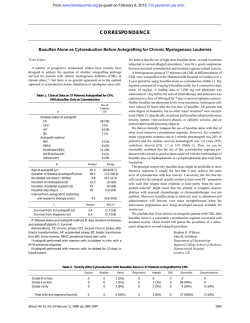
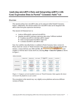
© Copyright 2026