
Targeted Gene Transfer to Human Hematopoietic Progenitor
From www.bloodjournal.org by guest on February 6, 2015. For personal use only.
RAPID COMMUNICATION
Targeted Gene Transfer to Human Hematopoietic Progenitor Cell Lines
Through the c-kit Receptor
By Paul Schwarzenberger, Sally E. Spence, John M. Gooya, Dennis Michiel, David T. Curiel, Francis W. Ruscetti,
and Jonathan R. Keller
In this report, we describe a novel gene therapy approach
was competed byexcess SLF orwith monoclonal antibodies
for hematopoietic stem/progenitor cells using aspecific rethat recognize c-kit and block
the binding ofSLF to its recepceptor-mediatedgene transfection procedure
to target c-kit+
tor. Maximum transfection efficiency (>go%) requires a 2cell lines. The vector consists of plasmid DNA containing a
hour incubation period the
of vector with the cells, and maxiluciferase reporter gene that is condensed by electrostatic
mum gene expression occurred 30 hours later. Removal of
forces with polylysine (PL) covalently linkedto streptavidin
the endosomalytic agent, AD, from the vector resulted in
(binds biotinylated ligand) and PL covalently linkedto adethe loss of gene expression. Vector targeting was versatile
novirus (AD;to achieve endosomal lysis)
with the final addiand could be changed by the addition of other biotinylated
tion of biotinylated steel
factor
(SLF-biotin).
Targeted
ligands.In principle,this vector shouldbe broadly applicable
to deliver genes to hematopoietic stemlprogenitor cells in
transfection of growth factor-dependent hematopoieticprogenitor cell lines
that express c-kit showed specific luciferase vitro and in vivo.
gene expression over cell lines that did not express c-kit.
0 1996 by The American Societyof Hematology.
This effect was dependent on the dose of SLF-biotin and
A
MAJOR GOAL OF GENE therapy has been to transfer
genes into hematopoietic stem cells,' because this
would create many therapeutic options, such as a potential
cure for inherited immunodeficiency diseases, blood cell disorders, acquired immunodeficiency syndrome, and caner.*-^ Thus far, the most efficient means to stably transfer
genes to human hematopoietic progenitor cells has been to
use retroviral vectors; however, use of these vectors results
in a low transfer efficiencytothemost
primitive human
In this regard, the presence of receptors for retroviruses on human hematopoietic stem cells hasneverbeen
shown. Furthermore, this method does not specifically target
hematopoietic cells or, in particular, primitive stem cells,
because retroviruses can infect many cell types.
From the Laboratory of Leukocyte Biology, Biological Response
Modijers Program, and Biological Carcinogenesis and Development Program, SAIC-Frederick; and the National Cancer InstituteFrederick CancerResearch and DevelopmentCenter, Frederick,
MD; and the Gene Therapy Program, University of Alabama, Birmingham, AL.
Submitted September I , 1995; accepted October 27, 1995.
The content of this publication does not necessarily reflect the
views or policiesof the Department of Health and Human Services,
nor does mention of trade names, commercial products, or organization imply endorsement by the U S . Government.
The work upon which this publication is based was performed
pursuant toContract #NOl-CO-56000 with the National Cancer
Institute, Department of Health and Human Services. The U.S. Government retains a non-exclusive, royalty-freelicense to use or duplicate this article in any manner and for any purpose whatsoever,
and to have or permit others to do so.
Address reprint requests to Jonathan R. Keller, Bldg 567, Room
252, PO Box B, Frederick Cancer Research and Development Center, Frederick, MD 21 702-1201
The publication costs of this article were defrayed in pari by page
charge payment. This article must therefore be hereby marked
"advertisement" in accordance with 18 U.S.C. section 1734 solely to
indicate this fact.
0 1996 by The American Society of Hematology.
0006-4971/96/8702-0046$3.00/0
472
A gene transfer system has been previously described that
uses asialo-orosomucoid (ligand) to specifically target asialoglycoprotein receptors expressed on hepatocytes.' The vector was constructed by covalently linking asialo-orosomucoid to polylysine (PL) that, by electrostatic forces, binds
and condenses DNA containing a reporter gene or other
genes of interest (molecular conjugate vector).8 In subsequent studies, transferrin (TF) was included in the vector as
the ligand instead of asialo-orosomucoid to target cells.9.'"
Furthermore, the molecular conjugate vector was greatly improved by including endosomalytic agents such as adenovirus (AD) and fusenogenic peptides from influenza virus that
mediate escape from endosomal lysis.""3 Endosomalytic reagents prevent endosomal degradation of entrapped DNA by
destroying the endosomal membrane.
Human hematopoiesis is supported by the proliferation
and differentiation of a small number of pluripotential stem
~ e 1 l s . l ~These
" ~ cells are responsible for maintaining sufficient numbers of committed hematopoietic cells for host
survival. Therefore, these cells are the optimal target population for stable expression of transferred therapeutic genes
(ie, adenine deaminase, glucocerebrosidase). In this regard,
these stedprogenitor cell populations have been shown to
express both c-kit (receptor for steel factor [SLF]) and CD34
(ligand for L-selectin).'7-24Thus, experiments presented here
were designed to determine whether a molecular conjugate
vector containing SLF could target gene transfer to c-kit'
hematopoietic cells. The experiments in this report describe
for the first time a versatile gene transfer vector that uses
SLF to target hematopoietic cell lines that express c-kit.
MATERIALS AND METHODS
Chemical linkage of AD-PL. W 162 cells were cultured to SO%
confluence in Dulbecco's modified Eagle's medium (GIBCO Life
Technologies, Grand Island, NY) supplemented with penicillin/
streptomycin (GIBCO) and 10% fetal calf serum (Atlanta Biologicals, Norcross, GA) at 37"C, S% CO2, and then were infected with
human type-S AD (d1014). Cells were harvested by centrifugation
when a cytopathic effect was observed, and the supernatant was
obtained from the infected W 162 cell pellet after 4 freeze-thaw
Blood, Vol 87, No 2 (January 15), 1996: pp 472-478
From www.bloodjournal.org by guest on February 6, 2015. For personal use only.
473
TARGETEDGENETRANSFER
cycles. Supernatants containing AD were further purified over cesium chloride by ultracentrifugation (in a gradient 1.33 m g h L to
1.45 mg/mL in 5 mmoUL HEPES [pH 7.81 at 15°C. at 18,000 rpm
for 90 minutes in an SW 28 rotor [Beckman, Palo Alto, CA]). The
lower band from this purification was harvested and repurified on a
second cesium chloride gradient purification (25,000 rpm for 18
hours in an SW 41 rotor). The apparent band was aspirated and
loaded onto a PD 10 column (Phannacia, Piscataway, NJ) equilibrated with HEPES-buffered saline (HBS). The eluate was resuspended in 3.6 mL of HBS and then mixed with2.4 mg of PL (Sigma,
40 mgof EDC (Pierce,
St Louis, MO) in 0.4 mL ofHBSand
Rockford, L)to achieve a final volume of 4 mL. This mixture was
allowed to react for 4 hours at 4°C and was then repurified over
CsCl (1.45 mg/mL) by ultracentrifugation as described above. The
opalescent band containing the ligated product was harvested and
diluted in viral preservation medium (0.01 m o m Tris [pH 81,0.1
m o m NaCI, 0.1% bovine serum albumin [BSA], 50 voUvol glycerol)
to achieve a final concentration of 5 X 10" virus particles/mL covalently linked to PL. Aliquots were stored at -70°C.
Plasmid DNA. The plasmid pluc4 was derived by cloning the
luciferase gene under the control of acytomegalovirus promotor into
the plasmid pstcxssc. The plasmid plc l, containing a P-galactosidase
reporter gene under cytomegalovirus promoter was a gift from Lin
Zhao Chen (ABL-Basic Research Program, Frederick, MD). Before
transfection, the plasmids were extracted with Triton X- 114 to remove endotoxin as d e s ~ r i b e d . ~ ~
Covalent linkage of streptavidin to PL (SA-PL). SA (Sigma) was
linked to PL (Sigma) and separated as described previously.'' The
SA and PL content were determined by optical density at 280 nm
and 223 nm. A total of 1 mol of PL was modified with 0.16 mol of
SA; the final concentration was 300 p g / m L PL and 40 pg/mL SA.
Cell lines and jZow cytometric analysis. HL-60, K562, MB02,
M0-7e,26.27and T F - I cells were maintained in RPM1 (GIBCO) supplemented with 10% fetal calf serum (Atlanta Biologicals), penicillidstreptomycin (GIBCO), and 2mmol L-glutamine (GIBCO).
Growth factor-dependent cell lines were maintained in cytokines as
follows: MB02 cells in 30 ng/mL granulocyte-macrophage colonystimulating factor (GM-CSF; Peprotech, Rocky Hill, NJ); and MO7e and W-l cells in 30 ngUmL interleukin-3 (L-3; Peprotech).
Expression of cell surface c-kit was determined by flow cytometric
analysis. Briefly, 1 X IO5 cells in 100 pL of phosphate-buffered
saline containing I% BSA were incubated with 10 ng of biotinylated
SLF (SLF-biotin; R&D Systems, Minneapolis, MN) for 30 minutes
at 4°C. washed, and then incubated with SA-fluorescein isothiocyanate (R&D Systems) for 30 minutes at 4°C. Cells were analyzed
immediately on a Coulter Profile (Hialeah, FL).
Construction of the molecular conjugate vector and transfection
procedure. The targeted vector was constructed by adding 100 pL
of AD-PL to 2 pg of reporter plasmid diluted in HBS for 30 minutes
at room temperature. Then, 2 pL of SA-PL diluted in HBS was
added and incubated for 30 minutes at room temperature. The control
vector was constructed by adding an equimolar amount of PL instead
of the SA-PL and incubated for 30 minutes at room temperature.
As a final step, biotinylated Steel factor (SLF-biotin; R&D Systems)
or biotinylated transferrin (TF-biotin; Sigma) was added and incubated for another 30 minutes at room temperature. A summary of
the vector construction is shown in Fig 1. Log-phase growing cells
(2 to 4 X lo6) were transfected by adding the complete vector or
the control vector to cells in a 1.5-mL Eppendorf tube and incubated
at 37°C for 2 hours. Cells were then returned to their normal growth
conditions in a 25-mL flask. Cells (2 x IO6) were harvested 48 hours
after transfection, washed 1 time with phosphate buffered saline,
and then lysed in 200 pL of lysis buffer (Promega, Madison, WI).
The lysate was pelleted by centrifugation, and the supernatant was
collected for assay. Aliquots of each sample were collected to determine protein concentration (Pierce, Rockford, IL) and luciferase
activity according to the procedures provided by Promega using a
luminometer (Berthold, Bad Wildung, Germany), with a measuring
time of 10 seconds. Each measurement was then adjusted to the
sample's protein concentration by dividing relative light units (IUU)
by protein (mg/mL).
RESULTS
SLF-targeted transfection of human hematopoietic cell
lines. To determine whether we could specifically target ckit+ hematopoietic cells, a molecular conjugate vector was
constructed by condensing plasmid DNA containing the luciferase reporter gene with PL covalently linked to incompetent AD (AD-PL; provides escape from endosomal lysis)
and PL covalently linked to SA (SA-PL; summarized in Fig
1). In the final step, SLF-biotin was added to the vector. The
control vector was similarly constructed except SA-PL was
replaced with unconjugated PL; thus, SLF-biotin could not
bind to the vector construct. Growth factor-dependent human
MO-7e and MB02 hematopoietic cell lines (positive for ckit expression) and HL-60 and TF-1 cells (negative for ckit expression) were exposed to the SLF-targeted vector
complexes or control vector complexes for 2 hours, returned
to their normal growth conditions for 48 hours, and then
harvested to determine luciferase reporter gene expression
(Fig 2). Whereas luciferase gene expression varied between
cell lines that were transfected with the control vector, transfection with the SLF-targeted vector resulted in increased
reporter gene expression in cell lines that expressed c-kit
(MB02 and MO-7e) but not in cell lines that did not express
c-kit (HL-60; see Fig 2) and TF-l (data not shown). It has
been previously shown that the reporter gene expression
obtained with the control vector is likely to be caused by
the entry of the vector via AD receptors on the cell lines or
through nonspecific ionic interactions with the PL.28
Because SLF-targeted transfection should be dependent
on the dose of SLF-biotin used in the molecular conjugate
vector, MO-7e, MB02, and HL-60 cells were transfected
with increasing amounts of SLF-biotin in the vector complex. Increasing the amount of SLF-biotin (maximum of 10
to 15 ng/mL) resulted in a dose-dependent increase in reporter gene expression (up to 12-fold) in MB02 (Fig 3A)
and MO-7e (Fig 3B) cells but not in HL-60 (Fig 3C) or TF1 cells (data not shown).
SpecGcity of SLF-targeted transfection. To show specific entry of the targeted vector through the kit receptor,
MB02 cells were preincubated with a 50-fold excess of
SLF or IL-3 or with SR-1 monoclonal antibodies (MoAbs)
that recognize c-kit and block the binding of SLF to its
receptor before the addition of the SLF-targeted vector.
The expression of luciferase is presented in Fig 4 as the
percentage of the RLU of the complete vector (100%; see
Fig 4). Whereas excess SLF effectively competed SLFtargeted transfection (luciferase gene expressionwas comparable with the control vector), IL-3,which does not bind
to the c-kit receptor, had no effect. Furthermore, MB02
cells preincubated with SR-1 monoclonal antibodies also
From www.bloodjournal.org by guest on February 6, 2015. For personal use only.
SCHWARZENBERGERET AL
474
Q-.
-
Extended
+
+ SA
+SA+ + + +
a
SW
SLF
Adenovirus
Steptavidin
Polylysine Conjugate
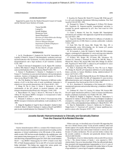
Fig 1. Summary of the molecular conjugate vector construction.
Briefly, the vector wasconstructed bycondensing DNA with PL covalently
linked t o AD and PL covalently linkedt o SA. SLF-biotin was added t o the vectorin the finalstep. For details refer t o the Materials and Methods.
inhibited SLF-targeted transfection, whereas the isotype
control antibodies had no effect (Fig 4). Thus, SLF-tar- for
geted transfection is specifically mediated through c-kit
receptorsexpressed on thehematopoieticprogenitorcell
lines.
Role of endosomalytic agents in SLF-targeted transfection. To show that replication incompetent AD was re-
quired in the conjugate vector to escape endosomal lysis and
subsequent gene expression, the effect of removing AD
from the vector was examined. As shown above and shown
here for comparison, the control vector gives 2.0 X lo4 5
7.2 X lo3 RLU that is increased to 5.4 X lo4 t 7.5 X lo3
by including SLF-biotin (Fig 5). However, substituting PL
for AD-PL (endosomalytic agent) in the molecular conjugate
Relative Light Units / rng Protein
Complete Vector
Control Vector
c- kit
Expression
CelI
Line
SLF
HL-60
619 S 0 3
913f964
0
MB02
21 4,000+14,000
38,00W4,000
69
MO-7e
155,000f32,OOO
52,00W27,000
88
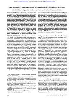
Fig 2. Hematopoietic cell lines were transfected
with the SLF-targeted molecular conjugate vector (column
2) or the controlvector (column
3). and luciferase activity was measured as described in Materials and Methods. Duplicate or triplicate determinations were made for each
cell line, and the data are presented as the mean RLU per mg/mL of cell lysate ? the SE. The data are representative of three separate
experiments. The expression of c-kit on the cell lines was determined by flow cytometric analysis using biotinylated SLF and SA-FITC as
described in Materials and Methods. Furthermore, the number of c-kit+ cells in M602 and MO-7e is notstatistically different.
From www.bloodjournal.org by guest on February 6, 2015. For personal use only.
475
TARGETEDGENETRANSFER
B MO-7e
6ol
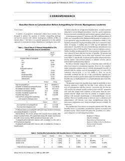
Fig 3. SLF dosdependent *kit-targeted transfection. hciferese gene expression was measured
in
MO-7e (A), M602 (B), and HL-80 (C) cells transfected
with the molecular conjugate vector containing increasing amounts of SLF-biotin as described in the
Materials and Methods. The data are presented as
the mean RLUof duplicate determinations f the SE
and are representative
of at least three experiments.
Asterix (*l i n d M e s statistically significant ( P < .05)
by Student's t-test.
e
+
+
SLF-biotln
+
-
SW-blotin
+
+
SLMlo(in
+
+
SLF-bloUn
I
SLF-blotln (ng)
vector construction resulted in loss of gene expression in
MO-7e cells, regardless of whether the conjugate vector contained SLF-biotin. Thus, AD-PL is required for efficient
SLF-targeted gene expression.
Versatility ofthe molecular conjugate vector. Inclusion
of SA linked to PL in the vector should generate a versatile
vector that is dependent only on the biotinylated ligand used.
Therefore, to show the versatility of the vector system, we
compared SLF-targeted transfection with the previously described W transfection on hematopoietic progenitor cell
lines using W-biotinto target the vector. As previously
shown?" inclusion of W-biotin in the conjugate vector
greatly increased (approximately 10-fold) the expression of
luciferase in K562 cells above the amount produced by the
control vector (Fig 6). Furthermore, as would be predicted
on the basis of c-kit expression, the SLF-targeted vector did
not enhance luciferase gene expression in K562 cells, which
do not express c-kit (Fig 6).
Kinetics of conjugate vector exposure and vector expression of SLF-targeted transfection. To maximize gene expression in hematopoietic cells, we examined the kinetics of
Aden-PL
Stmptllvldln-PL
C HL-60
conjugate vector exposure to cells during the transfection
period. MB02 cells were exposed to the molecular conjugate
vector from 10 to 240 minutes, then returned to their normal
growth conditions, and harvested 48 hours later to determine
the expression of luciferase. MB02 cells showed increased
gene expression when exposed to the conjugate vector up to
2 hours, after which gene expression decreased (Fig 7). No
loss in cell viability was observed after 2 hours of incubation
with the conjugate vector (data not shown). Thus, a 2-hour
incubation period results in maximum transfection efficiency.
Next, we examined the kinetics of gene expression in
SLF-targeted transfected MB02 and MO-7e (Figs 8B and
C) cells and in "F-targeted K562 cells (Fig 8A) and found
that maximum gene expression occurred after 30 hours and
decreased thereafter. This decrease was slightly more rapid
in MB02 and MO-7e cells, as compared with that in K562
cells. However, the maximum levels of gene expression in
SLF-targeted transfection of MB02 and MO-7e cells (1.4
X lo6 light units) exceeded the maximum levels of gene
expression in TF-infected K562 cells (5 X lo5 light units).
I
SR-l*
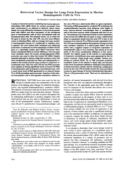
Fig 4. Specificii of SLF-targeted transfection.Luciferasegeneexpression(expressed
in RLU) was
measured in MB02 cells transfected with the indicated molucular conjugate vector components
as described in the Materials and Methods (top two bars
are the complete andcontrol vector results, respectively). Before the addition of the targeted vectors,
cells wereeither preincubated with anti-c-kit monoclonal antibody SR-l (3.34pg/mL), an irotvp. control antibody (3.34pg/mL; middle two bars) or were
preincubated with a 6O-fold oxc.01 of SLF ( 5 0 0 ng/
mL) or 11-3 ( 5 0 0 ng/mL). The d.t. are presented as
the mean RLUof dupllmte determinatitlonsf the SE
and are rqxesontative of at lswt thm experiments.
Asterix (*l indicates statistically signifiint ( P < .051
by Student's t-test.
I
20
40
60
80
100
120
0
ParccmtRLUotCompletalkctor
From www.bloodjournal.org by guest on February 6, 2015. For personal use only.
SCHWARZENBERGER ET AL
476
Adeno-PL
Streptavldin-PL
+
transfected
were
+
Ligand
-
SLF-biotin
+
SLF-biotin
Fig 5. Effect of the endosomalytic agent (AD-PL)
ongeneexpression
in SW-targeted transfection.
with the SLF-targeted
cells MO-7e
SLF-biotin
SLF-biotin
or the control vector, with or without PL linked to
AD, and measuredfor luciferase activity as described
prein the are
Materials
dataandThe
Methods.
sented as the mean RLU of duplicatedeterminations
the SE and are representative of three separate
experiments.Asterix ("1 indicates statistically s i g
RdativeLight Unib W@)I mghnl Proteinnfiicant
( p < .05) by Student'st-test.
*
Luciferase gene expression using the control vector was performed for each time point in the kinetic studies and results
showed specific transfection when compared with the targeted vector at all time points (data not shown). Thus, both
SLF- and TF-targeted transfection results in high levels of
transient gene expression.
Finally, to determine the frequency of gene transfer with
this vector, MB02 cells were transfected with the SLF-targeted vector containing a P-galactosidase reporter gene, as
described in the Materials and Methods. Cytocentrifuge
preparations of transfected MB02 cells were stained for pgalactosidase activity, and then individual cells were scored
for expression by transmission microscopy. Whereas untransfected MB02 cells showed no P-galactosidase activity,
cells transfected with control vector and SLF-targeted vector
were 9% and 97% positive, respectively (data not shown).
Thus, the frequency of SLF-targeted transfection is very high
and is comparable with the previously reported "F transfection in mouse hepatocytes."-13
DISCUSSION
As an alternative approach to widely used retroviruses for
gene therapy, we explored the use of molecular conjugate
vectors (PL-DNA complexes) to deliver genes to hematopoi-
etic cells via the receptor-mediated endocytosis pathway.
The molecular conjugate vector described here is made by
combining plasmid DNA containing a gene of interest with
PL to neutralize the charge on the DNA molecule that forms
DNA toroids of less than 200 nm in size (summarized in Fig
1). The PL contained in the vector is coupled to replication
incompetent AD and provides a mechanism to escape lysosomal degradation of the DNA, and PL coupled to SA provides a mechanism to target the DNA toroids to specific cell
types by the addition of biotinylated ligands. In this regard,
hematopoietic stedprogenitor cells have been shown to express numerous growth factor receptors. However, c-kit has
been shown to be expressed on murine pluripotential stem
cells capable of reconstituting all hematopoietic lineages in
irradiated recipient^:^." and c-kit is expressed on primitive
human hematopoietic progenitor
Furthermore, ckit expression is restricted to stedprogenitor cells and is
not expressed on their committed progeny except for mast
cells.22Thus, the studies presented here used SLF-biotin to
target the molecular conjugate vector to hematopoietic cell
lines that express c-kit. In addition, the studies presented
here used MO-7e cells because they, similar to their normal
counterpart in the bone marrow, express CD34 and c-kit and
are dependent on growth factors for their proliferation and
Adeno-PL
Straptavidin-PL
Ligand
i
t
+
-
TF-biotin
+
TF-b'oHn
Fig 6. Versatility of the moleculer conjugate vector. K562 cells were transfectedwith the molucular
conjugate
vector containing SLF-biotin or with TFbiotin, and luciferase activity was determined a described in the Metrials and Methods. The data are
presented as the mean RLU ofduplicate determinations the SE and are representativeof three separate experiments.
*
r
l
P*
+
a
i
2
3
i
5
Relative Light Unit. (x103 I mdml Protein
From www.bloodjournal.org by guest on February 6, 2015. For personal use only.
477
TARGETEDGENETRANSFER
T
1L
30
''
240
120
180
Time of Exposure (minutes)
Fig 7. Kinetics of conjugate vector expression after exposure to
cells during transfection period. MB02 cells were exposed to the
molecular conjugate complexesfrom 10 to 240 minuter, returnedto
their normal growth conditions, andthen analyzed for reporter gene
expression as described in the Materials and Methods. The data are
presented asthe mean RLU 2 the SE and are representative of three
separate experiments.
survival. The data presented in this report show that the
molecular conjugate vector can specifically target gene transfer to c-kit+ hematopoietic cell lines using SLF-biotin and
that transfer to these cells is highly efficient.
Future studies will determine whether this vector can efficiently transfer genes into normal human hematopoietic progenitor cells that express c-kit. In addition, we will extend
these studies to murine c-kit+ progenitor cells to develop an
in vivo gene transfer strategy using this vector. Furthermore,
because hematopoietic cells express numerous growth factor
receptors, we would like to compare targeted transfection
using other biotinylated cytokines such as GM-CSF and IL3. Because the targeted transfection described above is
highly efficient and expression is transient, this vector could
A
K562
be used to target human myeloid leukemias that variably
express growth factor receptors with suicide genes for bone
marrow purging ex vivo. Furthermore, therapeutic genes (cytokine genes) could be introduced into lymphoid cells using
biotinylated IL-2 or other ligands for tumor vaccines and
adoptive immunotherapy. Finally, SLF-targeted transfection
provides the framework to develop a better vector to enhance
stable integration into hematopoietic cells.
Because low levels of luciferase gene expression were
observed with the control vector, we would like to determine
in our system whether entry of the control vector into hematopoietic cells is nonspecific through ionic interactions via
PL or specific through AD receptors expressed on hematopoietic cells. In this regard, we will include tRNA during
the vector construction to neutralize any residual positively
charged PL or antibodies to the AD knob proteins known to
neutralize AD infection.
In summary, this gene therapy approach offers a number
of unique features including that: (1) uptake of DNA relies
on highly efficient receptor-mediated endocytosis, a physiologic pathway for macromolecular uptake not associated
with cellular toxicity; (2) the vector is versatile in that targeting can be changed using different biotinylated ligands;
(3) the vector can be prepared in large amounts and can
package up to 48 kb of DNAz9;and (4)the DNA does not
need to contain viral elements and obviates obvious potential
safety hazards associated with retroviral vectors.29In addition, although the efficiency of gene transfer via the receptormediated pathway is extremely high, the internalized DNA
has been shown to be degraded after fusion of the endosome
with lys~somes.'~
Because this effect is mediated by the viral
capsid proteins and is independent of viral gene expression,
AD genomic deletions, psoralen, and UV irradiation have
been used to eliminate any potential safety hazards.
ACKNOWLEDGMENT
TheauthorsthankDrDan
Longo (National Institute of Aging,
Baltimore, MD) for critical review of this manuscript, Dr Virginia
Broudy (University of Washington, Seattle, WA) for the generous
gift of SR-l monoclonal antibodies, Dr Dons Morgan (Hahnemann
B
c
MB02
M07
14-
161
1412-
10
10-
Fig 8. Kineticsofluciferasegeneexpression.
K562 cells were targeted with TF-biotin, or MB02
cells and MO-7e cells were targeted with SLF-biotin
and returned to their normal growth conditions according to theprocedures describedin the Materials
and Methods. The cella were harvested for luciferase
activity at the indicatedtimes, and the data are presented asthe mean RLU of duplicatedeterminations
f the SE and are representativeof three experiments.
0-
I 3-l
C L
40
Hours Incubation
From www.bloodjournal.org by guest on February 6, 2015. For personal use only.
SCHWARZENBERGER ET AL
University, Philadelphia, PA) for the generous gift of MB02 cells,
and Louise Finch for the fluorescence-activated cell sorting analysis
of the hematopoietic cell lines.
REFERENCES
1. Karlsson S: Treatment of genetic defects in hematopoietic cell
function by gene transfer. Blood 78:2481, 1991
2. Nienhuis AW, McDonagh KT, Bodine DM: Gene transfer into
hematopoietic stem cells. Cancer 67:2700, 1991 (suppl)
3. Baltimore D: Intracellular immunization. Nature 335:395, 1988
4. Miller DA: Human gene therapy comes of age. Nature 357:455,
1992
5. Apperley JF, Williams D: Gene therapy: Current status and
future directions. Br J Haematol 75:148, 1990
6. Brenner MK, Rill DR, Anderson WF, Ihle JN: Gene marking
to determine whether autologous marrow infusion restores long term
hemapoiesis in cancer patients. Lancet 342:1134, 1993
7. Schuening FG, Kawahara K, Miller DA, Storb R: Retrovirusmediated gene transduction into long term repopulating marrow cells
of dogs. Blood 78:2568, 1991
8. Wu G, Wu C: Receptor mediated in vitro gene transformation
by a soluble DNA carrier system. J Biol Chem 262:4429, 1987
9. Zenke M, Steinlein P, Birnstiel M: Receptor mediated endocytosis of transferrin-polycation conjugates: An efficient way to introduce DNA into hematopoietic cells. Proc Natl Acad Sci USA
87:3655, 1990
IO. Wagner E, Ztnke M, Birnstiel M: Transferrin-polycation conjugates as carriers for DNA uptake into cells. Proc Natl Acad Sci
USA 87:3410, 1990
1 1. Wagner E, Zatloukal K, Birnstiel M: Coupling of adenovirus
to transferrin-polylysine-DNA complexes greatly enhances receptor
mediated gene delivery and expression of transfected genes. Proc
Natl Acad Sci USA 89:6099, 1992
12. Curiel DT, Agarwal S, Wagner E, Cotton M: Adenovirus
enhancement of transferrin-polylysine mediated gene delivery. Proc
Natl Acad Sci USA 88:8850, 1991
13. Wagner E, Plank C, Zatloukal K, Cotton M, Birnstiel M:
Influenza virus hemagglutinin HA-2 N-terminal fusogenic peptides
augment gene transfer by transferrin-polylysine-DNA complexes:
Toward a synthetic virus-like gene-transfer vehicle. Proc Natl Acad
SciUSA 89:7934, 1992
14. Ogawa M: Differentiation and proliferation of hematopoietic
stem cells. Blood 81:2844, 1993
15. Metcalf D: The biology of hemopoiesis, in Metcalf D (ed):
The Hemopoietic Colony Stimulating Factors. Amsterdam, The
Netherlands, Elsevier Science B.V., 1984, p 1
16. Moore MAS: Clinical implication of positive and negative
hematopoietic stem cell regulators. Blood 78: I , 1991
17.Berenson RJ, Andrews RG, Bensinger WI, Buckner CD.
Bernstein ID: Antigen CD34+ marrow cells engraft lethally irradiated baboons. J Clin Invest 8 1:95 I , I988
18. Berenson RJ, Bensinger WI, Thomas ED: Engraftment after
infusion of CD34' marrow cells in patients with breast cancer or
neuroblastoma. Blood 77: 1717, 1991
19. Briddell R, Broudy V, Bruno E, Brandt JE, Hoffman R: Further phenotypic characterization and isolation of human hematopoietic progenitor cells using a monoclonal antibody to the c-kit receptor. Blood 79:3159, 1992
20. YamaguchiY,Gunji Y, Nakamura M, Suda T: Expression
of c-kit mRNAandprotein
during the differentiation of human
hematopoietic progenitor cells. Exp Hematol 21: 1233, 1993
21. Gunji Y, Nakamura M, Osawa H, Suda T: Human primitive
hematopoietic progenitor cells are more enriched in kit-low cells
than in kit-high cells. Blood 82:3283, 1993
22. Okada S, Nakauchi H, Nagayoshi K, Nishikawa S, Suda T:
Enrichment and characterization of murine hematopoietic stem cells
that express c-kit molecule. Blood 78:1706, 1991
23. Okada S, Nakauchi H, Nagayoshi K, Nishikawa S-I, Suda T:
In vivo and in vitro stem cell function of c-kit and Sca-l positive
murine hematopoietic cells. Blood 80:3044, 1992
24. Seiji 0, Nagayoshi K, Nakauchi H, Nishikawa S-I, Suda T:
Sequential analysis of hematopoietic reconstitution achieved by
transplantation of hematopoietic stem cells. Blood 81:1720, 1993
25. Manthorpe M, Cornefert-Jensen F, Hartikka J, Felgner J, Rundell A, Margalitch M, Dwarki V: Gene therapy by intramuscular
injection of plasmid DNA: Studies on firefly luciferase gene expression in mice. Hum Gene Ther 4:419, 1993
26. Avanzi GC, Brizzi MF, Giannotti J, Ciarletta A, Yang Y,
Pegoraro L, Clark SC: MO-7e human leukemic factor dependent cell
line provides a rapid and sensitive bioassay for the human cytokines
GMCSF and IL-3. J Cell Physiol 145:458, 1990
27. Avanzi GC, Lista P, Giovinazzo B, Pegoraro L: Selective
growth response to IL-3 of a human leukaemic cell line with megakaryoblastic features. Br J Haematol 69:359, 1988
28. Michael SI, Huang CH, Romer MU, Curiel DT: Binding incompetent adenovirus facilitates molecular conjugate-mediated gene
transfer by receptor-mediated endocytosis pathway. J BiolChem
268:6866, 1993
29. Cotton M, Wagner E, Birnstiel M: High efficiency receptormediated delivery of small and large (48 kb) gene constructs using
the endosome-disruption activity of defective or chemically inactivated adenovirus particles. Proc Natl Acad Sci USA 89:6094, 1992
From www.bloodjournal.org by guest on February 6, 2015. For personal use only.
1996 87: 472-478
Targeted gene transfer to human hematopoietic progenitor cell lines
through the c-kit receptor
P Schwarzenberger, SE Spence, JM Gooya, D Michiel, DT Curiel, FW Ruscetti and JR Keller
Updated information and services can be found at:
http://www.bloodjournal.org/content/87/2/472.full.html
Articles on similar topics can be found in the following Blood collections
Information about reproducing this article in parts or in its entirety may be found online at:
http://www.bloodjournal.org/site/misc/rights.xhtml#repub_requests
Information about ordering reprints may be found online at:
http://www.bloodjournal.org/site/misc/rights.xhtml#reprints
Information about subscriptions and ASH membership may be found online at:
http://www.bloodjournal.org/site/subscriptions/index.xhtml
Blood (print ISSN 0006-4971, online ISSN 1528-0020), is published weekly by the American
Society of Hematology, 2021 L St, NW, Suite 900, Washington DC 20036.
Copyright 2011 by The American Society of Hematology; all rights reserved.


© Copyright 2026