Controls for Reverse Transcriptase-Polymerase Chain
From www.bloodjournal.org by guest on February 6, 2015. For personal use only.
3984
CORRESPONDENCE
Controls for Reverse Transcriptase-Polymerase Chain Reaction Amplification of BCR-ABL Transcripts
To the Editor:
The validity of reverse transcriptase-polymerase chain reaction
(RT-PCR) results as evidence of specific gene expression depends
on use of appropriate controls. Negative controls are designed to
demonstrate absence of PCR products in amplifications of cDNA
from cells that do not express the gene and in reactions that do not
contain DNA template (“water blanks”). Positive controls should
provide evidence that the cDNA template is of good quality for the
desired amplification, if the gene in question is actually expressed
by the tested cells.
Many RT-PCR-based studies include parallel amplifications of
the P-actin gene as proof of amplifiable cDNA template. Reports
from our laboratory’ and others2have shown the inadequacy of these
positive controls, partly because this gene is expressed at high levels,
and partly because the 0-actin-amplified products may be also derived from genomic DNA, which is invariably present as a contaminant inRNA preparations. In thecase of RT-PCRtests for expression
of the BCR-ABL hybrid gene of chronic myeloid leukemia (CML)
and Philadelphia-positive acute lymphoblastic leukemia, some
groups use amplifications of a small fragment on the ABL gene to
show the presence and quality of the cDNA template. However, we
think that these so-called “ABL controls” are inadequate, because
the primers3used for these PCRs can promote amplification of ABL
sequences present in ( l ) both cDNA and genomic DNA templates,
and (2) both the normal ABL allele and the translocated BCR-ABL
gene (Fig 1A). The PCR products from these amplifications do not
derive necessarily or exclusively from the normal ABL gene transcripts, and therefore are not a suitable control for tests designed to
detect or exclude BCR-ABL expression.
This problem is particularly relevant when a two-step or “nested”
RT-PCR strategy is needed to show “ABL” amplification,’ as illustrated on Fig 1B. For these experiments, RNA was extracted“ from
IO5 cells from cell line cultures and from peripheral blood leukocytes
from a CML patient (P384), and reverse-transcribed into cDNA as
de~cribed.~
Genomic DNAwas extracted from the same samples
and purified to 25 ng/pL by standard methods. PCR amplifications
were performed as described6 in a 20-pL reaction from 1 pL DNA
From www.bloodjournal.org by guest on February 6, 2015. For personal use only.
CORRESPONDENCE
3985
A
Genomic DNA
In+ Cl+ C2-
"
C
cDNA
c3-
c
4
cDNA
Gen. DNA
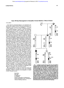
Fig 1. (A) Schematic representation of the 5' end
of the ABL gene locus (genomic DNA), and of normal
ABL and BCR-ABL transcripts IcDNAI. The relative
positions of the PCR primers are indicated by arrows.
The sequences of these primers are: In' = (5')TCAGAAlTCCCCCCmCTClTCCAG; C l + = (5')lTCAGCGGCCAGTAGCATCTGACTT; C I (5')TAACACTCTAAGCATAACTAAAG; C T = 15')CCAAGCAACTACATCACGCCAGTCAACA. Ethidium-bromide stained
agarose gels show products of PCR amplifications
CT,(C) CT'
CI,and (D)
with primers (B) Cl'
In'
CI. M, 123-bp DNA ladder lGlBC0 BRL, Paisley, Scotland); N1 and N2 are no-DNA negative controls for first- and second-step PCRs, respectively.
+
+
++
++
(for first-step PCR) or 1 pL neat or diluted products from the firststep (for nested PCR) as templates.
PCR with primers C l + (in ABL exon 2) and C3- (in ABL exon
3) produced a 275-bp fragment from the cDNA templates (Fig IB,
lanes 1 through 4). including that from KYO-I cells which, like the
EM-2 cell line used by others as control for BCR-ABL and ABL
expression,' lack a normal chromosome 9,' and do not express the
normal ABL gene? Therefore, in this case the amplified exon 2-
exon 3 ABL sequence must be. derived exclusively from the BCRABL transcripts. The same primer combination was able to pronwte
amplification also of genomic DNA templates (Fig lB, lanes 5
through 8) in the form of an 875-bp fragment containing a 600-bp
intron' between ABL exons 2 and 3. Heminested PCRs of a 1 : 4 0 0
dilution of first-step products with primers within ABL exon 2 (Cl'
C 2 3 produced a 172 bp-fragment in all samples (Fig IC), regardless
of their original template preparation @DNA v genomic DNA) or
-
From www.bloodjournal.org by guest on February 6, 2015. For personal use only.
CORRESPONDENCE
3986
of the number of genes and transcripts contributing to the ABL exon
2 sequence amplification (HL60: only normal ABL; KYO-1: only
BCR-ABL; K562 and P384: both normal ABL and BCR-ABL).
The fact that cDNA preparations contain amplifiable amounts of
genomic DNA was confirmed by positive amplifications of these
preparations using a 26-mer sense primer (In’) located at the 3’ end
of the intron’ preceding ABL exon 2 and the antisense C2- primer
(not shown). Furthermore, positive amplifications were also obtained
C2-)
with this primer pair when using the diluted first-step (Cl’
PCRs as templates, confirming the presence of amplifiable amounts
of genomic DNA carried over from the cDNA preparations even
after extensive dilution (Fig ID).
Identical results for all the PCRamplificationsdescribed here were
observed whenusingABL
antisense C3- primer-specific cDNA
synthesis,’ and whencellular RNA was extracted by two other methods, 10.1 I This suggests that “cDNA preparations” that are actually
devoid of any cDNA (because of RNA loss or degradation, or failure
of reverse transcription), but are contaminated with genomic DNA
during theRNA extraction process, could still produce an “ABL
positive” amplification product with no RT-PCR control value.
In conclusion, we suggest caution in the interpretation of negative
RT-PCR results for BCR-ABL when the “positive control” amplification test is not cDNA specific and single-gene specific. Demonstration of normal ABL or normal BCR expression as evidence of
cDNA integrity requires amplification with primers framing a normal
sequence that is disrupted in the formation of the BCR-ABL gene,
ie, the junction of exons Ib-a2 or Ia-a2 for ABL, and the fragment
comprising exons b2 to b4 for BCR.
++
Junia V. Melo
Natasha S. Kent
Xiu-Hua Yan
John M. Goldman
LRF Centre for Adult Leukaemia
Departmeni of Haematology
RPMS, Hammersmith Hospital
London, UK
REFERENCES
1. Cross NCP, Lin F, Goldman JM: Appropriate controls for
reverse transcription polymerase chain reaction (RT-PCR). Br J
Haematol87:218, 1994
2. Taylor JJ, Heasman PA: Control genes for reverse transcriptase/polymerase chain reaction (RT-PCR). Br J Haematol
86:444, 1994
3. Keating A,Wang XH, Laraya P: Variable transcription of
BCR-ABL byPh’ cells arising from hematopoietic progenitors in
chronic myeloid leukemia. Blood 83:1744, 1994
4. Chomczynski P, Sacchi N: Single-step method of RNA isolation by acid guanidinium thiocyanate-phenol-chloroformextraction.
Anal Biochem 162:156, 1987
5 . Cross NCP, Melo JV, Feng L, Goldman JM: An optimized
multiplex polymerase chain reaction (PCR) for detection of BCRABL fusion mRNAs in haematological disorders. Leukemia 8: 186,
1994
6 . Melo JV, Goldman JM: Specific point mutations that activate
v-ab1 are not found in Philadelphia-negativechronic myeloid leukaemia, Philadelphia-negative acute lymphoblastic leukaemia or blast
transformation of chronic myeloid leukaemia. Leukemia 6:786, 1992
7. Keating A: Ph positive CML cell lines. Baillieres Clin Haemato1 1:1021, 1987
8. Melo JV, Gordon DE, Cross NCP, Goldman JM: The ABLBCR fusion gene is expressed in chronic myeloid leukemia. Blood
81:158, 1993
9. Grosveld G, Venvoerd T, van Agtboven T, de Klein A, Ramachandran KL, Heisterkamp N. Stam K, Groffen J: The chronic myelocytic cell line K562 contains a breakpoint in bcr and produces a
chimeric bcr/abl transcript. Mol Cell Biol 6607, 1986
10. Sambrook J, Fritsch EF, Maniatis T Molecular Cloning: A
Laboratory Manual. Cold Spring Harbor, NY, Cold Spring Harbor
Laboratory, 1989
11. Higuchi R: Simple and rapid preparation of samples for PCR,
in Erlich HA (ed): PCR Technology. New York, NY, Stockton.
1989, p 31
From www.bloodjournal.org by guest on February 6, 2015. For personal use only.
1994 84: 3984-3986
Controls for reverse transcriptase-polymerase chain reaction
amplification of BCR-ABL transcripts [letter]
JV Melo, NS Kent, XH Yan and JM Goldman
Updated information and services can be found at:
http://www.bloodjournal.org/content/84/11/3984.citation.full.html
Articles on similar topics can be found in the following Blood collections
Information about reproducing this article in parts or in its entirety may be found online at:
http://www.bloodjournal.org/site/misc/rights.xhtml#repub_requests
Information about ordering reprints may be found online at:
http://www.bloodjournal.org/site/misc/rights.xhtml#reprints
Information about subscriptions and ASH membership may be found online at:
http://www.bloodjournal.org/site/subscriptions/index.xhtml
Blood (print ISSN 0006-4971, online ISSN 1528-0020), is published weekly by the American
Society of Hematology, 2021 L St, NW, Suite 900, Washington DC 20036.
Copyright 2011 by The American Society of Hematology; all rights reserved.
© Copyright 2026
