
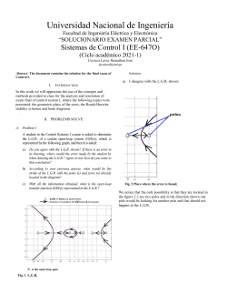
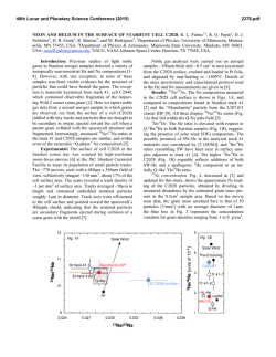
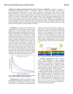
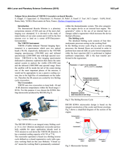
1-s2.0-S1529943014000552-main
The Spine Journal 14 (2014) 1070–1071
Cervical osteoid osteoma
progression to osteoblastoma
A 16-year-old boy presented with 6 months of moderate
neck pain, more severe at night, and temporarily relief
by nonsteroidal anti-inflammatory drugs. Radiographs were
normal. Magnetic resonance imaging showed edema in C6
vertebral body and surrounding C5–C6 posterior element
(Fig. 1). Computed tomography (CT) scan findings were
consistent with osteoid osteoma located on the right pedicle
of C6 (Fig. 2). Intralesional excision was proposed to the
patient and parents who did not accept. One year later,
the patient reported a worsening cervical pain no longer
responsive to analgesics with root irritation in the right
arm. A new CT scan showed considerable expansion of
the lesion involving C6 right pedicle and articular process
surrounding the C6 nerve root and vertebral artery, highly
Fig. 2. Computed tomography scan showed the typical feature of osteoid
osteoma (axial view).
suspicious for osteoblastoma (Fig 3, Left and Right). An intralesional excision and posterior C4–C7 fusion provided
immediate pain relief. Histologic examination of the specimens confirmed the diagnosis of osteoblastoma. Five years
after surgical treatment, the patient was asymptomatic; CT
scan (Fig. 4) and radiographs (Fig. 5) showed no local recurrence and complete fusion from C4 to C7 at the left side.
This case demonstrates that although rare, spinal osteoid
osteoma can progress to osteoblastoma [1].
Reference
[1] Bruneau M, Polivka M, Cornelius JF, George B. Progression of an osteoid osteoma to an osteoblastoma. Case report. J Neurosurg Spine
2005;3:238–41.
Fig. 1. Sagittal magnetic resonance imaging demonstrated bone and soft
tissue edema.
1529-9430/$ - see front matter Ó 2014 Elsevier Inc. All rights reserved.
http://dx.doi.org/10.1016/j.spinee.2013.12.021
Michele Cappuccio, MDa
Federico De Iure, MDa
Luca Amendola, MDa
Alessandro Corghi, MDa
Alessandro Gasbarrini, MDb
a
Department of Orthopaedics and
TraumatologydSpine Surgery
Maggiore Hospital ‘‘C.A. Pizzardi’’
M. Cappuccio et al. / The Spine Journal 14 (2014) 1070–1071
1071
Fig. 3. Computed tomography scan showed progression of the lesion: (Left) axial view and (Right) sagittal view.
Largo Nigrisoli 2
40100 Bologna, Italy
b
Department of Oncologic and
Degenerative Spine Surgery
Rizzoli Orthopedics Institute
Via G.C. Pupilli 1
40100 Bologna, Italy
Fig. 4. Computed tomography scan 5 years after the surgery.
FDA device/drug status: Not applicable.
Author disclosures: MC: Nothing to disclose. FDI: Nothing to disclose.
LA: Nothing to disclose. AC: Nothing to disclose. AG: Nothing to disclose.
Fig. 5. Radiograph 5 years after the surgery.


© Copyright 2026