
Characterization of Grb2-Binding Proteins in Human
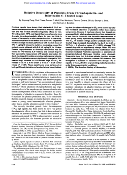
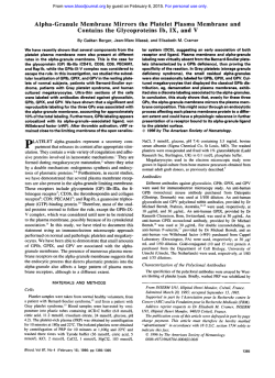
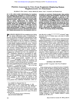
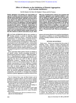
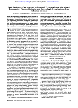
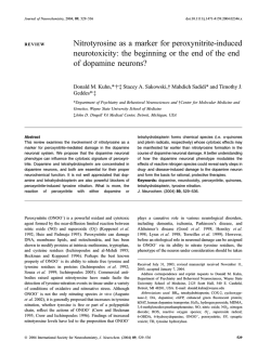
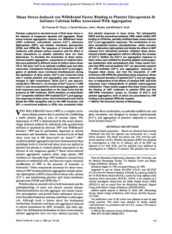
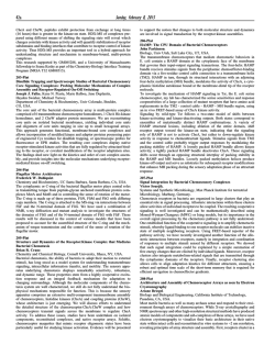
From www.bloodjournal.org by guest on February 6, 2015. For personal use only.
Characterization of Grb2-Binding Proteins in Human Platelets Activated
by FcyRIIA Cross-Linking
By Anne Robinson, Jonathan Gibbins, Belen Rodriguez-Liiiares, Pete M. Finan, Lynn Wilson, Stuart Kellie,
Paul Findell, and Steve P. Watson
Glutathione-S-transferase(GST)-GrbZfusion proteinshave
been used to identify the potential role of GrbZ-binding
proteins in platelet activation by the platelet low-affinity
IgG receptor, FcyRIIA. Two tyrosine phosphoproteins of 38
and 63 kD bind to the SHZ domain of Grb2 following
FcyRllA stimulation of platelets. Both are located in the
particulate fraction following platelet activation and are
also able to bind to a GST-construct containing the SH2
and SH3 domains of phospholipase C y l . p38 also forms a
complex with the tyrosine kinase csk in stimulated cells
and is a substrate for the kinase. The SH3 domains of Grb2
form a stable complex with SOSl and two proteins of 75
kD and 120 kD, which undergo tyrosine phosphorylation
in FcyRIIA-stimulatedcells. The 75-kD protein is recognized
by antibodies to SLP-76, which has recently been isolated
from T cells and sequenced. Tyrosine phosphorylation of
p38 and p63 is also observed in platelets stimulated by the
tyrosine kinase-linkedreceptor agonist collagen and by the
G protein-coupled receptor agonist thrombin, although
phosphorylation of SLP-76 is only observed in collagenstimulated platelets. p38 and p63 may provide a docking
site for Grb2, thereby linking GrbZ SH3-binding proteins
SOS1, SLP-76, and p120 to downstream signalling events.
0 1996 by The American Society of Hematology.
A
sisting of an SH2 domain flanked by two SH3 domains.”
The SH3 domains of Grb2 have been shown to interact with
a number of proteins including the carboxy-terminal proline
rich domain of SOS, a guanine nucleotide exchange factor
for ras,” a novel SH2 domain-containing protein SLP-76,
which is expressed in hematopoietic cell^,'^,'^ the guanosine
triphosphate hydrolase (GTPase) dynamin’‘ and the 120kD protein product of the c-cbl proto-oncogene.” The SH2
domain of Grb2 binds directly to specific phosphotyrosine
sequences in receptors such as the epidermal growth factor
receptor (EGF-R)I3,l8,’’and the protein tyrosine phosphatase
R-PTPa.” It also binds to nonreceptor tyrosine phosphoproteins including the tyrosine phosphatase syp (PTPLD)’’ and
the adaptor proteins shc’’ and insulin receptor substrate-1
(IRS-l).23Binding of Grb2 to membrane-localized proteins
via its SH2 domain provides a mechanism for the translocation of Grb2 SH3-binding proteins to the membrane. For
example, ras activity is regulated by translocation of a Grb2/
SOS complex to the membrane where ras is located.’’
It is becoming increasingly clear that Grb2 is regulated
by multiple proteins through its SH2 domain and that it is
capable of translocating a number of proteins through its
SH3 domains. For example, a human T-cell 36- to 38-kD
protein (p36-38), which becomes tyrosine-phosphorylated
and membrane-localized following TCR-mediated stimulation, is able to form a stable complex with the SH2 domain
of Grb2.24-’hAn equivalent protein has recently been cloned
in rat and has been named Lnk.” p36-38 and Lnk have also
been shown to bind to PLCyl and phosphatidylinositol 3kina~e.’~.’~
It has been proposed that p36-38Lnk may act as
a bridging protein, coupling the TCR to both the Grb2/ras
signalling pathway and activation of PLCy 1 .25
In human platelets, we have noted that following
FcyRIIA-mediated stimulation, major bands of 38-, 4 5 , and
72-kD are tyrosine-phosphorylated independent of protein
kinase C (PKC) activation and Ca’+ elevation.’ The 72- and
45-kD bands have been shown to contain syk and FcyRIIA,
respectively,’ but the 38-kD band is as yet unidentified. In
view of the homology between syk and ZAP 70 and their
similar associations with the ITAMs of the FcyRIIA and the
TCR, respectively, we have investigated whether platelet
p38 is analogous to the Grb2-binding protein p36-38Lnk in
T cells and have also characterized other Grb2-associating
proteins.
LOW-AFFINITY Fc receptor, FcyRIIA (CD32) mediates the interaction of platelets with immune complexes.’ Cross-linking of Fc yRIIA using specific antibodies
mimics the effect of the immune complex and leads to tyrosine phosphorylation of the recepto? and activation of
phospholipase Cy2 (PLCy2): The receptor contains a sequence motif that closely resembles the immunoreceptor tyrosine-based activation motif or ITAM (also known as
ARAM and TAM) found in the cytoplasmic domain of
chains of the T-cell receptor (TCR), B-cell receptor, the
high-affinity IgE receptor (FcdU), FcyI, and FcyIII receptor~.’*~
In these receptor complexes, two Y X X M sequences
are separated by 6 to 8 intervening residues resulting in the
characteristic ITAM motif. In the FcyRIIA, there are 12
residues separating the YXXL sequences, forming an extended ITAM-like motif. The ITAM of FcyRIIA associates
with the tandem SH2 domains of ~ y k . ’Syk
. ~ is a homolog
of the T-cell tyrosine kinase ZAP-70, which interacts with
the tyrosine-phosphorylated ITAMs in the 4 chains of the
TCR complex.8
Src homology 2 and 3 (SH2 and SH3) domains are recognized as protein modules that play an important role in signal
transduction mediated by receptor tyrosine kinases and tyrosine kinase-linked receptors (reviewed in Pawson’). SH2 domains bind to short phosphotyrosine-containing sequences
in specific phosphoproteins,” and SH3 domains bind to sequences of approximately 10 amino acids that are rich in
proline residues.’’ Mammalian Grb2 (Cuenorhabditis eleguns Sem-5 and Drosophila Drk) is an adaptor protein conFrom the University Department of Pharmacology, Oxford; the
Yamanouchi Research Institute, Littlemore Hospital, Oxford, U K ;
and Syntex Research, Palo Alto, CA.
Submitted September 14, 1995; accepted March I , 1996.
Supported by the Wellcome Trust (London, UK). S.P.W. is a Royal
Society Research Fellow.
Address reprint requests to Steve Watson, PhD, University Department of Pharmacology, Mansfield Road, Oxford OX1 3QT, UK.
The publication costs of this article were defrayed in part by page
charge payment. This article must therefore be hereby marked
“advertisement” in accordance with 18 U.S.C. section 1734 solely to
indicate this fact.
0 1996 by The American Society of Hematology.
0006-4971/96/8802-0023$3.00/0
522
Blood, VOI 88, N O 2 (July 15), 1996: pp 522-530
From www.bloodjournal.org by guest on February 6, 2015. For personal use only.
523
GRE-BINDINGPROTEINS IN HUMAN PLATELETS
aPY
12389 67 -
Fig 1. Rapid and transient tyrosine phosphorylation of a 38-kD molecule. Platelets were stimulated
by addition of anti-FcyRIIA MoAb IV.3 (1 pglmL) for
1 minute followed by cross-linker IF(ab’I230 pglmLl
for the indicated time or left untreated. Whole cell
lysateswere resolved on 10% SDS-PAGE and immunoblotted using antiphosphotyrosine MoAb 4610.
Results are representative of 10 experiments.
4
50 37.5 34-
MATERIALS AND METHODS
Antibodies and reagents. Monoclonal antibody (MoAb) 4G10
specific for phosphotyrosine, PLCy 1 MoAb, polyclonal anti-SOS
antibody, and mitogen-activated protein (MAP) kinase MoAb (recognizing p42 MAP kinase) were purchased from Upstate Biotechnology Inc (TCS Biologicals Ltd, Bucks, UK). Polyclonal anti-Grb2
antibody and p62 MoAb were purchased from Affiniti Research
Products Ltd (Nottingham, UK). FcyRll specific MoAb was purchased from Madarex Inc (Annandale, NJ). CD3 MoAb, OKT3 was
a kind gift from Denis Alexander (Babraham, Cambridge, UK).
Polyclonal csk antibody was purchased from Santa CNZ Biotechnology (Autogen Bioclear UK Ltd, Devizes, Wilts, UK). Polyclonal
anti-SLP-76 antibody was raised by Paul Findell. Anti-src MoAb
was a gift from Joan Brugge (Ariad Pharmaceuticals Inc, Cambridge,
MA). Sheep F(ab’)2 raised against mouse IgG (M-1522), Nonidet
P-40 (NP-40). thrombin, and Triton-X-I00 were purchased from
Sigma (Poole, Dorset, UK). Collagen (native collagen fibrils from
equine tendons) was from Nycomed (Munich, Germany). [Y-~*P]
adenosine triphosphate (ATP specific activity [s.a.], 3,000 Ci/mmol)
was purchased from N.E.N.-DU Pont (Stevenage, Herts, UK).
Peptide. The EGF receptor peptide with the sequence PVPE-Y
(phosphate)INQS, corresponding to the autophosphorylation site of
the receptor, has been described previously” and was kindly provided by Julian Downward (Imperial Cancer Research Fund, Lincoln’s Inn Fields, London, UK). The equivalent nonphosphorylated
peptide was purchased from Zinsser Analytic (Maidenhead, Berkshire, UK).
Fusion proteins. Fusion proteins encoding glutathione-s-transferase (GST)-Grb2 myc and double SH3 mutant GST-Grb2 myc
P47UG203R have been described previou~ly’~
and were kindly provided by Sean Egan (Whitehead Institute for Biomedical Research,
Massachusetts Institute of Technology, Cambridge, MA).
PIatelet preparation. Human platelets were isolated from drugfree volunteers on the day of the experiment according to the guidelines in the Helsinki Doctrine. They were resuspended at a concentration of 0.8 to 1.6 x 10⁹/mL.Platelets were suspended in a modified
Tyrodes-HEPES buffer containing indomethacin (10 pmol/L) and
EGTA (1 mmoVL). and incubated for 15 minutes before the experiment.’ All experiments were performed at 37°C with continuous
stirring at 1,200 rpm.
T-cell culture. The human leukemic Jurkat T-cell line was provided by the Department of Pathology, Oxford University and was
cultured in RPMI 1640 supplemented with 10% fetal calf serum.
Cell activation. Platelets were stimulated via FcyRIIA using
MoAb IV.3 ( I pglmL) and the cross-linker [F(ab’)2 30 pg/mL],
which was added after 1 minute (the latter time was taken at the
beginning of the experiment). Platelets were stimulated by collagen
70kDa
4 FcyFUIA
4- 38kDa
10” 20” W W 2’
5’
1(Y
FcyIUIA cross-linking time
at 100 pg/mL and by thrombin at 1 U/mL. Reactions were stopped
for precipitation after 2 minutes of stimulation by addition of an
equal volume of cold lysis buffer (NaCI. 300 mmolk: Tris, 20
mmol/L; phenylmethylsulfonyl fluoride [PMSF], 1 mmolk, EDTA,
10 mmolk, Na3VO4, 2 mmol/L; pH 7.3 containing Triton X-100,
2%); in some experiments NP-40 (1%) was used instead of Tritox
X-100. Following lysis, the insoluble fraction was removed by centrifugation at 13,000 rpm for 10 minutes at 4°C. For Western blotting
of whole cell lysates, reactions were stopped after the indicated
time by addition of nonreducing Laemmli sodium dodecyl sulfatepolyacrylamide gel electrophoresis (SDS-PAGE) sample buffer.
T cells were washed once in phosphate-buffered saline before
stimulation via the TCR by cross-linking CD3 using OKT3 (1 pg/
mL). Cross-linker [F(ab’), 8 pg/mL] was added after 1 minute, and
reactions stopped after 2 minutes by pelleting the cells in a microfuge
at 13,000 rpm for 5 seconds and adding cold lysis buffer to the
pellet. Following lysis, the Triton insoluble fraction was removed
by centrifugation at 13,000 rpm for 10 minutes at 4°C. For Westem
blotting of whole cell lysates, reactions were stopped after the indicated time by addition of nonreducing Laemmli SDS-PAGE sample
buffer.
Precipitation and depletion. Lysates were precleared for 1 hour
at 4°C using glutathione beads and then incubated with 25 pg of
GST fusion protein immobilized on glutathione-coated beads for 2
hours at 4°C. Precipitates were washed twice in 500 pL of lysis
buffer and three times in 500 pL Tris-buffered saline containing
0.1% Tween (TBS-T). If depletion of 38 kD was required, the 2hour precipitation step was replaced by three 45-minute incubations
with 25 pg of GST-Grb2 (mut) fusion protein immobilized on glutathione beads. The resulting depleted cell lysates were used for precipitation.
Immunoprecipitation. Lysates were precleared for I hour at 4°C
using protein A-Sepharose and then incubated with 5 pg of p5WSk
MoAb, immobilized on protein A-sepharose, for 2 hours at 4°C.
Precipitates were washed twice in 500 pL of lysis buffer and three
times in 500 pL TBS-T.
In vitro kinase assav. p5W“ complexes were precipitated from
cell lysates as described above. Assays were performed in 20 pL of
kinase buffer (5 mmol/L MgClz. 5 mmol/L MnCI2, 100 mmolR.
NaCI, 20 mmol/L HEPES, and 5 pCi [y-”PIATP). Reactions were
performed for 10 minutes at room temperature and stopped by addition of 0.5 mL of ice cold 100 mmol/L EDTA (pH 7.4).
SDS-PAGE. Samples were boiled in nonreducing SDS-PAGE
sample buffer for 15 minutes. Samples were then resolved by SDSPAGE (10%) and transferred to polyvinylidene fluoride (PVDF). [y”P]-labeled phosphoproteins were visualized by autoradiography.
Altematively, the membranes were immunoblotted using the indi-
From www.bloodjournal.org by guest on February 6, 2015. For personal use only.
ROBINSON ET AL
524
A 123-
D
aGrb2
8938kDa -b
6750-
37.534-
+ - + +
-
u
t
50
-
-
GST
GST-Grb;
platelet whole cell lysate
B
63kDa
aPY
aPY
+
+ - + +
"lu
,
[M
W38 kDa
+
GST GSTGrb2
(wt)
123-
EGF-Rpeptide
-
E
34- , + , , - + , FqRIIA
C
-
FcyRnA
but)
t 7 5 kDa
t 6 3 kDa
50 37.5
-~
I
- I
GST GST-Grb2
C120 kDa
123 89 -.
67 -
FcyRlIA
EGF-R peptide
[ILM1
-
+
+
+
-
S
SO
U-
FcyRllA
EGF-R peptide
GST
GST-Grb2
W )
platelets JurkatT-cells
"
aPY
89 -
67-
a" 63kDa
50-
63kDa
+
+
+ FcyRIlA
+
5
GST
GST-Grb2
*38kDa
37.534-
+
U-
50 EGF-R peptide
W )
+ - + + - +
"
-U
GST
GSTGSTGST
Grb2
(mut)
Grb2
(mut)
7SkDa
aPY
+
+
" -
-
+
GST
GST-Grb2
+
FcyRlIA
50 EGF-R peptide
[PM1
W )
cated antibody followedby secondary antibody conjugated to horseradish peroxidase and Enhanced Chemiluminescence (ECL) detection.
Subcellular fracrionarion. Subcellular fractionationwasperformed after activation of platelets. Cells were resuspended inan
equalvolume of hypotonicbuffer (10 mmol/LTris. 1 mmol/L
EDTA, 1 mmolL PMSF, I mmolL Na3V04) and sonicated on ice
for 1 minute. The lysate was centrifuged at 1,OOOg for 10 minutes
to remove whole cells and large debris. The supernatant was centrifuged at lO0,OOOg for 1 hour and the supernatant (cytosolic fraction)
From www.bloodjournal.org by guest on February 6, 2015. For personal use only.
GRB-BINDING PROTEINS IN HUMAN PLATELETS
525
Fig 2. Grb2 binds to a 38-kD tyrosine phosphoproteinvia its SH2 domain. (A) Unstimulated platelets were lysed in nonreducing SDS-PAGE
sample buffer, resolved on 10% SDS-PAGE, and immunoblotted using Gib2 antibody. (61Platelets were left untreated or stimuleted by addition
of anti-FcyRWA MoAb IV.3 (1pglmL) for 1 minute followed by cross-linker [F(ab'Iz 30 pglmL1 for 2 minutes. Cdls were then lysed in buffer
containing 1% Triton X-100. A 25-pglatsay of GST alone or GST-GrbZltwt)fusion protein was immobilized on glutathione-coated agarose
beads and used for protein precipitation from pletdet lysates. Bound proteins were resolved on 10% SDS-PAGE and immunoblotted using
antiphosphotyrosine MoAb 4610. (C) Platelets were left untreated or stimulated as described above. Jurkat T cells were left untreated or
stimulated by addition of anti-CD3 MoAb OKT3 (1 pg/mL) for 1 minute followed by addition of cross-linker [F(ab'), 8 pglmL1 for 2 minutes.
Stimulation is indicated by the (+Isign below the correaponding lane. Cells were lysed in buffer containing 1% Triton X-100. A 25-pglatsay
of GST alone or GST-GrbZ(mut)fusion protein was used for protein precipitation from platelet and T-cell lysates. Bound proteins were resolved
on 10% SDS-PAGE and immunoblotted with antiphosphotyrosineMoAb 4610. (D) Platelets were left untreated or stimulated as described
above and lysed in buffer containing 1% Triton X-100. A 25-pglasray of GST alone and GSTGrb2(mut)fusion protein was used to precipitate
from platelet lysates. EGF-R peptide was added to the cell lysates at the indicatedconcentrations along with the fusion protein. Bound proteins
were resolved on 10% SDS-PAGE and immunoblotted with antiphosphotyrosineMoAb 4610. The three parts of the figure were taken from
separate ECL exposures because of differing intensities in phosphorylation of each protein. (E) Platelets were left untreated or stimulated as
described above and lysed in buffer containing 1% Triton X-100. A 25-pglassay of GST alone and GSTGrb2(wt) fusion protein was used to
precipitate from platelet lysates. EGF-R peptide was added to the cell lysates at the indicated concentrations along with the fusion protein.
Bound proteins were resolved on 10% SDS-PAGE and immunoblotted with antiphosphotyrosineMoAb 4610. Representativeresults from two
separate experiments are shown.
i
used directly for precipitation, The pellet (particulate fraction) was
dissolved with 1% Triton X-100 and used for precipitation.
RESULTS
p38 is tyrosine-phosphorylated in a rapid, but transient,
manner in FcyRIIA-stimulated platelets. Fc yRIIA crosslinking induces tyrosine phosphorylation of multiple proteins
(Fig 1). Of particular interest are rapidly phosphorylated
bands at 72, 45, and 38 kD. We have shown that the 45and 72-kD bands contain FcyRIIA and syk, respectively,
and that tyrosine phosphorylation of these three bands is
independent of PKC activity and Ca2+release.* This is consistent with these proteins playing a role in linking the
Fc yRIIA receptor to a signal transduction pathway upstream
of PLCy2. Tyrosine phosphorylation of the 72-kD protein
occurs within 10 seconds of stimulation, peaks at 60 seconds,
and is sustained for the 10-minute time course of the experiment. Phosphorylation of the FcyRIIA and p38 is rapid, but
transient, peaking at 1 minute and returning to near basal
levels after 10 minutes. The time course of phosphorylation
of p38 is similar to that of T cell p36-38Lnk following CD3mediated s t i m u l a t i ~ n implying
,~~
that both proteins play a
role in early activation events.
Tyrosine-phosphoryhtedGrb2-binding proteins in FcyRIL4stimulated platelets. The 2.54) adaptor protein Grb2 was
detected in platelet cell lysates by Western blotting (Fig 2A).
Precipitation from platelet cell lysates using a wild-type (wt)
GST-Grb2 fusion protein followed by antiphosphotyrosine
Western blotting showed the presence of several coprecipitating tyrosine phosphoproteins. These included a 38-kD
doublet, 63-, 7 5 , and 120-kD proteins and a broad band
above 123 kD (Fig 2B). The 63-kD tyrosine phosphoprotein
was coprecipitated with GST-Grb2(wt) in both basal and
activated platelets. There was considerable donor variability
in relative intensity of phosphorylation of the above proteins
in stimulated cells. In particular, marked increases in tyrosine
phosphorylation of the 63-kD band were observed on stimulation in some, but not all, experiments.
Grb2 SH2-binding proteins. A mutated form of GSTGrb2 [GST-Grb2(mut)], in which the SH3 domains have
been made nonfunctional by single point m ~ t a t i o n ,precipi'~
tates 38- and 63-kD tyrosine phosphoproteins in Fc yRIIAstimulated platelets (Fig 2C) providing evidence that they
bind to the SH2 domain of Grb2. It is likely that both SH2binding proteins interact independently with Grb2, as there
is no consistent ratio of Grb2 association between p38 and
p63 from experiment to experiment. Indeed, in a number of
experiments, each of the two bands has been seen to associate with GST-Grb2(mut) despite barely detectable levels of
the other. This variability may represent differences between
blood donors in the kinetics or extent of phosphorylation of
p38 and p63 or in expression of these two proteins. p63
does not bind to GST-Grb2(mut) under basal conditions in
contrast to results seen with wild-type GST-Grb2. This suggests that binding of p63 to Grb2 under basal conditions
requires functional SH3 domains or that there is more than
one 63-kD Grb2-binding protein.
A Grb2 SH2-binding phosphopeptide corresponding to the
autophosphorylation site of the EGF-R inhibited completely
p38 binding to GST-Grb2(mut) (Fig 2D) and GST-Grb2(wt)
(Fig 2E); in contrast, the nonphosphorylated EGF-R peptide
did not inhibit binding of p38 (not shown). These observations confirm that p38 binds to the SH2 domain of Grb2.
The phosphorylated EGF-R peptide inhibited completely the
binding of p63 to GST-Grb2(mut) (Fig 2D), but only partially competed out the binding to GST-Grb2(wt) (Fig 2E).
The nonphosphorylated EGF-R peptide did not inhibit binding of p63 (not shown). These results are consistent with
those described above and strengthen the argument that p63
is an SH2/SH3 binding protein or that there are two proteins
of this molecular weight, one of which is SH2-binding and
the other, which is SH3-binding. In some experiments, the
63-kD band was resolved as a doublet.
We investigated the subcellular localization of p38 and
p63 following platelet activation. Particulate and cytosolic
fractions were made from platelets stimulated by FcyRIIA
cross-linking. GST-Grb2 was added to both fractions, and
the bound tyrosine phosphoproteins analyzed by SDS-PAGE
followed by Western blotting. Figure 3 shows that both p38
and p63 were found exclusively in the particulate fraction
following Fc yRIIA-mediated stimulation of platelets. Antibodies raised against these molecules are required to deter-
From www.bloodjournal.org by guest on February 6, 2015. For personal use only.
ROBINSON ET AL
526
mine localization of their unphosphorylated forms. To confirm that fractionation had separated cytosolic from
particulate proteins, we probed for MAP kinase, which is
known to be located in the cytosol. As shown in Fig 3,
MAP kinase partioned to the cytosolic fraction. It is possible,
therefore, that p38 and p63 may act to recruit Grb2 and its
associated proteins to the particulate fraction following cell
activation.
Characterization of p38. Platelet p38 comigrates with
the T-cell GrbZbinding protein p36-38Lnk (Fig 2C). This
protein was originally characterized as a 36- to 38-kD molecule that associates with the SH2 domain of Grb2 following
TCR-mediated stimulation of Jurkat T ~ e l l s . ~It~is. ~not
’ clear
whether platelet p38 and T cell p36-38 are related, although
both are found in the particulate fraction following cell activation.
T cell p36-38Lnk has been shown to form a trimeric
complex with Grb2 and PLCy 1.25.2h We have confirmed the
PLCyVGrb2 interaction in Jurkat T cells by Western blotting GST-Grb2(mut) complexes for PLCyl (not shown), but
have been unable to show a similar interaction between GSTGrb2(mut) and platelet PLCyl (not shown). It is possible
that this result reflects a low level of PLCyl expression in
platelets. This is supported by the observation that a 38-kD
tyrosine phosphoprotein from FcyRIIA-activated platelets
associates (together with 63- and 75-kD tyrosine phosphoproteins) with a GST fusion protein consisting of the SH2
and SH3 domains of PLCyl (not shown).
It has been reported that following platelet stimulation via
FcyRIIA, it is PLCy2 and not PLCy 1 that becomes tyrosine
phosphorylated: To assess whether a GST-Grb2PLCy2 interaction might occur in platelets, we used GST-Grb2(mut)
to precipitate lysates from FcyRIIA-stimulated platelets.
However, Westem blotting the GST-Grb2(mut) complexes
for PLCy2 did not show an interaction (not shown).
The tyrosine kinase csk catalyzes phosphorylation of srclike protein tyrosine kinases on a negative regulatory tyrosine residue located near the carboxy terminu~.~’
A recent
report has shown that in response to cross-linking of
FcyRIIA receptors on human erythroleukemia (HEL) cells
and human platelets, csk becomes tightly associated with a
tyrosine phosphorylated protein of 36 kD, which is found
exclusively in the particulate fraction of the HEL celk2*To
clarify the relationship between the platelet Grb2-binding
p38 and the p36, which interacts with csk, we performed in
vitro kinase assays on csk immunoprecipitates from platelet
cell lysates with or without depletion of the p38 GrbZbind-
aMAPkinase
aPY
kinase
m
aPY
463kDa
P+38ma
4-
c
ing protein. Figure 4 shows that a molecule with a molecular
weight of 38 kD becomes phosphorylated in immunoprecipitated csk complexes in agreement with the results of Ford
et a12xin HEL cells.
This molecule was absent in in vitro kinase assays performed on csk immunoprecipitates from cell lysates depleted
of p38 GrbZbinding protein by repeated GST-Grb2(mut)
precipitation. This result strongly suggests that the 38-kD
protein, which becomes phosphorylated in platelet csk complexes, is the same as the platelet p38 Grb2-binding molecule. We also considered the possibility that p38 is itself a
kinase and that phosphorylation in csk complexes may be
due to autophosphorylation. However, in vitro kinase assays
of GST-Grb2(mut) precipitates do not show kinase activity
associating with the fusion protein (data not shown) suggesting that p38 and other associated proteins, eg, p63, do
not have intrinsic kinase activity.
Characrerizarion ofp63. Platelet p63 comigrates with a
T-cell tyrosine phosphoprotein that also binds to the SH2
domain of Grb2(mut) following TCR stimulation of Jurkat
T cells (Fig 2C). It is not clear whether these two proteins
are related. We have not determined whether the T-cell 63kD GrbZbinding protein described here is ras GAP binding
p62, which has been shown to interact with the SH2 domain
of Grb2 in T cells.2g
Proteins that are candidates for platelet p63 include the
ras GAP binding protein p62, the adaptor protein shc, and
c-src kinase. Western blotting of platelet whole cell lysates
indicates that while ras GAP binding protein p62 is clearly
expressed in T cells, it is undetectable in platelets (not
shown). The adaptor protein shc interacts with the SH2 domain of Grb2 and is expressed in three forms of 46, 52, and
62 kD.We were unable to detect the 62-kD form of shc in
platelets by Western blotting (data not shown) demonstrating
that the p63 GrbZbinding protein is not shc. We also considered that the p63 Grb2-binding protein might be the abundant
platelet kinase c-src, but were unable to show an association
between c-src and GST-Grb2(wt) (not shown).
Grb2 SH3-binding proteins. Several recent studies have
demonstrated that SOS and Grb2 form a stable complex via
the proline rich C-terminal region of SOS and the SH3 domains of Grb2.’*.I9GST-Grb2(wt) fusion protein was used
to precipitate lysates of basal and FcyRIIA-activated platelets, and the Grb2 complexes were then Western blotted
using mSOSl antibody. Figure SA shows that SOSl is present in platelets and that a low level of SOSl binds to GSTGrb2(wt), but not to GST-Grb2(mut) (not shown), in both
c
m
c
m
Fig 3. The 38-kD tyrosine phosphoprotein is found only in the particulate fraction. Platelets were stimulated by addition of anti-FcyRIlA
MoAb IV.3 (1 pg/mL) for 1 minute followed by cross-linker [F(ab‘)* 30 pg/mL] for 2 minutes. A 25-pglassay of GST-GrbZ(wt) fusion protein
was used to precipitate from both cytosolic (e) and membrane (m) fractions prepared as described in Materials and Methods. Associated
proteins were resolved on 10% SDS-PAGE and immunoblotted using antiphosphotyrosine MoAb 4610. Unprecipitated cytosolic and particulate
fractions were immunoblotted using anti-MAP kinase MoAb.
From www.bloodjournal.org by guest on February 6, 2015. For personal use only.
527
GRB-BINDING PROTEINS IN HUMAN PLATELETS
iuka
123 -
.
A
aSOSl
89 -
675037.5 ~~
4-38kDa
34-
-
+ +
+
FcyRIIA
GST-Grb2
depletion
123 89 67 -
50 37.5 34-
a csk mAb
Fig 4. p38 associates with and is a substrate for csk. Platelets
were stimulated by addition of anti-FcyRIIA MoAb IV.3 (1 pglmL) for
1 minute followed by cross-linker IF(ab’)* 30 pglmL1 for 2 minutes.
Cells were lysed in buffer containing 1%Triton X-100. Cell lysates
were depleted of the 38-kD Grb2-binding protein by sequential precipitation using GST-Grb2lmut) as described in Materials and Methods. Cell lysates were immunoprecipitated using polyclonal anti-csk
antibody (5 pglassay). In vitro kinase assays were performed on the
csk precipitates as described in Materials and Methods. The upper
panel shows an autoradiogram of the in vitro kinase assay and the
lower panel an immunoblot of the same membrane using polyclonal
anti-csk antibody. The results are representative of t w o experiments.
resting and activated cells. The apparently low level of SOSl
binding to GST-Grb2 may reflect a constitutive association
between endogenous Grb2 and SOSl or the promotion of
other Grb2 SH3 interactions by platelet Grb2 SHZbinding
proteins.”
The 75-kD and 120-kD tyrosine phosphorylated proteins
that bind to GST-Grb2(wt) did not bind to GST-Grb2(mut)
(Fig 2C). The binding of p75 to GST-Grb2(mut) was partially (<10%) reduced by a relatively high concentration of
the phosphorylated EGF-receptor peptide (50 pmol/L) (Fig
2E), but not by the nonphosphorylated peptide (50 pmol/L)
(not shown). This may reflect a nonspecific action of the
phosphorylated peptide or that the SH2 domain of Grb2
contributes to the interaction of p75 with Grb2. These results
strongly suggest that the 75-kD and 120-kD proteins bind
to one or both of the SH3 domains of Grb2.
A novel T cell Grb2-binding protein of 75 kD has been
cloned and is an SH2 domain-containing protein named SLP76 (SH2 domain-containing Leukocyte Protein of 76 kD).I5
Western blotting of GST-Grb2 (wt) precipitates using anti
SLP-76 antisera indicates the presence of a 75-kD band that
comigrates with the 75-kD tyrosine-phosphorylated Grb2binding protein. SLP-76 binds to GST-Grb2(wt) (Fig 5B).
but not GST-Grb2(mut) (not shown) in lysates from both
basal and activated platelets, demonstrating that it interacts
with one or both of the SH3 domains of Grb2.
Grb2-binding proteins in collagen- and thrombin-stimuluted cells. Stimulation of platelets via the tyrosine kinaselinked FcyRIIA and collagen receptors or the G-protein-
-
wcl + -
--u+=k
+ :FcyRIIA
GST GST-Grb2
(wt)
B
OrSLP-76
123 89 -
4SLP-76
67 50 37.5 34 -
+ - +
:FcyRIIA
GST GST- wcl
Grb2
Fig 5. SOSl and SLP-76 associate with Grb2 in basal and activated
platelets. (AI Platelets were left untreated or stimulated by addition
of anti-FcyRllA MoAb IV.3 (1 pglmL) for 1 minute followed by crosslinker 1F(ab’l230 p g l m L l for 2 minutes. Cells were then lysed in buffer
containing 1% Triton X-100. A 25-pglassay of GST alone or GSTGrb2(wild type Iwtl)fusion protein was immobilized on glutathione
coated agarose beads and used for protein precipitation from p l a t e
lets lysates. Bound proteins were resolved on 10% SDS-PAGE and
immunoblotted using polyclonal anti-SOS antibody. Results are representative of three experiments. IB) Platelets were left untreated or
stimulated by addition of anti-FcyRIIA MoAb IV.3 (1 pglmL) for 1
minute followed by cross-linker [Flab‘)* 30 pglmL1 for 2 minutes.
Cells were then lysed in buffer containing 1% Triton X-100. A 25-pgl
assay of GST alone or GST-GrbZtwt) fusion protein was immobilized
on glutathione-coated agarose beads and used for protein precipitation from platelets lysates. Bound proteins were resolved on 10%
SDS-PAGE and immunoblotted using polyclonal anti-SLP-76 antibody. The right side of the figure shows the presence of SLP-76 in
whole cell lysate Iwcl). Results are representative of t w o experiments.
From www.bloodjournal.org by guest on February 6, 2015. For personal use only.
ROBINSON ET AL
528
A
I!
aPY
1 2 3-
4 12OkDa
89 -
-7SkDa
463kDa
37.5 34 -
438kDa
67 SO -
-+
-
aI’Y
B
i
+ Collagen
GST GSTGrb2
(wt)
123 89 67 -
4 120kDc1
D
so -
37.5 31 -
463kDa
438kDn
, + , , - +,Thrombin
GST GSTGrb2
(M’t)
Fig 6. GST-Grb2 (wild-type [wtll-binding tyrosine phosphoproteinsin FcyRIIA, collagen- and thrombin-stimulated platelets. Plateletswere
left untreated or stimulated by addition of collagen (100 pg/mL) for 2 minutes or thrombin (10 U/mLl for 2 minutes. Cells were then lysed in
buffer containing 1% Triton X-100. A 25-pglassay of GST alone or GST-Grb2(wtl fusion protein was immobilized on glutathione-coatedagarose
beads and used for protein precipitation from platelets lysates. Bound proteins were resolved on 10% SDS-PAGE and immunoblotted using
antiphosphotyrosine MoAb 4610. The positions of 38-, 63-, 75-, and 120-kD tyrosine-phosphorylatedproteins are shown (p75 was not detected
in thrombin-stimulated platelets and is not hidden by the 63-kD band). Results are representative of five experiments.
linked thrombin receptor induces tyrosine phosphorylation
of distinct, but overlapping, sets of proteins. We, therefore,
investigated whether stimulation via collagen and thrombin
receptors induced phosphorylation of p75 and pl20 and association between Grb2, p63, and p38. Figure 6 shows that
both stimuli induced comparable GST-Grb2(wt) association
with p63 and p38 and phosphorylation of p120. However,
while p75 was tyrosine phosphorylated following collagenmediated stimulation, it remained unphosphorylated following thrombin-induced activation.
DISCUSSION
The present study has demonstrated that two tyrosine
phosphorylated proteins of 38 kD and 63 kD associate with
the SH2 domain of Grb2 following FcyRIIA-mediated stimulation of platelets. Tyrosine-phosphorylated p38 and p63
were both found exclusively in the particulate fraction of
platelets. Their association with Grb2 may therefore function
to relocate cytosolic Grb2 and its associated SH3-binding
proteins to the particulate fraction following cell activation.
p63 and p38 might dock Grb2 to different sites within the
cell, bringing SH3-binding proteins into contact with a different range of substrates. In this way, platelet p63 and p38
may link the FcyIIA receptor to at least two different signal
transduction pathways via Grb2. A recent report has shown
that following stimulation of mast cells via the ITAM-containing FceRI, a 33-kD tyrosine phosphoprotein binds to the
SH2 domain of Grb2.3’ It is possible that mast cell p33, T
cell p36-38Lnk. and platelet p38 form a group of small
molecular weight proteins that link ITAM-containing receptors to Grb2.
Seih et a125have shown that p36-38Lnk in T cells mediates an interaction between Grb2 and PLCyl and propose
that p36-38 links the TCR to both the ras pathway and the
generation of second messengers by PLCy 1. We have not
detected an association between GST-Grb2 and PLCyl in
platelets. This result may indicate that T cell p36-38Lnk
and platelet p38 are structurally different or that the level
of PLCyl expression in platelets is not sufficient for this
association to occur at detectable levels. Although we cannot
differentiate between these two possibilities at present, we
have noted that a 38-kD tyrosine phosphoprotein in
FcyRIIA-stimulated platelets associates with a GST fusion
protein containing the SH2 and SH3 domains of PLCyI.
In HEL cells and platelets Ford et aI2’ have described a
36-kD molecule that associates with and is phosphorylated
by csk following FcyRIIA-mediated stimulation. This association is dependent on the SH2 domain of csk. The 38-kD
Grb2-binding molecule that we describe here also associates
with and is phosphorylated by csk strongly suggesting that
p38 may be the same molecule as that described by Ford et
The association between p38 and csk may function to
relocate the cytosolic kinase to the particulate fraction bringing it into contact with its substrate, ie, a member of the src
family of tyrosine kinases. There is considerable evidence
for a role of src-like tyrosine kinases in signaling by immune
receptors containing ITAM motifs.’ The src-like kinase is
believed to associate with the nonphosphorylated receptor
and to mediate phosphorylation of the ITAM. In this model,
csk might take part in a negative feedback pathway acting
to inhibit or switch off the FcyRIIA-mediated signal. Srclike kinases have been shown to associate with the FcyRIIA
in monocytic THP-1 cells (p56/53’”’ and p59h‘L)3Zand in
neutrophils
We have also described the association
between FcyRIIA and an unidentified tyrosine kinase in unstimulated platelets.’
Three Grb2 SH3-binding proteins have been described in
this study, two tyrosine-phosphorylated proteins of 75 and
120 kD and SOS1, the exchange factor for ras. Following
TCR-induced stimulation of T cells, Grb2 links the TCR to
the ras signalling pathway via SOS resulting in activation
of ras downstream effector molecules such as MAP kinase.’
The presence of ras in platelets is however controversial.
Manning and Brass34have reported that it is absent from
From www.bloodjournal.org by guest on February 6, 2015. For personal use only.
GRB-BINDINGPROTEINS IN HUMAN PLATELETS
platelets, while Bhullar and H a ~ l a mhave
~ ~ observed a low
level of expression. Although stimulation of platelets via
collagen or thrombin results in activation of MAP kinase,
this is blocked by a PKC inhibitor indicating that it is independent of ras or that the ras-MAP kinase pathway requires
coactivation of PKC.36
By analogy with SOS, it is likely that p120 and p75 are
Grb2 effectors and that their relocalization brings them into
contact with their substrates. p75 is recognized by antibodies
to T-cell SLP-76, an SH2-containing protein that has no
apparent enzymatic activity, but contains a number of SH2and SH3-binding regions. The function of p75 is not known.
The pl20 tyrosine phosphoprotein may be related to the
proto-oncogene product c-cbl, which has been shown to interact with Grb2 in T ~ e l l s ' ~and
, ~ 'which becomes phosphorylated following FcyRIIA-mediated stimulation of HL-60
The tyrosine kinase-linked collagen receptor and the Gprotein-linked thrombin receptor also appear to link to Grb2mediated signaling pathways. As with activation of platelets
by FcyRIIA-cross-linking, both thrombin and collagen stimulate phosphorylation of p120 and binding of tyrosine phosphorylated p38 and p63 to the SH2 domain of GST-Grb2.
However, while collagen-induced platelet activation results
in tyrosine phosphorylation of p75, this protein is not phosphorylated in thrombin-stimulated cells. This may indicate
that the role of Grb2 differs between platelets stimulated by
G-protein and tyrosine kinase-linked receptors.
It is important to emphasize that although the present
study has provided evidence for association of proteins with
the SH2 and SH3 domains of Grb2 in platelets activated
by FcyRIIA cross-linking and other agonists, the functional
significance of these interactions is not known. Indeed, the
interaction of SOS 1 with the SH3 domain of Grb2 may have
little or no physiological relevance in platelets, possibly due
to a low level of expression of ras as discussed above. Determination of the importance of these interactions in platelet
activation will require the initial identification of p38, p63,
and p120 and an understanding of their role and that of p75
in cell signaling. The importance of these interactions may
become apparent through studies on platelets isolated from
animals that lack the relevant protein or its downstream target.
ACKNOWLEDGMENT
We would like to thank Sean Egan for his gift of GST-Grb2
myc and GST-Orb2 myc P47L/G203R, Julian Downward for useful
advice and his gift of EGF-R peptide, and Denis Alexander for his
gift of OKT3 MoAb.
REFERENCES
1. Chacko GW, Duchemin A, Coggeshall KM, Osbome JM,
Brand JT, Anderson CL: Clustering of the platelet Fcy receptor
induces noncovalent association with the tyrosine kinase ~ 7 2 ~J' .
Biol Chem 269:32435, 1994
2. Blake RA, Asselin J, Walker T, Watson SP: Fcy receptor I1
stimulated formation of inositol phosphates in human platelets is
blocked by tyrosine kinase inhibitors and associated with tyrosine
phosphorylation of the receptor. FEBS Lett 342:15, 1994
3. Huang M, Indik 2 , Brass LF, Hoxie JA, Schreiber AD, Brugge
529
JS: Activation of FcyRII induces tyrosine phosphorylation of multiple proteins including FcyRII. J Biol Chem 2675467, 1992
4. Blake RA, Schieven GL, Watson S P Collagen stimulates tyrosine phosphorylation of phospholipase C y2 but not PLC y l in
human platelets. FEBS Lett 355:212, 1994
5. Weiss AT, Littman DR: Signal transduction by lymphocyte
antigen receptors. Cell 76:263, 1994
6. Ravetch JV: Fc receptors: Rubor redux. Cell 78553, 1994
7. Yanaga F, Poole A, Asselin A, Blake R, Schieven GL, Clark
EA, Law C-L, Watson SP: Syk interacts with tyrosine phosphorylated proteins in human platelets activated by collagen and crosslinking of the FCy-IIA receptor. Biochem J 311:471, 1995
8. Gaven LK, Zhu Y,Letourneur F, Hu Q, Bolen JB, Matis LA,
Klausner RD, Shaw AS: Interactions of p 5 p and ZAP-70 with Tcell receptor activation motifs: Defining the nature of a signalling
motif. Mol Cell Biol 14:3729, 1994
9. Pawson T: Protein modules and signalling networks. Nature
373:573, 1995
IO. Songyang 2, Shoelson SE, Chaudhuri M, Gish G, Pawson P,
Haser WG, King F, Robets T, Ratnofsky S, Lechleider RJ, Nee1
BG, Birge RB, Fajardo JE, Chou MM, Hanafusa H, Schamausen
B, Cantley JC: SH2 domains recognize specific phosphopeptide sequences. Cell 72:767, 1993
11. Ren R, Mayer BJ, Cicchetti P, Baltimore D: Identification of
a ten-amino acid proline-rich SH3 binding site. Science 259: 1157,
1993
12. Downward J: The GRB2ISEM-5 adaptor protein. FEBS Lett
338:113, 1994
13. Egan SE, Giddings BW, Brooks M W ,Buday L, Sizeland
AM, Weinberg RA: Association of Sos Ras exchange protein with
Grb2 is implicated in tyrosine kinase signal transduction and transformation. Nature 363:45, 1993
14. Reif K, Buday L, Downward J, Cantrell DA: SH3 domains
of the adapter molecule Grb2 complex with two proteins in T cells:
The guanine nucleotide exchange protein Sos and a 75kDa protein
that is a substrate for T cell antigen receptor-activated tyrosine kinases. J Biol Chem 19:14081, 1994
15. Jackman JK, Motto DG, Sun Q. Tanemoto M, Turck CW,
Peltz GA, Koretzky GA, Findell PR: Molecular cloning of SLP-76,
a 76kDa tyrosine phosphoprotein associated with Grb2 in T cells. J
Biol Chem 270:7029, 1995
16. Gout I, Dhand R, Hiles ID, Fry MJ, Panayotou G, Das P,
Troung 0, Totty NF, Hsuan J, Booker GW, Campbell D, Waterfield
MD: The GTPase dynamin binds to and is activated by a subset of
SH3 domains. Cell 75:25, 1993
17. Donovan JA, Wange RL, Langdon WY, Samelson LE: The
protein product of the c-cbl protooncogene is the 120-kDa tyrosinephosphorylated protein in Jurkat cells activated via the T cell antigen
receptor. J Biol Chem 269:22921, 1994
18. Li N, Batzer A, Daly R, Yajnik V, Skolnik E, Chardin P,
Bar-Sagi D, Margolis B, Schlessinger J: Guanine-nucleotide-releasing factor hSOSl binds to Grb2 and links receptor tyrosine kinases
to Ras signalling. Nature 36335, 1993
19. Lowenstein EJ, Daily RJ, Batzer AG, Li W, Margolis B,
Lammers R, Ullrich A, Skolink EY, Bar-Sagi D, Schlessinger J:
The SH2 and SH3 domain-containing protein Grb2 links receptor
tyrosine kinases to ras signaling. Cell 70:431, 1992
20. der Hertog J, Tracy S, Hunter T Phosphorylation of receptor
protein-tyrosine phosphatase a on Tyr789, a binding site for the
SH3-SH2-SH3 adaptor protein Grb2 in vivo. EMBO J 13:3020, 1994
21. Li W, Nishimura R, Kashishian A, Batzer AG, Kim WJH,
Cooper JA, Schlessinger J: A new function for a phosphotyrosine
phosphatase linking Grb2-SOS to a receptor tyrosine kinase. Mol
Cell Biol 14:509, 1994
22. Rozakis-Adcock M, Mc Glade J, Mbamalu G, Pelicci G, Daly
From www.bloodjournal.org by guest on February 6, 2015. For personal use only.
530
R, Li W, Batzer A, Thomas S, Brugge J: Association of the Shc
and Grb2lSem5 SH2-containing proteins is implicated in activation
of the Ras pathway by tyrosine kinases. Nature 360:689, 1992
23. Skolnik EY, Lee C-H, Batzer A, Vicentini LM, Zhou M, Daly
R, Myers MG Jr, Backer JM, Ullrich A, White MF, Schlessinger J:
The SH2/SH3 domain-containing protein Grb2 interacts with tyrosine-phosphorylated IRS 1 and SHC. Implications for insulin control
of Ras signalling. EMBO J 12:1929, 1993
24. Buday L, Egan SE, Rodriguez Viciana P, Cantrell DA, Downward J: A complex of Grb2 adaptor protein, SOS exchange factor,
and a 36-kDa membrane-bound tyrosine phosphoprotein is implicated in ras activation in T cells. J Biol Chem 269:9019, 1994
25. Seih M, Batzer A, Schlessinger J, Weiss A: Grb2 and phospholipase C-yl associate with a 36- to 38 kilodalton phosphotyrosine
protein after T-cell receptor stimulation. Mol Cell Biol 14:4435,
1994
26. Huang X, Li Y, Tanaka K, Moore G, Hayashi JI: Cloning
and characterization of Lnk, a signal transduction protein that links
T-cell receptor activation signal to phospholipase Cy I , Grb2 and
phosphatidylinositol 3-kinase. Proc Natl Acad Sci USA 92: 11618,
1995
27. Nada S, Yagi T, Takeda H, Tokunaga T, Nakagawa H, Ikawa
Y, Okada M, Aizawa S: Constitutive activation of Src family kinases
in mouse embryos that lack Csk. Cell 73:1125, 1993
28. Ford CE, Furlong MT, Geahlen RL, Harrison ML: Signalinginduced association of a tyrosine-phosphorylated 36kDa protein with
p50csk. J Biol Chem 269:30378, 1994
29. Richard S, Yu D, Blumer KJ, Hausladen D, Olszowy MW,
Connelly PA, Shaw AS: Association of p62, a multifunctional SH2-
ROBINSON ET AL
and SH3-domain-binding protein, with src family tyrosine kinases,
Grb2, and phospholipase Cy-1. Mol Cell Biol 15:186, I995
30. Ravichandran KS, Lorenz U, Shoelson SE, Burakoff SJ: Interaction of shc with Grb2 regulates association of Grb2 with mSOS.
Mol Cell Biol 15593, 1995
3 1. Turner H, Reif K, Rivera J, Cantrell DA: Regulation of the
adapter molecule Grb2 by the FctRI in the mast cell line RBL2H3.
J Biol Chem 2709500, 1995
32. Gazzadeh S, Bolen JB, Fleit HB: Physical and functional
association of src-related protein tyrosine kinases with FCyRIIA in
monocytic THP-1 cells. J Biol Chem 269:8878, 1994
33. Hamada F, Aoki M, Akiyama T, Toyoshima K: Association
of immunoglobulin G Fc receptor I1 with src-like protein tyrosine
kinase Fgr in neutrophils. PNAS 90:6305, 1993
34. Manning DR, Brass LF: The role of GTP-binding proteins in
platelet activation. Thromb Haemost 66:393, 1991
35. Bhullar RP, Haslam R J Gn-proteins are distinct from ras p21
and other known low molecular mass GTP-binding proteins in the
platelet. FEBS Lett 237:168, 1988
36. Borsch-Haubold AG, Kramer RM, Watson SP: Cytosolic
phospholipase A2 is phosphorylated in collagen- and thrombin-stimulated human platelets independent of protein kinase C and mitogen
activated protein kinase. J Biol Chem 270:25885, 1995
37. Meisner H, Conway BR, Hartley D, Czech MP: interactions
of cbl with Grb2 and phosphatidylinositol 3'-kinase in activated
Jurkat cells. Mol Cell Biol 15:3571, 1995
38. Marcilla A, Rivero-Lezcano OM, Agarwal A, Robbins KC:
Identification of the major tyrosine kinase substrate in complexes
formed after engagement of Fcy receptors. J Biol Chem 270:9115,
1995
From www.bloodjournal.org by guest on February 6, 2015. For personal use only.
1996 88: 522-530
Characterization of Grb2-binding proteins in human platelets
activated by Fc gamma RIIA cross-linking
A Robinson, J Gibbins, B Rodriguez-Linares, PM Finan, L Wilson, S Kellie, P Findell and SP
Watson
Updated information and services can be found at:
http://www.bloodjournal.org/content/88/2/522.full.html
Articles on similar topics can be found in the following Blood collections
Information about reproducing this article in parts or in its entirety may be found online at:
http://www.bloodjournal.org/site/misc/rights.xhtml#repub_requests
Information about ordering reprints may be found online at:
http://www.bloodjournal.org/site/misc/rights.xhtml#reprints
Information about subscriptions and ASH membership may be found online at:
http://www.bloodjournal.org/site/subscriptions/index.xhtml
Blood (print ISSN 0006-4971, online ISSN 1528-0020), is published weekly by the American
Society of Hematology, 2021 L St, NW, Suite 900, Washington DC 20036.
Copyright 2011 by The American Society of Hematology; all rights reserved.

© Copyright 2026