
Alpha-Granule Membrane Mirrors the Platelet Plasma
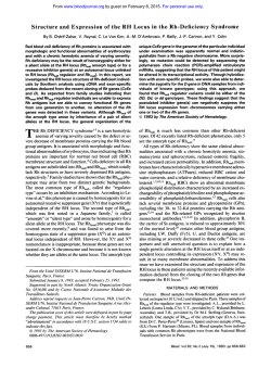
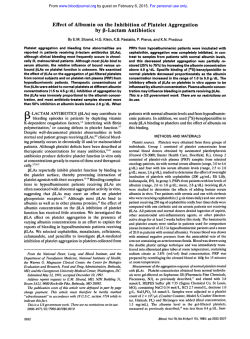
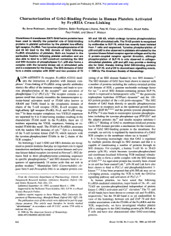
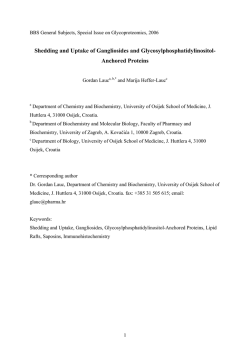
From www.bloodjournal.org by guest on February 6, 2015. For personal use only.
Alpha-Granule Membrane Mirrors the Platelet Plasma Membrane and
Contains the Glycoproteins Ib, IX, and V
By Gaetan Berger, Jean-Marc Mass& and Elisabeth M. Cramer
We have recently shown thatseveral components from the
platelet plasma membrane were also present at different
rates in the alpha-granule membrane. This is the case for
the glycoprotein (GP) Ilb-llla (CD41).CD36,CD9,
PECAMI,
and Rap1b. while theGPb-IX-V complex wasconsidered t o
subcelescape the rule. In this investigation, we studied the
lular localization of GPlb, GPIX, and GPV in the restingplatelets of normalsubjects, patients with Bernard-Soulier syndrome, patients with Gray platelet syndrome, and human
cultured megakaryocytes. Ultra-thin sectionsof the cells
were labeled with antibodies directed against glycocalicin,
GPIb, GPIX, and GPV. We have shown thata significant and
reproducible labelingfor the three
GPs was associated with
the alpha-granule membrane, accounting for approximately
10% of the total
labeling. Furthermore, GPlb labeling appears
colocalized with its alpha-granule-associated ligand, von
Willebrand factor (vWF). After thrombin activation, vWF remained close to the limiting
membrane ofthe open canalicu-
lar system (OCS), suggesting an early association of both
receptor and ligand. Plasma membrane and alpha-granule
labeling was virtuallyabsent from theBernard-Soulier platelets (characterized by a GPlb deficiency), thus proving the
specificity of the reaction. In Gray platelets (storage granule
deficiency syndrome), the smallresidual
alpha-granules
were also occasionally labeled forGPlb, GPIX, and GPV. Cultured megakaryocytes that displayed the classical GPlb distribution, eg, demarcation and plasma membranes, exhibited also a discrete labelingassociated t o t halpha-granules.
e
In conclusion, this study showsthat, evenly for these three
GPs, the alpha-granule membrane mirrors theplasma membrane composition. This
might occur through an endocytotic
process affecting each plasma membrane protein t o a different extent and could
have a physiologic relevance in further
presentation of a receptor bound t o i t alpha-granule
s
ligand
t o t h eplatelet surface.
0 1996 by The American Societyof Hematology.
P
NaCl, 5 mmoVL glucose, pH 7.4) containing 3.5 mg/mL bovine
serum albumin (Sigma Chemical CO,St Louis, MO). The washed
platelets were resuspended and fixed with 1 % glutaraldehyde (Ladd
Research Inc, Burlington, UK) in 0.1 mom phosphate buffer.
Megakaryocytes used in the electron microscopic study were
grown in liquid culture from bone marrow precursors obtained from
normal adult graft donors, as previously described?
LATELET alpha-granules represent a secretory compartment that releases its content after appropriate stimulation. They contain a wide variety of coagulation and adhesive proteins involved in hemostatic mechanisms.*They are
formed during megakaryocyte maturation,’ where they arise
by a double mechanism: endogenous synthesis and endocytosis of plasmatic proteins.”6 Furthermore, in recent studies,
we have demonstrated that several plasma membrane receptors are also present in the alpha-granule limiting membrane.
These receptors include glycoprotein (GP) IIb-IIIa, the fibrinogen receptor’; CD36, the thrombospondin and collagen
receptor*;CD9; PECAM19;and Raplb, a guanosine triphosphate (GTP)-binding protein.’” Therefore, most of the studied proteins seemed to follow the rule, except the GPIb-IXV complex, which was considered until now to be restricted
to the plasma membrane, possibly because of its cytoskeletal
association.” In this study, we have tried to document this
statement using an immunoelectron microscopic approach
performed on normal andpathologic platelets and megakaryocytes. We have been able to demonstrate that small amounts
of GPIb, GPIX, and GPV are associated with the alphagranule membrane. The presence of numerous plasma membrane receptors on the alpha-granule membrane suggests that
the endocytic process that directs plasmatic proteins into the
alpha-granule also affects a large pattern of plasma membrane receptors, although to a different extent.
Antibodies
Different antibodies against glycocalicin, GPIb, GPIX, and GPV
were used for immunoelectron microscopy study. An anti-human
GPIb monoclonal mouse antibody purchased from Dakopatts
(Glostmp, Denmark) was used at 11100 dilution. An anti-human
glycocalicin and GPV polyclonal rabbit antibodies, provided by Dr
Michael Berndt, Prahran, A~stralia,’~.’~
were used, respectively, at
10 p g h L and 30 &mL.An
anti-human GPIX, provided by Dr
Kenneth Clemetson, Bern, Switzerland, was used at 30 pg/mL. An
anti-human GPIX monoclonal antibody, provided by Dr Michael
Berndt,16wasused at 30 pg/mL. For double immunolabeling, an
anti-human P-selectin,” provided by Dr Michael Berndt, andan
anti-human von Willebrand factor (vWF) purchased from Cappel
Laboratory (Downington, PA) were used, respectively, at 30 pg/
mLand 1/50 dilution. Gold-conjugated (10 and15 nm) protein A
purchased from the Department of Cell Biology, University of
Utrecht (Utrecht, The Netherlands) were used, respectively, at 1/80
and1/35 dilutions.
Characterization of the Poiyclonal Antibodies
The specificities of the polyclonal antibodies were assayed
by Western blotting of platelet lysate. Briefly, washed PRP was solubilized by
MATERIALS AND METHODS
Cells
Platelet samples were taken from normal healthy volunteers, from
a patient with Bernard-Soulier syndrome,’* and from a patient with
Gray platelet syndrome.” Blood samples were harvested by venipuncture into plastic tubes containing ACD-C buffer (6.8 mmol/L
citric acid, 11.2 mmoVL trisodium citrate, 24 mmoVL glucose, pH
4.2). The platelet-rich plasma ( P m ) was obtained by centrifugation
for 10 minutes at 180g and 22°C.The isolated platelets were obtained
by centrifugation of PRP for IO minutes at 1,100g and22°C and
washed three times with Tyrode buffer (36 mmol/L citric acid, 5
mmol/L KCI, 2 mmol/L CaC12, 1 mmol/L MgC12, 103 mmol/L
Blood, Vol 87,No 4 (February 15).
1996:
pp 1385-1395
From INSERM (191, Hdpital Henri Mondor, Crkteil, France.
Submitted March 20, 1995; accepted September 15, 1995.
Supported in part by I’Association pour la Recherche contre le
Cancer (ARC) and la Fondation pour laRecherche Medicab (FRM).
Address reprint requests to Dr Elisabeth M. Cramer, INSERM
U91, Hapita1 Henri Mondor. 94010 Crkteil, France.
The publication costs of this article were defrayed in part by page
charge payment. This article must therefore be hereby marked
“advertisement” in accordance with 18 U.S.C. section 1734 solely to
indicate this fact.
0 1996 by The American Society of Hematology.
0006-4971/96/8704-0004$3.00/0
1385
From www.bloodjournal.org by guest on February 6, 2015. For personal use only.
BERGER, MAS&,
'*
AND CRAMER
. .
.
Fig 1. (A and B) lmmunogold
localization of GPlb with a
monoclonal antibody on thin
frozen sections of restingplatelets.
lmmunolabeling is found t o be
associated with
the
plasma
membrane (pm) and open canalicular system (ocs).Some gold
particles are also associated
with thealpha-granule (a) membrane. whilemitochondria(m)
are devoid of labeling. Bars, 250
nm.
n
45
Fig 2. Biochemical characterization of the polyclonal antibodiesused in this studyby Western blotting of plateletlysates. (A) The anti-glycocalicin antibody recognizes a unique protein of approximately
145 kD molecular weight. (B) The anti-GPV antibody
recognizes a protein of approximately 80 kD molecu(C) the anti-GPIX antibody recognizes
lar weight, and
a major protein of approximately 22 kD molecular
weight.
koa
80 kDa
22 kDa
U
From www.bloodjournal.org by guest on February 6, 2015. For personal use only.
GPh, IX, V IN PLATELET ALPHA-GRANULES
1387
..
Fig 3. (A) lmmunogold localization of GPlb on thin frozen sections of resting platelets with the polyclonal anti-glycocalicin antibody. A
strong labeling is observed to be associatedwith the plasma membrane(pm) and alpha-granules(al. (B) Double immunolabelingusing different
sizes of gold particles (for P-selectin: 15 nm, arrows; glycocalicin: 10 nm, arrowheads) allows identification of labeled granules fa) as alphagranules, while mitochondria (m) are devoid of labeling. (inset) The same results are obtained with a double immunolabeling using anti-vWF
(15 nm, arrows) and anti-glycocalicin(10 nm, arrowheads). Bars, 250 nm.
the addition of 2% sodium dodecyl sulfate
(SDS) and 1 mmoVL EDTA
and separated on SDS-polyacrylamide gel electrophoresis (PAGE) with
or without a reducing condition with 5% 2-p-mercaptoethanol. using
a 7% resolving gel and 3% stacking gel. Gels were electroblotted on
niwcellulose filtermembrane by semidrytransfer. The nonspecific
binding was blockedin 5% low fat powder milk, and membranes were
probed with polyclonal antibodiesfor 1 hour at m m temperature. The
labeled proteins were revealed after incubation with sheep anti-rabbit
immunoglobulins bound to peroxidase.
Electron Microscopy
Normal platelets and megakaryocytes were prepared for immunoelectron microscopy as follows: they were fixed in 1% glutaralde-
hyde in 0.1 mol/L phosphate buffer, pH 7.4, for 1 hour at 22°C.
washed three times with the same buffer, embedded in sucrose, and
freezed in liquid nitrogen.'* Pathologic platelets were also embedded
in glycol methacrylate according to the method described by Leduc
and Bernhard'"; this technique permitted a more lasting storage for
precious samples. Then, the immunochemical reactions wereperformed on thin sections collected on copper grids according to the
method of Slot et al.'x Briefly, the sections were labeled by a first
incubation with the antibodies diluted in phosphate-buffered saline
(PBS) containing I % bovine serum albumin (Sigma) for 20 minutes
at 22°C. washed, and then incubated with protein A-gold ( I O nm)
for 20 minutes at room temperature. The sections were counterstained with 2% uranyl acetate, pH 7, and methyl cellulose uranyl.
From www.bloodjournal.org by guest on February 6, 2015. For personal use only.
1388
BERGER, MAS&, AND CRAMER
Fig 4. lmmunolocalization of GPIX (A) and GPV (B) with polyclonal antibodieson thin frozen sections of resting platelets. As expected for
these GPlb-associated proteins, immunolabeling is foundto beassociated with the plasma membrane (pm), the open canalicular system (ocs)
membrane, and also the alpha-granule membrane (a), while mitochondria(m) are devoid of labeling. For GPIX, the same results are obtained
using a monoclonal anti-GPIX instead of the polyclonal antibody (A, inset). Bars, 250 nm.
From www.bloodjournal.org by guest on February 6, 2015. For personal use only.
1389
GPIB, IX, V IN PLATELET ALPHA-GRANULES
For double immunolabeling,a short time fixation with 1% glutaraldehyde in phospate buffer was realized after the incubation with the
gold conjugate, and a second round of labeling with the second
antibody was realized usinga different size of gold particle protein
A
conjugate. Samples were observed on a Philips 450 CM 10 electron
microscope.
Quantitative Estimation
Membrane labeling intensity was evaluated by counting the gold
particles per micrometer of membrane. The alpha-granule pool was
with thealphaquantified by countingthegoldbeadsassociated
granule membrane on one hand and the plasma membrane on the
other: the ratio of intracellular versus plasma membrane pool per
equatorial platelet section was calculated.
RESULTS
Resting Platelets
Immunolocalization of GPIb. To ensure the specificity
of the observed immunolabeling reaction, a monoclonal antibody against GPIb was used as a first instance. Immunolabeling on thin sections allows marking of plasma membrane
proteins as well as intracellular proteins. In this experiment,
labeling for GPIb antigen with a monoclonal antibody was
detected on the plasma membrane, and at the luminal face
of the open canalicular system (OCS). Careful examination
led to the observation that some gold particles were also
bound on alpha-granule membranes, while other structures
such as mitochondria were devoid of labeling (Fig 1A and
B). Using a monospecific polyclonal antibody (Fig 2A)
raised against purified glycocalicin, we obtained a stronger
labeling on the same structures; eg, plasma membrane, OCS;
and alpha-granule membrane (Fig 3A).
Due to their high number, large size, and dark nucleoids,
alpha-granules could be identified. In addition to morphologic criteria, double immunolabeling with both anti-glycocalicin and an anti-P-selectin polyclonal antibody (Fig 3B)
or an anti-vWF polyclonal antibody (Fig 3B, inset) as alphagranule markers confirmed that the labeled granules were,
indeed, alpha-granules because of the codistribution of the
proteins.
Immunolocalization of GPIX and GPV. GPIb has been
described to form a noncovalent complex in the platelet
membrane with GPIX and GPV?’ We have investigated the
localization of both proteins in normal resting platelets and
found that their distributions were similar to GPIb (eg,
plasma membrane, OCS) and that a small proportion of gold
probes were also present on the alpha-granule membrane
(Fig 4A and B).
The characterization of the polyclonal antibody anti-GPIX
is shown in Fig 2C; we have also confirmed our results using
a monoclonal antibody and have obtained a weaker labeling
but an identical distribution (Fig 4A, inset).
Pathologic Platelets
Gray platelet syndrome is a rare congenital bleeding disorde?’ in which the platelets are markedly deficient in morphologically recognizable alpha-granules.” The cause of the abnormality affecting the alpha-granules is unknown, butit
appears that the alpha-granule membrane is normally consti-
tuted because its proteic components, such as P-selectin,23
GPIIb-IIIa,’ and CD36,* are present. On such platelets, immunolabeling for GPIb was found on the plasma membrane
and also on abnormal alpha-granule-like structures (small
residual alpha-granules and small or large vacuoles identified
as empty pathologic alpha-granules; Fig 5A). Similar localization was also obtained for both GPIb-associated glycoproteins, GPIX and GPV (Fig 5C and E). These results suggest
that this pool storage deficiency is not due to an alphagranule membrane composition defect.
Bernard-Soulier syndrome is an inherited bleeding disorder characterized by a deficiency in the GPIb-IX-V comp l e ~ . These
~ ~ . platelets,
~~
embedded in glycol methacrylate
or in sucrose, displayed a severely decreased immunolabeling for GPIb including the alpha-granule labeling. The same
results were obtained for GPIX and GPV and attested for
the specificity of the labeling observed on normal platelets
(Figs 5B, D, and F and 6A).
Colocalization Between GPIb and vWF
To assess that alpha-granule GPIb could be associated
with vWF, we performed a double immunolabeling for GPIb
and vWF combined with quantitative estimations of associated labeling. We found that more than 80% of doublelabeled alpha-granules showed an apparent association between GPIb and vWF labeling (Fig 3B, inset; Fig 6B, inset),
whereas less than10% presented this characteristic when
P-selectin was used as the alpha-granule marker (Fig 3B).
Moreover, less than20%of
such an association between
vWF and the other alpha-granule membrane-associated receptors, CD9 and PECAM1, was found (Table l ) . After
thrombin activation, most vWF labeling was located along
the OCS membrane and was codistributed with GPIb labeling (Fig 6B).
Megakaryocytes
We further investigated the localization of GPIb in the
platelet precursors, the megakaryocytes. Mature human cultured megakaryocytes displayed the classical membrane distribution for GPIb (eg, plasma and demarcation membranes),
but also some alpha-granules showed immunolabeling for
this glycoprotein (Fig 7). Similar results were found for
GPIX and GPV (not shown).
Control
When the primary antibody was either replaced by a nonrelevant polyclonal antibody or omitted from the immunolabeling, staining was completely negative.
Quantitative Estimation
When quantitative measurements of the GPIb labeling
were realized on normal platelets, they showed an association of approximatly 10% of total labeling with the alphagranule membrane (Fig 8). On Bernard-Soulier platelets, a
decrease of 90% of total normal platelet labeling was observed. In these semiquantitative estimations, immunolabeling on glycol methacrylate-embedded Bernard-Soulier plate-
From www.bloodjournal.org by guest on February 6, 2015. For personal use only.
From www.bloodjournal.org by guest on February 6, 2015. For personal use only.
GPIE, IX, V IN PLATELET ALPHA-GRANULES
1391
Fig 5. lmmunolocalization of GPlb, GPIX, and GPV in glycol methacrylate-embedded pathologic platelets. In the Gray platelets, GPlb (A),
GPlX (B), and GPV (C) are present and localized on the same structures, eg, plasma membrane tpm), small residual granules (a), and also
vacuolar structure (a'), usually considered as empty granules. In the Bernard-Soulier platelets, which do notexpress the GPlb-IX-V complex,
the immunolabeling forGPlb (D), GPlX (E), and GPV (F) is seriously decreased. This finding also attests for the specificity
of the reaction. Bar,
250 nm.
A
Fig
6. Sucrose-embedded
Bernard-Soulier platelets immunolabeled for GPlb: As in glycol
methacrylate-embedded cells,
membrane labeling for GPlb is
virtually absent, close t o the
background staining. (B) Double
immunolabeling forGPlb, 10 nm
gold particles(arrowheads), and
gold
particles
vWf, 15 nm
(arrows). On resting platelets,
double-labeled alpha-granules
tinset, A) show
frequently
a
close association of both labeling. Afterthrombin activation,
most of the
vWf labeling appears
to be associated with the open
canalicular system (OCS) membraneandcodistributed
with
GPlb. Bar, 250 nm.
.
From www.bloodjournal.org by guest on February 6, 2015. For personal use only.
1392
BERGER,MASSE,
Table 1. Colocalization of GPlb and vWF
Double Labeling
% of Alpha-Granules
Showing Colocalirationof Both Markers (n)
GPlbIvWF
GPlbP-selectin
CD9IvWF
PECAMlIvWF
(20)
9 (15)
(15)
14 (20)
On normal platelets, 82% of double-labeled alpha-granules show a
colocalization of GPlb and vWF. When P-selectin is used as an alphagranule marker, only 9% of double-labeled granules present such a
colocalization. As a control, double labeling between vWF and two
alpha-granule receptors, CD9 and PECAM1, show, respectively, 19%
and 14% of marker association. These data suggest that GPlb and
vWF are specifically associated.
lets was compared with glycol methacrylate-embedded
control platelets. In the same way, immunolabeling for GPIb
on cryosectioned control and Bernard-Soulier platelets were
compared (Fig 9).
AND CRAMER
DISCUSSION
The ultrastructural localization of membrane receptorscell
in
organization may reflect the dynamic feature of these membranes within secretory and endocytotic pathways. The alphagranules are the main secretory organelles of platelets and megakaryocytes. They arise from a dual mechanism: endogenous
synthesis occumng in megakaryocytes and endocytosis from
the surrounding extracellular medium.'26 The first mechanism
as thrombospondin,
involvesmanyhemostaticfactors,such
beta-thromboglobulin, and vWF, whose correspondingmRNAs
are present in megakaryocytes.'Thesecondpathwayisthe
route of several plasmatic proteins such as immunoglobulins,
albumin,andfibrinogen,which
are acquiredexclusivelyby
endocytosis?' Fibrinogen endocytosis appearsto be a receptormediated process, under the control ofGPIIbIIIaz8This fibrinogen endocytosis takes place at theofend
megakaryocyte maturation" and continues during platelet life?9
The proteins of the alpha-granule membrane can also be
categorized in two groups. On the one hand, some receptors
Fig 7. lmmunogold localization of GPlb with a polyclonal anti-glycocalicin antibody on cultured megakaryocytes embedded in glycol
methacrylate. In these cells, the GPlb labeling is widely present on demarcation membranes (dm), and some alpha-granules (a1 are positive.
Bar, 250 nm.
From www.bloodjournal.org by guest on February 6, 2015. For personal use only.
GPIB, IX, V IN PLATELET ALPHA-GRANULES
are restricted to the alpha-granule limiting membrane and
are absent from the plasma membrane, such as P-~electin,~'
osteonectin,31 and GMP33?* On the other hand, further
studies have shown that some proteins that are normal components of the plasma membrane are also present on the
alpha-granule membrane at different rates; eg, GPIIbIIIa,
with approximately 50% of the total platelet pool of GPIIbIIIa present in the alpha-granule membranes'; CD36, with
35%'; CD9 and PECAM1, with 25%9; and the small GTPbinding protein RapIb, with 15%.1° Until now, GPIb, which
is the platelet receptor that mediates the adhesion of unstimulated platelets to v W F , ~was
~ considered to be restricted to
the plasma membrane of resting platelets, probably because
of its cytoskeletal association.*'In the present report, we
show that even for this membrane-associated protein, a consistent amount of GPIb, and also associated glycoproteins
IX and V, is present in the platelet alpha-granule membrane.
In previous report^,'^,^^ we proposed that GPIb was absent
from the alpha-granule membrane because the observed scattered labeling associated with this structure was considered
as background staining. Technical improvements-first, in
the marked decrease of background staining using glycin as
the saturation agent and phosphate buffer rather than Tris
buffer, and second, using protein-A-gold instead of goat antirabbit gold-have increased technical sensitivity and permittedto
demonstrate the observed labeling for GPIb
associated with the alpha-granule membrane as a specific
Sucrose embedded cells
~ o m platelets
d
n=10
Bernard-Soulier
platelets n=10
Fig 9. Comparison of GPlb labelingon normal and Bernard-Soulier
platelets. On Bernard-Souliar platelets, the totallabeling appears seriously decreased.
0
Alpha-granuleassociatedlabeling
plasma membrane andocs associated labeling
0
labeled mitochondria: background staining.
Fig 8. Distribution of the GPlb labeling on normal platelets (n
= 25). On normal platelets, approximately 109'0 of total labeling is
associatedwith thealpha-granules. Mitochondria representthe background staining.
association. Moreover, this result has been definitely confirmed using a monoclonal antibody.
Labeling for the three glycoproteins Ib, IX, and V was
dramatically decreased on platelets from a patient with Bernard-Soulier syndrome, attesting for the specificity of the
reaction. On platelets from a patient with the storage disease
Gray platelet syndrome, in which platelets lacknormal
alpha-granules, the residual pathologic granules were labeled
for these three associated glycoproteins (Ib, IX, and V). This
finding shows that GPIb-IX-V localization is unaltered in
From www.bloodjournal.org by guest on February 6, 2015. For personal use only.
1394
BERGER,MASSE,
these patients and confirms and supplements previous observations describing this pathology as soluble protein storage
deficiency in which the alpha-granule membrane is normal.
Relative to previous findings in fibrinogen endocytosis
that implicate GPIIb-IIIa~*and also to the presence in the
alpha-granule membrane of numerous plasma membrane receptors, these results raise the question of a protein alphagranule targeting signal. A cDNA P-selectin transfection
study in a pituitary cell line has led to the description of a
23-amino acid cytoplasmic domain of P-selectin responsible
for its direct transport to secretory g r a n ~ l e s .However,
~~.~~
concerning proteins like GPIIb-IIIa, CD36, or GPIb, which
display a double localization in platelets and megakaryocytes, it appears more delicate to consider the existence of
a specific signal leading the protein in two different compartments. On the other hand, the hypothesis of plasma membrane targeting followed by a specific and regulated endocytosis to the alpha-granules, as it has been proposedas
targeting mechanism of a lysosomal pr~tein,~’
could appear
more suitable. Numerous receptors, included some 8-integrins, have been shown to contain in their cytoplamic domain
a specific endocytotic motif, NPXY.38.39
Other specific signals, such as the di-leucin motif and tyrosine-based motif,
or secondary structures in the cytoplasmic tail of membrane
receptors are also commonly proposed as endocytotic and
targeting signal^.^'.^' The description of such a specific signal
governing a differential rate of endocytosis from the platelet
plasma membrane to the alpha-granules, perhaps through
differential coated pits affinity, could explain our results but
remains to be determined. Such a process has been described
for the transferrin
and for the low density lipoprotein receptor.43
Furthermore, a preliminary study of GPLIb-IIIa expression
during megakaryocyte maturation shows the appearance of
the first protein expression on the plasma membrane, on
demarcation membranes, andthen on the alpha-granule
membrane. These observations hinge on the hypothesis of
an indirect targeting, implicating first a plasma membrane
expression.
Concerning the physiologic relevance of the presence of
GPIb on the alpha-granule membrane, the observation of an
apparent codistribution of GPIb and its ligand vWF on resting and thrombin-activated platelets raises the possibility for
a specific role of GPIb in the redistribution and the presentation of functional adhesive complex (GPIb-vWF) during
platelet activation. Such a phenomenon has already been
proposed for GPIIb-IIIa and f i b r i n ~ g e nand
~ . ~might
~
begeneralized to other alpha-granule receptors.
In conclusion, this report documents the original composition of the limiting membrane of alpha-granules, the platelet
secretory organelles, which qualitatively mimicks the plasma
membrane, and proposes a functional interpretation for this
composition.
ACKNOWLEDGMENT
We thank Valerie Arqanuthurry for biochemical characterization
of thepolyclonal antibodies and Josette Guichard and Tayebeh Youssefian for helpful comments.
AND CRAMER
REFERENCES
1. Harrison P, Cramer EM: Platelet a-granules. BloodReview
7:52, 1993
2.KiefferN,
Guichard J, Farcet JP, Vainchenker W, BretonGorius J: Biosynthesis of major platelet proteins in human blood
platelets. Eur J Biochem 164:189, 1987
3. Cramer EM, Debili N, Martin JF, Galdwin AM, Breton-Gorius
J, Harrison P, Savidge GF, Vainchenker W: Uncoordinated expression of fibrinogen compared with thrombospondin and von Willebrand factor in maturing human megakaryocytes. Blood 73:1123,
1989
4. George JN, Saucerman S , Levine SP, Knieriem LK, Bainton
DF: Immunoglobulin G is a platelet alpha granule-secreted protein.
J Clin Invest 76:2020, 1985
5. Louache F, Debili N, Cramer EM, Breton-Conus J, Vainchenker W: Fibrinogen is not synthesized by humanmegakaryocytes. Blood 77:311, 1991
6. Harrison P, Wilbourn B, Debili N, Vainchenker W, Lawrie
AS, Breton-Gorius J, Masse JM, Savidge GF, Cramer EM: Uptake
of plasma fibrinogen into the alpha-granules of human megakaryocytes and platelets. J Clin Invest 84:1320, 1989
7. Cramer EM, Savidge GF, Vainchenker W, Berndt MC, Pidard
D, Caen JP, Masse JM, Breton-Gorius J: Alpha-granule pool of
GPIIb-IIIa in normal and pathologic platelets and megakaryocytes.
Blood 75:1220, 1990
8. Berger G, Caen JP, Berndt MC, Cramer EM: Ultrastructural
demonstration of CD36 in the alpha-granule membrane of human
platelets and megakaryocytes. Blood 823034, 1993
9. Cramer EM, Berger G, Berndt MC: Platelet alpha-granule and
plasma membrane share two new components: CD9 and PECAMl .
Blood 84:1722, 1994
10. Berger G, Quarck R, Tenza D, Levy-Toledano S , deGunzburg
J, Cramer EM: Ultrastructural localization of the small GTP-binding
protein Rap1 in human platelets and megakaryocytes. Br J Haematol
88:372, 1994
11. Fox JEB: The platelet cytoskeleton. Thromb Haemost 70:884,
1993
12. Cramer EM, Meyer D, le Menn R, Breton-Gorius J: Eccentric
localization of von Willebrand factor in an internal structure of
platelet alpha-granule resembling that of Weibel-Palade bodies.
Blood 66:710, 1985
13. Cramer EM, Vainchenker W, Vinci G, Guichard J, BretonGorius J: Gray platelet syndrome: Immunoelectron microscopic localization of fibrinogen and von Willebrand factor in platelets and
megakaryocytes. Blood 66:1309, 1985
14. Cramer EM, Lu H, Caen JP, Soria C, Berndt MC, Tenza D:
Differential redistribution of platelet glycoproteins Ib and IIb-IIIa
after plasmin stimulation. Blood 77:694, 1991
15. Berndt MC, Phillips DR: Purification and preliminary physicochemical characterization of human platelet membrane glycoprotein V. J Biol Chem 25659, 1981
16. Berndt MC, Gregory C, Chong BH, Zola H, Castaldi PA:
Additional glycoprotein defect in Bernard-Soulier syndrome: Confirmation of genetic basis by paretal analysis. Blood 62:800, 1983
17. Gamble JR, Skinner MP, Berndt MC, Vadas MA: Prevention
of activated neutrophil adhesion to endothelium by soluble adhesion
protein GMP140. Science 249:414, 1990
18. Slot JW, Geuze JJ, Weerkamp AJ: Localization of Macromolecular Components by Application of the Irnmunogold Technique
on Cryosectioned Bacteria. New York, NY, Academic, 1988
19. Leduc EH, Bernhard W: Recent modification of the glycol
methacrylate embedding procedure. J Ultrastruct Res 10:196, 1967
20. Modderman PW, Admiraal LC, Sonnenberg A, vonder Borne
AEGKr: Glycoproteins V and Ib-IX form a noncovalent complex in
the platelet membrane. J Biol Chem 267:364, 1992
From www.bloodjournal.org by guest on February 6, 2015. For personal use only.
GPIE, IX, V IN PLATELET ALPHA-GRANULES
21. Raccuglia G Gray platelet syndrome. A variety of qualitative
platelet disorder. Am J Med 51:818, 1971
22. White JG: Ultrastructural studies of Gray platelet syndrome.
Am J Path01 95445, 1979
23. Rosa JP, George JN, Bainton DF, Nurden AT, Caen JP,
McEver RP:Gray platelet syndrome. Demonstration of alpha granule
membranes that can fuse with the cell surface. J Clin Invest 8 0 1 138,
1987
24. Bernard J, Soulier JP: Sur une nouvelle vari6t6 de distrophie
thrombocytaire htmorragipare cong6nitale. Semin H6p Paris
24:3217, 1948
25. Clemetson KJ, McGregor JL, James E: Characterization of
platelet membrane glycoprotein abnormalities in Bernard-Soulier
syndrome and comparison with normal by surface-labeling techniques and high-resolution two dimensional gel electrophoresis. J
Clin Invest 70:304, 1982
26. George JN: Immunoglobulin G: Its significance for theevaluation of thrombocytopenia and for understanding the origin of alphagranule proteins. Blood 76859, 1990
27. Handagama PJ, Shuman MA, Bainton D F Incorporation of
intravenously injected albumin, IgG and fibrinogen in guinea pig
megakaryocyte granules. Proc NatlAcad Sci USA 84:861-865.
84:861, 1989
28. Handagama PJ, Scarborough RM, Shuman MA, Bainton DF:
Endocytosis of fibrinogen into megakaryocyte and platelet alphagranules is mediated by aIIb-p3 (glycoprotein IIb-IIIa). Blood
82:135, 1993
29. Heilmann E, Hynes LA, Friese P, George JN, Burstein SA,
Dale GL: Dog platelets accumulate intracellular fibrinogen as they
age. J Cell Physiol 161:23, 1994
30. McEver RP: Properties of GMP140, an inducible granule
membrane protein of platelets and endothelium. Blood Cells 1673,
1990
31. Metzelaar MJ, Heijnen HFG, Sixma JJ, Nieuwenhuis HK.
Identification of a 33-Kd protein associated with the alpha-granule
membrane (GMP-33) that is expressed on the surface of activated
platelets. Blood 79:372, 1992
32. Breton-Gorius J, Clezardin P, Guichard J, Debili N, Mamaval
L, Vainchenker W, Cramer EM, Delmas P Localization of platelet
osteonectin at the internal face of the alpha-granule membrane in
platelets and megakaryocytes. Blood 79:936, 1992
33. George JN, Torres MM: Thrombin decrease von Willebrand
factor binding to platelet glycoprotein b.Blood 71:1253, 1988
1395
34.LuH, Menashi S, Garcia I, Cramer EM, Li H, Tenza D,
de Romeuf C, Soria J, Soria C: Reversibility of thrombin-induced
decrease in platelet glycoprotein Ib function. Br J Haematol 85: 116,
1993
35. Koedam JA, Cramer EM, Briend E, Furie B, Furie BC,
Wagner DD: P-selectin, alpha granule membrane protein of platelets
and endothelial cells, follows the regulatory secretory pathway in
AtT-20 cells. J Cell Biol 3:617, 1992
36. Disdier M, Morrissey JH, Fugate RD, Bainton DF, McEver
R P Cytoplasmic domain of P-selectin (CD62) contains the signal
for sorting into the regulated secretory pathway. Mol Biol Cell 3:309,
1992
37. Braun M, Waheed A, von Figura K Lysosomal acid phosphatase is transported to lysosomes via the cell surface. EMBO J 8:3633,
1989
38. Trowbridge IS: Endocytosis and signals for internalization.
Curr @in Cell Biol 3:634, 1991
39. Vignoud L, Usson Y, Balzac F, Tarone G, Block M R Internalization of the alpha 5 beta 1 integrin does not depend on “NPXY”
signals. Biochem Biophys Res Commun 199:603, 1994
40. Letoumeur F, Klausner RD: A novel di-leucin motif and a
tyrosine-based motif independently mediate lysosomal targeting and
endocytosis of CD3 chains. Cell 69:1143, 1992
41. Collawn JF, Lai A, Doming0 D, Fitch M, Hatton S, Trowbridge IS: YTRF is the conserved internalization signal of the transferrin receptor, and asecond YTRF signal at position 3 1-34 enhances
endocytosis. J Biol Chem 268:21686, 1993
42. Collawn JF, Stangel M, Kuhn LA, Esekogwu V, Jing S, Trowbridge IS, Tainer JA: Transferrin receptor internalization sequence
YXRF implicates a tight turn as the structural recognition motif for
endocytosis. Cell 63:1060, 1990
43. Chen WJ, Goldstein JL, Brown MS: NPXY, a sequence often
found in cytoplasmic tails, is required for coated pit-mediated internalization of low density lipoprotein receptor. J Cell Biol 265:3116,
1990
44. Suzuki H, Baba I, Tanoue K, Yamazaki H: A possible role
of a-granule GPIIb-IIIa as acarrier of intragranular fibrinogen during
release reaction. Thromb Haemost 65:698, 1991 (abstr)
45. Nurden P, Humbert M, Bihour C, Piotrowicz R, Kunicki TJ,
Nurden AT:A MAb (AP6) recognizing the sequence 209-222 of
GPIIIa shows an activation-dependent binding to the surface membrane of platelets yet binds to alpha-granule membranes of unstimulated platelets. Thromb Haemost 73:1194, 1995 (abstr)
From www.bloodjournal.org by guest on February 6, 2015. For personal use only.
1996 87: 1385-1395
Alpha-granule membrane mirrors the platelet plasma membrane and
contains the glycoproteins Ib, IX, and V
G Berger, JM Masse and EM Cramer
Updated information and services can be found at:
http://www.bloodjournal.org/content/87/4/1385.full.html
Articles on similar topics can be found in the following Blood collections
Information about reproducing this article in parts or in its entirety may be found online at:
http://www.bloodjournal.org/site/misc/rights.xhtml#repub_requests
Information about ordering reprints may be found online at:
http://www.bloodjournal.org/site/misc/rights.xhtml#reprints
Information about subscriptions and ASH membership may be found online at:
http://www.bloodjournal.org/site/subscriptions/index.xhtml
Blood (print ISSN 0006-4971, online ISSN 1528-0020), is published weekly by the American
Society of Hematology, 2021 L St, NW, Suite 900, Washington DC 20036.
Copyright 2011 by The American Society of Hematology; all rights reserved.


© Copyright 2026