
Platelets Generated In Vitro From Proplatelet-Displaying
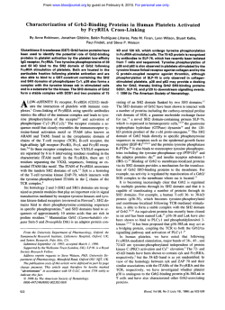
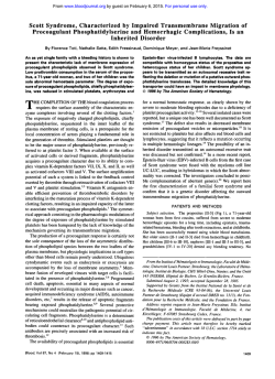
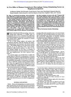
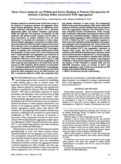
From www.bloodjournal.org by guest on February 6, 2015. For personal use only.
Platelets Generated In Vitro From Proplatelet-Displaying Human
Megakaryocytes Are Functional
By Esther S.Choi, Janet L. Nichol, Martha M. Hokom, Alex C. Hornkohl, and Pamela Hunt
An in vitro culturesystemdemonstrating the transitions
from megakaryocyte progenitors to functional platelets is
described. CD3Cselected cells from normal human peripheral blood are cultured under conditions
that promote megakaryocyte formation. After 8 to 11 days, enriched populations of mature megakaryocytes
are
replated under
conditions that favor the development of proplatelets. Proplatelets express the platelet-specificproteins,glycoproteins Ib and Ilb (GPlb and GPllb), and fibrinogen and also
contain microtubule coils equal in size to those found in
plasma-derived platelets. In addition, proplatelets have ultrastructuralfeatures in common with plasma-derivedplatelets. Platelet-sized particlesfrom the proplatelet culture su-
pernatants are examined. Ultrastructurally, these particles
are identicalto plasma-derivedplatelets. Functionally, these
culture-derived platelets aggregate in response to both
thrombin and adenosine diphosphate (ADP) plus fibrinogen.
This aggregation is specifically inhibited by the addition of
a function-blocking anti-GPllbllla antibody. Culture-derived
platelets stimulated with agonists also express the activation-dependent antigens P-selectin
and functional fibrinogen
receptor. This is the first description of an in vitro culture
system that sequentially
demonstrates
megakaryocyte
growth, development, and platelet production.
0 1995 by The American Societyof Hematology.
P
LATELETS ORIGINATE from megakaryocytes during
tems that rely on rodent10~14~15~18~23,24"29
or bovine" megathe terminal differentiationphase of
karyocytes may be of limited value to the study of human
thrombopoiesis.
However, the precise mechanism by which platelets are shed
This report describes a model system in which functional
from megakaryocytes is not fully understood. Studies of this
platelets are generated in vitro from human megakaryocytes.
process are hampered by the low frequency of megakaryoPlatelet formation appears to occur via proplatelet intermedicytes in bone marrow as well as by a lack of in vitro systems
ate structures. The system exploits earlier observations that
in which platelet shedding reproducibly occurs. Theoretic
human megakaryocytes can be generated from peripheral
models of platelet shedding have been developed from obserblood leukocytes stimulated with plasma from thrombocytovations of fixed cells. Megakaryocytes displaying membrapenic ani mal^.^' In the present study, the frequency of culnous structures outlining platelet-fields form the basis of
ture-generated megakaryocytes was improved by preenrichthe demarcation membrane model of platelet shedding.'.'
ing the leukocyte population for CD34+ megakaryocyte
Observations of long cytoplasmic processes emanating from
progenitors. Mature megakaryocytes that appear within 8 to
megakaryocyte cell bodies have led to the proplatelet con1l days of culture are then available for studies of proplatelet
cept?"' Proplatelets, dependent on microtubules for stabildevelopment and, subsequently, for platelet fragmentation.
ity,l2-I5display constrictions along their lengths that define
This in vitro system is relevant to clinical conditions that
platelet-sized area^.",^^"^ The areas between the attenuation
affect the last phase of thrombopoiesis, platelet formation
points contain platelet-specific organelles and proand release.
t e i n ~ . ' " ' ' ~Micrographs
~ ~ ~ , ~ ~ . of
~ ~bone marrowfixed in situ
show proplatelets projecting through the endothelial cell
MATERIALS AND METHODS
layer of marrow venous sinusoids into the c i r ~ u l a t i o n . ~ ~ " ~ ' ~ ~ ' ~
It has been postulated that the circulatory shear force within
Isolation of CD34+ Progenitor Cells From Peripheral
the m a r r ~ w ' . ~or~possibly
.~~
the
aids
in
the fragBlood
mentation of proplatelets into platelets.
Written informed consent was obtained from donors of leukapherThe study of proplatelet development has been advanced
esis units, normal AB plasma, and plasma-derived platelets. Leuthrough the use of tissue culture systems that permit this
kapheresis units from a pool of normal healthy donors committed to
differentiation process to occur in vitro.L4~15'1',1y,23-30
Although
the program were purchased from HemaCare (LeukaPAK; Sherman
these culture systems have been useful for the study of proOaks, CA). Peripheral mononuclear cells were isolated by FicollPaque (Pharmacia, Piscataway, NJ), rinsed, and adherence-depleted
platelet development, they have been of limited value for
overnight. Nonadherent cells were collected and fractionated with
the study of platelet fragmentation as observations of platecounterflow centrifugal elutriation in a Beckman (Fullerton, CA) J2let-like particles in these cultures are reported as rare
M1 centrifuge with a JE-6B elutriation rotor to enrich for megakaryevents14.15.18,25,26.31
and in no case has the identity and function
ocyte progenitor^.'^" Rotor speed was 2,020 rpm, and the flow rate
of the particles been established. Furthermore, culture syswas increased by 0.5 W m i n from 8.5 W m i n to 13 W m i n over
From AMGEN, Inc Thousand Oaks, CA.
Submitted March 17, 1994; accepted September 13, 1994.
Address reprint requests to Pamela Hunt, PhD, AMGEN,Inc,
Amgen Center, mail stop T-1A-206, Thousand O a b , CA 91320.
The publication costs of this article were defrayed in part by page
charge payment. This article must therefore be hereby marked
"advertisement" in accordance with 18 U.S.C. section 1734 solely to
indicate this fact.
0 1995 by The American Society of Hematology.
0006-4971/95/8502-0002$3.00/0
402
63 minutes. Cells of interest were collected in 50-mL fractions at
flow rates from 11 to 13 mUmin and frozen in 90%fetal bovine
serum (FBS) and 10% dimethylsulfoxide (DMSO) in the cell freezer
(Gordinier 9000) using Cryopack Software Version 3.2 (Gordinier
Electronics Inc. Roseville, MI) until use. In some experiments, leukapheresis units were processed without elutriation. Mononuclear
cells were isolated by Ficoll-Paque in Hank's buffered saline solution
(HBSS) containing phenol red (GIBCO, Grand Island, NY). After
rinsing in HBSS, cells were either processed immediately or after
overnight incubation in Iscove's Modified Dulbecco's medium
(IMDM) and 10% FBS at 4°C.
Blood, Vol 85, No 2 (January 15). 1995: pp 402-413
From www.bloodjournal.org by guest on February 6, 2015. For personal use only.
403
FUNCTIONALCULTURE-DERIVED HUMAN PLATELETS
CD34" progenitor cells were isolated using a magnetic cell sorting
system, Mini-MACS (Miltenyi Biotec, Auburn, CA) by one of two
methods. One method used anti-CD34-biotinylated antibody from
AMAC (Westbrook, ME) followed by stxptavidin microbeads (Miltenyi Biotec). The other method used the CD34 isolation kit (Miltenyi Biotec) in accordance with the manufacturer's recommendations. When elutriated, frozen/thawed cells were processed; an
average of 1.4 X 10⁶ CD34' cells were obtained per leukapheresis
unit, with a range of 2.0 X 10" to 4.0 X 10⁶ (n = 9). When nonelutriated, fresh cells were processed, an average of 4.8 X lo6 CD34+
cells were obtained per leukapheresis unit, with a range of 2.9 X
lo6to 6.2 X lo6 (n = 4). Elutriated cell populations contained fewer
CD34" cells than nonelutriated cell populations as they had been
preenriched for megakaryocyte progenitors. The wide range in the
number of CD34+ cells obtained was attributed to donor variation3&
The purity of CD3Cselected cells was determined for each isolation by using a combination of anti-CD34 antibodies [HPCA-l and
HPCA-2, each at 1:lO in phosphate-buffered saline (PBS); Becton
Dickinson, Lincoln Park, NJ] and the HistoMark Streptavidin-P-Gal
system (Kirkegaard and Perry Labs, Gaithersburg, MD). Immunopositive blue cells were counted microscopically. On the average,
cells selected with anti-CD34 biotinylated antibody followed by
streptavidin microbeads were about 40% CD34' (range, 15% to
93%; r = 19). Similarly, cells selected with the CD34 isolation kit
averaged about 50% CD34+ (range, 22% to 90%; n = 8). When
CD34-selected cells were reapplied to Mini-MACS columns, the
percentage of CD34+ cells increased to an average of 90% (range,
83% to 93.5%; n = 4). The percentage of CD34+ cells obtained by
this staining method and by flow cytometric analyses were virtually
identical in every case studied.
Culture of Human Megakaryocytes
CD34+ enriched cells were stimulated to form megakaryocytes in
the culture system described by Mazur et a l . 3 2 All of the reagents
were purchased from GIBCO unless indicated otherwise. Briefly,
the cells were plated at 5 X 10' cells per milliliter in growth medium
(Meg GM) that consisted of IMDM with 1 X penicillin/streptomycin/
glutamine (P/S/G), 1 X sodium pyruvate, 1 X minimum essential
medium vitamins, 1 X nonessential amino acids, 0.1 mmoVL 3mercapto l-propanediol, 2 mg/mL L-asparagine, and 0.2% deionized
bovine serum albumin (BSA; Calbiochem, San Diego, CA). Fresh
Meg GM was prepared every 2 weeks. Meg GM was supplemented
with 10% normal human platelet-poor heparinized (33 U/mL) AB
plasma, and 10% aplastic canine
Cells were incubated in
96-well or 24-well tissue culture plates (Falcon, Lincoln Park, NJ)
in volumes of 100 pL or 5 0 0 pL, respectively, at 37°C with 5%
CO2 for 8 to 11 days.
Enrichment of Human Megakaryocytes
Megakaryocytes were enriched from the cultured cell population
according to the procedure of Levine and F e d ~ r k o ?with
~
some
, OO
modifications. Cultured cells were collected, centrifuged at 1O
rpm for 15 minutes, resuspended in CATCH buffer at 5 X lO%nL,
and subjected to velocity ~edimentation~'.~~
that consisted of a twostep BSA-gradient, 2.41% (450 pL) and 4.83% (900 pL), at lg for
1.5 hours. No more than 2.5 X 10' cells were applied to onevelocity
sedimentation tube (Falcon no. 2063). Cells that entered the bottom
layer were collected. Megakaryocytes were identified by immunostaining with an anti-glycoprotein Ib/Ib/IIb antibody cocktail (antiGPIb/Ib/IIb; anti-Ib purchased from DAKO Corp, Carpenteria, CA;
anti-Ib and anti-IIb purchased from Biodesign, Kennebunkprt, ME)
and the HistoMark Streptavidin-b-Gal System. Immunopositive blue
cells were microscopically counted. For morphologic analyses, the
cells were cytocentrifuged onto slides (Shandon Inc, Pittsburgh, PA)
at 400 rpm for 5 minutes and stained with a modification of WrightGiemsa stain using Wescor's automatic cell stainer (Wescor Inc,
Logan, UT; model no. 7120). Megakaryocytes were staged according
to the criteria of vin ne."
Induction of Proplatelets From Mature Megakaryocytes
The megakaryocyte-enriched population was replated in Meg GM
at 5 X 1cv' cells per milliliter in 96-well plates (Falcon no. 3072)
supplemented with 10% heparinized human AB plasma but without
aplastic canine serum. Some experiments were conducted in the
absence of 10% AB plasma to determine the necessity of plasma
for proplatelet induction. The cultures were examined daily for emergence of proplatelets. A megakaryocyte bearing one or more cytoplasmic processes was considered a proplatelet-displaying megakaryocyte. The processes were at least twice the length of the cell body
diameter. The percentage of megakaryocytes displaying one or more
cytoplasmic processes was determined by visual examination of
video prints taken of the culture wells.
Detection of Platelet-Specijc GPIbIIbIIIb on Proplatelet
Membranes
Megakaryocytes displaying proplatelets were fixed in situ by adding glutaraldehyde (1.6% final concentration) to the culture wells.
The culture plates were then centrifuged at 800 rpm for 15 minutes.
The cells were rinsed and blocked with 10% normal goat serum
(NGS) for 20 minutes at room temperature. Anti-GPIb/Ib/IIb cocktail
and the HistoMark Streptavidin-P-Gal system was used as described
above.
Detection of Fibrinogen Within Proplatelets by Indirect
Imrnunojluorescence
Megakaryocytes displaying proplatelets were stabilized with
0.38% formaldehyde and 100 mmoVL EDTA and sedimented onto
silanized slides (Oncor, Gaithersburg, MD) by cytocentrifugation.
These cells were fixed for 20 minutes in 4% paraformaldehyde and
1% Triton-X 100, rinsed three times in PBS, and then blocked for
20 minutes in 10% FBS-PBS at room temperature. Mouse antifibrinogen monoclonal antibody (MoAb; American Diagnostics,
Greenwich, CT) was used at 1:50 in 10% FBS-PBS for 1 hour at
room temperature. Goat anti-mouse-fluorescein isothiocyanate
(FITC; Cappel, Durham, NC) was used at 1:1,OOO in 10% FBSPBS for 30 minutes at room temperature. Slides were mounted in
Immumount (Shandon) and visualized with an Olympus BH2 microscope (Olympus, Chatsworth, CA).
Detection of Microtubules Within Proplatelets by Indirect
Imrnunojluorescence
For microtubule staining, megakaryocytes displaying proplatelets
were treated as above except that 4% paraformaldehyde, 1% TritonX 100, and 10%methanol was used as a fixative. Cells were blocked
with 5% NGS in PBS for 20 minutes at room temperature. The antitubulin antibody cocktail (anti-a-tubulin MoAb, Amersham, Buckinghamshire, England; anti-P-tubulin MoAb, Amersham; and rabbit
anti-tubulin antibody, Biomedical Technologies, Stoughton, MA, at
manufacturers' recommended dilutions) was incubated with cells at
37°C for 1 hour. Goat anti-mouse IgG-FITC and goat anti-rabbit IgGFITC (Cappel; at 1:1,OOO each) were used as secondary antibodies at
37°C for 30 minutes. Cells were rinsed and mounted in Immumount
and visualized with a Nikon MICROPHOT-FXA microscope (Nikon, Irvine, CA).
Electron Microscopy of Proplatelets and Platelets
Megakaryocytes displaying proplatelets were stabilized with
0.38% formaldehyde and 100 mmoVL EDTA, centrifuged at 800
From www.bloodjournal.org by guest on February 6, 2015. For personal use only.
CH01 ET AL
404
.
Y
. "-
Fig l. Mature megakaryocyte selection, proplatelet development, and presence of platelet-specific proteins detected on the surface and
within proplatelets. (AI A representative Wright-Giemsa staining of megakaryocytes after 8 days of culture time enriched by velocity sedimentation 153% megakaryocytes; bar, 40 pm). (B) Phase micrograph of proplatelet development 3 days after replating (bar, 40 pm). (C1 Anti-GPlbl
Ib/llb antibody staining on proplatelets detected by HistoMark, bright-field micrograph (bar, 15 pm). (D) Control panel for HistoMark, in which
the primary antibody cocktail was omitted (bar, 13 pm). (E) Anti-fibrinogen antibody within proplatelets detected by indirect immunofluorescence (bar, 40 pm).
rpm for 1 S minutes, resuspended in 1% glutaraldehyde, and allowed
to settle on poly-L-lysine coated aclar film for 4.5 minutes. The cells
were rinsed with PBS, treated with I % OsOI for I hour, rinsed with
PBS,and sequentially dehydrated with ethanol (30%. SO%, 70%.
and 90%). Uranyl acetate (2%) was included during the 70% ethanol
dehydration step. The cells were infiltrated with 50% L.R. White
plastic (EM Sciences, Fort Washington, PA: medium grade) in ethanol for IS minutes, then treated to two changes of 1 0 0 % L.R. White
over 24 hours. Fresh L.R. White was polymerized onto aclar film
in aluminum dishes at 52°Cfor 24 hours. Proplatelets were identified
under the light microscope on polymerized plastic, sectioned, and
viewed under the electron microscope (Phillips 300, Mahwah, NJ).
Plasma-derived platelets were obtained as reported previo~sly.'~
Platelet-sized fragments (putative culture-derived platelets) were isolated from culture supernatantsin which significant numbersof megakaryocytes displaying proplatelets had been visible 24 hours earlier
(proplatelet cultures). These culture supernatants were centrifuged
at 1,OOO rpm (Sorvall RT6000, DuPont, Newtown, C T ) for IS minutes at roomtemperature. Half ofthe culture volumes (approximately
S0 pL per well) was taken from the top of each well and pooled to
one tube. The pooled material was centrifuged at 1,200 rpm (Sorvall
RT6000) for IS minutes to pellet cells carried over from culture.
The supernatant containing platelet-sized fragments was placed in
fresh 1.5-mL microfuge tubes and centrifuged at 7,000 rpm (Beckman Microfuge 12) for S minutes at room temperature. The supernatant from each tube was aspirated, leaving about 20 pL of medium
in each tube. The pellets containing platelet-sized fragments were
resuspended in the residual 20 pL ofmedium of each tubeand
pooled in one microfuge tube. Plasma-derived and culture-derived
platelets were fixed, dehydrated, and infiltrated as above in microfuge tubes that were centrifuged for each change of solution. Polymerization was also performed in the microfuge tubes, which were
cut away before sectioning.
Platelet Aggregation Studies
Thrombin aggregation. Plasma-derived (citrated blood) or culture-derived platelets were rinsed three times withPBSandincubated with or without 0.8 UlmL thrombin (Sigma, St. Louis, MO)
at 37°C for 10 minutes. Platelet preparations were then cytocentrifuged onto glass slides at 1,200 rpm for 5 minutes. The slides were
air-dried and stained in a slide stainer.
Adenosine diphosphate (ADP)/fibrinogenaggregation. Plasmaderived or culture-derived platelets were prepared bygel filtration
according to Shattil et al'" on Sepharose-2B (Pharmacia) in gel filtration buffer (l37 mmol/L NaCI, 2.7 mmol/L KCI, 1 mmol/L MgCI2,
5.6 mmol/L glucose, I mglmL BSA, and 20 mmol/LHEPES: pH
7.4). For aggregation studies, I X 10' platelets were incubated for S
minutes in 600 pglmL of human fibrinogen (American Diagnostica,
Greenwich, CT) and 2 mmol/L ADP (Calbiochem) in a final volume
of IS0 pL. Ten microliters of each platelet suspension were pipetted
into wells of Terasaki-style microtiter plates (Vanguard International, Neptune, NJ) and fixed with glutaraldehyde (1.6% final concentration) for 15 minutes with low speed centrifugation (800 rpm:
Sorvall6OOO).Preliminary experiments were performed todetermine
the optimal concentrations of ADP and fibrinogen necessary to cause
From www.bloodjournal.org by guest on February 6, 2015. For personal use only.
405
FUNCTIONAL CULTURE-DERIVED HUMAN PLATELETS
Fig 2. Microtubule coils within proplatelets detected by anti-tubulin antibody cocktail indirect immunofluorescence. (AI Platelet-sizedcoils
(3 to 5 p m ) are shown at thetips of fully developed proplatelets. Arrow indicates a proplatelet tip that ends in a tapered fashion. (B1 Plateletsized coils are also present along the lengths of proplatelets. (C) Negative control in which the primary antibody cocktail was omitted to
demonstrate the specificity of fluorescence. All panels contain images of the same magnification (bar, 10 p m ] .
aggregation of platelets at this concentration. Photographs ofthe
platelet aggregates were taken through an inverted microscope.
Inhibition of Platelet Aggregation by Anti-GPIIhIIIa
Antibody
Culture- and plasma-derived platelets were washed by gel filtration.'" Platelets were diluted to 2,000 platelets per microliter in the
gel filtration buffer. To 10' platelets, 50 pglmL (final concentration)
of either isotype control antibody (anti-CD34; AMAC) or anti-GPIIbIlIa antibody (AMAC; no. 0145) was added and allowed to incubate
for 5 minutes at room temperature. 200 pglml of Human fibrinogen
(200 pglmL; American Diagnostica) and 900 pmol/L of ADP
(Calbiochem) were then added with gentle pipetting and allowed to
incubate for an additional S minutes. Platelet suspensions were pipetted into microtiter wells to observe for aggregation. Platelets were
tixed and photographed as described above. Video prints were taken
of each field containing aggregates, and the longest diameter of each
aggregate was measured.
Detection of Activation-Dependent Antigens on Platelets
P-selectin. Culture- and plasma-derived platelets were washed
by gel filtration." A set of platelets from each population was fixed
immediately (resting platelets) withan equal volume of cold 1%
paraformaldehyde, pH 7.4. for at least 2 hours. Another set of platelets was stimulated with I UlmL thrombin for 1 minute and then
fixed as above (activated platelets). Preliminary experiments determined the minimal concentration of thrombin necessary for activationof platelets at this concentration. Fixed platelets were rinsed
and incubated for I hour with MoAb against CD62 (Serotec, Oxford,
UK) or the isotype control mouse IgG, (Becton Dickinson) at final
incubated
concentrations of 10 pglmL. Platelets wererinsedand
with goat anti-mouse-FITC (Cappel; 1:500) for 30 minutes, rinsed,
and analyzed for fluorescence intensity by flow cytometry (FACScan
Flow Cytometer, Becton Dickinson). The FACScan was calibrated
using 2-pm beads (Becton Dickinson). Events were collected, without gating, on a log scale for forward side scatter (FSC) and side
scatter (SSC; FSC, EOO; SSC, 326 V; fluorescence I [FLI 1. 686 V;
FSC threshold, 1 0 0 V). An analytical gate was drawn on the FSC/
SSC dot plot ofresting platelets, which excluded a few large contam-
inating cells and small debris. The same gate was transposedonto the
FSClSSC plot of the activated platelet populations. The histograms
shown represent gated events. For plasma-derived platelets, 50.000
events were collected from resting and activated populations, of
which 48.821 and 48.010 respective events were analyzed. For culture-derived platelets, 7.000 events were collected from resting and
activated populations, of which 4,068 and 3.503 respective events
were analyzed. Specific fluorescence-positive platelets werethose
with higher fluorescence intensity than seen with the isotype control.
Percent specific fluorescence intensity was obtained by comparing
specific fluorescence intensity to total fluorescence intensity.
F~tnctir~nd
Jihrinogenreceptor (nctir~ntdGPlIhllIn). Culturederived and plasma-derived platelets wereprepared bygel filtration." ADP stimulation and identification of activated GPllbllla by
thePAC1 antibody was performed according to Shattil et al.'".'*
Platelets were incubated with SO pglmL PACI-FITC with or without
40 pmol/L ADP andwithout stirring at room temperature for 15
minutes. Preliminary experiments determined the concentration of
ADP necessary for significant activation. The platelets were diluted
with equal volume of the gel elution buffer and analyzed byflow
cytometry without fixation. Platelets were collected through a live
FCSlSSC gate preset on the platelet population within whole
blood.2x
This gate excluded white blood cells. red blood cells, and debris.
For each histogram. 25,000 gated events were collected.
RESULTS
Proplatelet Induction From Mature Megc~kan~ocytes
CD34-enriched human peripheral blood cells were stimulated to form megakaryocytes by the addition 10%
of aplastic
canine plasma to Meg
GM. After 8 days in culture, 82%.
8%. 7%. and 3% of the megakaryocytes were classified as
stage I, 11, 111, or IV, respectively. After 12 days in culture,
the relative proportions of megakaryocytes within each stage
had shifted to 48%, 19%, 138, and 20%. respectively.
Megakaryocyteswereenrichedfromthepopulationof
cultured cells by velocity sedimentation (Fig IA). Cells
selected by this procedure averaged S4% megakaryocytes (n
= 13). with a range of 31 % to 82%. An average of 2 X IO6
From www.bloodjournal.org by guest on February 6, 2015. For personal use only.
CH01 ET AL
406
Fig 3. Ultrastructural similarities between proplatelets and normal platelets. (A, B) Electron micrographs of megakaryocytes displaying
proplatelets showing platelet-sized structures that are linked by thin cytoplasmic bridges (arrows in B). (C) Electron micrograph of plasmaderived (normal) platelets shown for size andultrastructuralcomparison with (A) and (B).All panels contain images of the same magnification
(bar, 3 pm).
mature megakaryocytes was obtained per leukapheresis unit,
with a range of 4 X IOJ to 3 X IO". The megakaryocyte
percentage values obtained from immunostaining closely
correlated with those obtained by morphologic analysis.
Other cell types frequently present in the megakaryocytecnriched population includedmyelomonocytic cells and blast
cells of unknown origin. Veryfew lymphoid andno erythroid cells were detected (F. Naeim. UCLA Medical Center,
University of California, Los Angeles, CA: July 1993).
Enriched megakaryocytes were replated in Meg GM supplementedwith
10% heparinized human AB plasma but
without aplastic canine serum. Within 2 to 3 days. megakaryocytes displaying proplatelets were observed (Fig 1 B). Constrictions delineating platelet-sized areas were seen along the
length of the proplatelets. The frequency of cells displaying
proplatelets increased with time after replating. At the peak
of proplatelet development. generally occurring after 3 to 4
days postreplating, an average of 40% 5 16% of the megakaryocytes exhibited proplatelets. In some experiments, enriched megakaryocytes were replated on Matrigel (Collaborative Biomedical Research, Bedford, MA) to determine
if the
presence of extracellular matrix proteins and proteoglycansenhanced proplatelet development. Megakaryocytes replated on
plastic or on Matrigel-coated surfaces both developed proplatelets with a similar time course. The percentage of megakaryocytes undergoing differentiation was approximately60% 5 S%
in each case by day 3 postreplating (data not shown).To determine if plasmaproteinsaffectedproplateletdevelopment,
megakaryocyte-enriched populations were replatcdin Mcg GM
in theabsenceofheparinizedhumanABplasma.
In these
plasma-freeexperiments.proplateletdevelopmentstill
occurred. although ata decreased frequency, which was generally
30% to 50% ofthatobservedwhenheparinizedhumanAB
plasma was included in the medium. To determine if serum
proteins affected proplatelet development, megakaryocyte-enriched populations were replated in Meg GM in the presence
of 10% serum. The presence of serum significantly inhibited
proplatelet development (data not shown).
Detection of Platelet-Specjfic Proteins o n the S ~ r j a c eqf
and Within Proplatelets
GPIb and IIb are platelet-specific plasma membrane proteins found on the surface of megakaryocytes.""" Figure 1C
shows that GPlb and IIb arepresent on thecellbody
of
megakaryocytes and throughout proplatelets. all the way to
the tip. Figure I D shows the control experiment in which
the primary antibody cocktail was omitted. The use of antiGPllla was avoided due to its tendency for non-specific
binding in the presence of serum or plasma.
Fibrinogen is a protein found in alpha-granules of mature
megakaryocytes and resting
Fibrinogen is localized within proplatelets as seen by indirect immunofluorescence of detergent-permeabilized megakaryocytes displaying proplatelets (Fig IE). There was no detectable
staining when detergent was omitted during the fixation step
(data not shown), indicating thatthefibrinogenmolecules
are not cell surface-associated.
Microtubule Coil Detection Within Proplatelets
The presence of microtubule coils circumferentially located within platelets has beenwell documented andis a
From www.bloodjournal.org by guest on February 6, 2015. For personal use only.
_"
Fig 4. Observation ofplatelet-sizedfragments
from culture supernatants of megakaryocytes displaying proplatelets by phase microscopy. (A) The
arrow indicates a row of platelet-sized fragments
(putative culture-derived plateletsl, as if it had just
been released from a proplatelet.(B)The arrow indicates platelet-sized doublets. Both panels contain
images of the same magnification (bar, 40 pm).
-
_."I.
*,*
(4
a
1
Fig 5. Electron micrographs of culture-derived platelets(A,B) and plasma-derived platelets (C). (A) A culture-derived platelet
with a smooth
contour, resembling an inactivated platelet.
(B)A culture-derived plateletwith ruffledcontour, resembling an activated platelet. (Cl A plasmaderived (normal) plateletwith activated morphology. Allpanels contain images of the same magnification (bar, l pm).
From www.bloodjournal.org by guest on February 6, 2015. For personal use only.
408
CH01 ET AL
Fig 6. Cukure-derived platelets aggregate in response t o thrombin or ADP/fibrinogen. Phase micrographs of cukurederived platelets before
agonist addition (A), after thrombin addition(C), and after ADPlfibrinogen addition (E). Plasma-derivedplatelets before agonist addition (B), after
thrombin addition (D), and after ADP addition (F). All panels contain images of the same magnification (reference bars in B and F, 20 pm).
structural characteristic by which platelets are defined..'".''
Figure 2 demonstrates the presence of microtubule coils
within proplatelets. The diameter of these microtubule coils
iswithin the same range as those within plasma-derived
platelets (approximately 3 to 5 pm). Some proplatelets displayed microtubule coils along the processes, whereas some
displayed microtubule coils only at the tips (Fig 2A and B).
Some proplatelet tips were rounded with a microtubule coil
enclosed and some were not, in which case the microtubule
strands ended in a
fashion
Fig
Ultrastructure Similarities Between Proplatelets and
Platelets
Electron micrographs demonstrate ultrastructural similarities between proplatelets and plasma-derived platelets. Pro-
From www.bloodjournal.org by guest on February 6, 2015. For personal use only.
409
FUNCTIONAL CULTURE-DERIVED HUMAN PLATELETS
ments in the culture supernatant was approximately 3.2 X
106/mL(range, 1.4 X 10⁶/mLto 5.3 X 10⁶/mL;n = 10).
Table 1. Platelet Aggregation is Blocked
With Anti-GPllbllla Antibody
Aaareaate Diameter (urn)
Culture-derived platelets
lsotype control
GPllbllla antibody
Plasma-derived platelets
lsotype control
GPllbllla antibody
0-10
11-20
21-30
31-40
41-50
250
0
0
8
22
0
19
0
8
0
14
0
19
0
27
0
15
0
4
0
0
0
6
0
0
Specific inhibition of platelet aggregation by anti-GPllbllla antibody.
Gel-filtered culture- and plasma-derived plateletswere incubated with
50 pg/mL of either isotype control antibody (anti-CD34) or anti-GPllbllla antibody in the presence ofhuman fibrinogen and ADP asdetailed
in Materials and Methods. From each population, 20,000 platelets
were aliquoted into microtiter wells, where they formed a monolayer
suspension. Platelet aggregate diameters were measured from video
prints.
platelets contain electron-dense alpha granules (Fig 3A and
B) similar to those found in normal platelets (Fig 3C). In
the proplatelet sample shown in Fig 3B, platelet-sized areas
defined by constrictions (arrows) can be seen that are similar
in size (approximately 3 to 5 pm) and morphologic appearance to plasma-derived platelets.
Observation of Platelet-Size Fragments
Examination of proplatelet cultures showed the presence
of platelet-sized fragments (putative culture-derived platelets; Fig 4). These fragments were observed in every proplatelet culture examined. Generally, these particles were
first observed within 24 hours after proplatelet development.
If the culture vessel was handled gently to minimize agitation, the putative culture-derived platelets could be seen
lined-up, as if they had just undergone the transition from
proplatelets to platelets (arrow, Fig 4A). Sometimes, two
or three putative culture-derived platelets were seen linked
together by thin cytoplasmic bridges (arrow, Fig 4B). The
mean platelet volume (MPV) of the putative culture-derived
platelets was similar to that ofnormal platelets (6 to 10
H L ) , according
~~
to analyses with an electronic cell counter
(data not shown). The concentration of platelet-sized frag-
Fig 7. P-selectin expression on thrombin-stimulated platelets. Culturederived(AI or plasma-derived (B1 platelets were fixed before (unshadedl or
after (shaded) 60 secondsof activation with 1 U/
mL thrombin. Platelets were incubated with anti-Pselectin (anti-CDWI antibody followed by goat antimouseFlTC.
Ultrastructural Similarities Between Culture-Derived and
Plasma-Derived Platelets
Putative culture-derived platelets were examined by transmission electron microscopy. The size and ultrastructural
composition of the culture-derived fragments (Fig 5A and
B) were virtually identical to those of normal platelets derived from plasma (Fig 5C). The diameter of the culturederived platelets was approximately 3 pm. A culture-derived
platelet shown inFig5A
displays a smooth contour and
spherical shape. Another culture-derived platelet shown in
Fig 5B displays a ruffled and irregular contour, similar in
shape to the plasma-derived platelet shown in Fig 5C. The
activated appearance of the platelets in Fig 5B and C is most
likely due to centrifugation before fixation. In addition to
the similarities in size and granular components of culturederived and plasma-derived platelets, careful examinations
of culture-derived platelet micrographs showed circumferential microtubule strands, although they were not as prominent
as those found in plasma-derived platelets. Sometimes microfibrils were also observed in culture-derived platelets.
Culture-Derived Platelets Aggregate in Response to
Thrombin and ADP/Fibrinogen
The function of culture-derived platelets was investigated.
Culture-derived or plasma-derived platelets incubated in the
absence of agonists do not aggregate, as shown in Fig 6A
and B, respectively. After addition of the strong platelet
agonist thrombin, both populations of platelets aggregated
(Fig 6C and D). Because of the concern that the cytocentrifugation involved in the assay may have caused the aggregation, all these slides were examined with extreme caution,
and the aggregates were found to be unique to platelet preparations treated with thrombin. Addition of the weak platelet
agonist ADP together with fibrinogen also induced aggregation of gel-filtered, culture-derived and plasma-derived platelets (Fig 6E and F). ADP/fibrinogen-induced aggregation
was dependent on an interaction between fibrinogen and the
fibrinogen receptor as aggregation could be blocked with a
neutralizing anti-GPIIbIIIa antibody. Culture- and plasmaderived platelets incubated with either anti-GPIIbIIIa or an
isotype-control antibody (anti-CD34) were stimulated with
m
From www.bloodjournal.org by guest on February 6, 2015. For personal use only.
CH01 ET AL
Fig 8. Activation-dependent GPlbllla expression
on ADP-dmulated pletelets. Culturederived (AI or
plaame-derived (B1 platelets were incubated with
PAC1-FKCantibody ss eitherrestingpopulations
(unshadd) or as activatedpopulations (shadedd).
Platelets were activated with 40 pmol/L ADP for 15
minutes.
ADP and fibrinogen. Whereas both culture-derived and
plasma-derived platelets aggregated in the presence of the
control antibody, neither population aggregated in the presence of anti-GPIIbIIIa (Table I).
Expression of Activation-Dependent Antigens on CultureDerived Platelets
Normal functional platelets, when activated, express certain cell surface antigens not associated with resting platelets.
Two antigens of this type are P-selectin (CD62, GMP-140,
or PADGEM)37*49
and the functional fibrinogen receptor (activated GPIIbIIIa).36~50
P-selectin, present on the alpha granules of resting platelets, becomes mobilized to the plasma
membrane after activation. As shown in Fig 7A, P-selectin
(CD62) expression on culture-derived platelets increased
significantly after activation with thrombin. The percentage
of specific CD62' platelets increased from 15.2% to 61.5%
with activation. Figure 7B shows an identical experiment
performed with plasma-derived platelets where the percentage of specific CD62' platelets increased from 20.6% to
58.6% with activation. The presence of a CD62+ subpopulation of resting platelets is attributed to accidental activation
occurring during the platelet isolation procedures. The platelet surface protein complex GPIIbIIIa changes conformation
after platelet activation in its conversion to a functional fibrinogen receptor. PAC 1, a MoAb against the activated form
of GPIIbIIIa, has been previously described to be specific
for activated platelet^.^^'^^ As shown in Fig SA, PAC-1labeling of culture-derived platelets increased significantly
after activation with ADP. The percentage of specific PAC1 platelets increased from 7.04% to 95.1 % with activation.
When this experiment was performed with plasma-derived
platelets (Fig 8B), the percentage of specific PAC-1+ platelets increased from 2.13% to 97.9% with activation.
+
DISCUSSION
This report describes an in vitro culture system in which
to study human megakaryocyte development and platelet
formation. CD34+ megakaryocyte progenitors are selected
from leukapheresis units that are now commonly prepared
by manyblood banks. The number of CD34' cells recovered
per leukapheresis unit is sufficient to generate as many as 3
X IO6 megakaryocytes in culture. This fact, along with the
observations that megakaryocytes generated in culture from
peripheral blood progenitor cells are morphologically, antigenically, and endomitotically normal,51make this an ideal
system for the study of in vitro human platelet formation.
Human megakaryocytes will spontaneously form proplatelet structures in culture. While normal human plasma
enhances the phenomenon, it is not required. Matrigel has
no apparent effect on the frequency or the rate of in vitro
proplatelet development. These data suggest that direct interactions between megakaryocytes and the extracellular matrix
are perhaps not necessary for proplatelet development, in
agreement with somez3but not al119*25*26
reports.
The developmental point at which megakaryocytes become capable of spontaneously producing proplatelets is not
known but obviously occurs within the time frame of the
culture system. Although at any given time 40% to45%
of the megakaryocytes display proplatelets, the possibility
remains that all the megakaryocytes could eventually do so
upon reaching the critical maturation point. One explanation
for less than 100% of the megakaryocytes developing proplatelets at any given time may be due to the wide range in
cytoplasmic and nuclear maturity displayed by the culturegenerated megakaryocytes. Megakaryocyte differentiation is
not synchronous in culture, despite the fact that the starting
cell population is enriched for CD34+ cells. Another explanation maybe related to the use of CATCH buffer. The
metabolic inhibitors adenosine and theophylline inthe
buffer, necessary to maintain maximum megakaryocyte integrity:3 may differentially inhibit proplatelet development
depending on cell cycle or maturation stages of the megakaryocytes.
Microtubules are essential structural elements of proplatelets and of platelets. In proplatelets, longitudinal microtubules may guide or direct the membrane and organelle flow
of the developing processes.12~1s~38~5*~53
In platelets, microtubule coils provide structural support and may also harbor
microtubule-associated proteins important for platelet function, analogous to the situation found in neurites." In this
report both longitudinal microtubules and circumferential
From www.bloodjournal.org by guest on February 6, 2015. For personal use only.
41 1
FUNCTIONALCULTURE-DERIVED HUMAN PLATELETS
microtubule coils are observed in human proplatelets. Other
investigators using rodent systems have not observed microtubule coils in proplatelets and have proposed that the structures may not form until after platelet fragmentation." The
differences in these results may be due to species variation
or to the possibility that the described culture system allows
for additional maturation of proplatelets during the culture
period. Human proplatelets also display the platelet antigens
GpIb and 11t,41.55-57 and electron-dense alpha granules containing fibrin~gen!~"*~~*~~
Taken together, these antigenic
and structural data emphasize the platelet-like nature of proplatelets and support the notion that proplatelets may be the
structural precursors to platelets.
There have been scattered reports suggesting that plateletlike fragments can be found in megakaryocyte cultures. Radley and Hatshom" observed fragments consisting of 2 to 10
linked, putative platelets migrating out of mouse marrow
explants. The presence of platelet strings suggests that platelet release may not occur sequentially from the distal proplatelet tip. More recently, the same group has identified
proplatelet-fragments within murine long-term bone marrow
cultures. The presence of fragments was reported as occumng rarely.B Leven and Yed4also reported fragmentation
of guinea pig proplatelets, but only in response to thrombocytopenic rabbit plasma and cytochalasin D in combination.
Although elaborate proplatelet-like networks have been seen
on cultured human megakaryocytes, evidence of detached
platelet-sized particles in culture has not been presented.=
This report presents the first formal identification and
functional analysis of platelets most likely arising from proplatelet cultures of any species. These are de novo synthesized platelets and not residual platelets from leukapheresis
units, as the average culture time before platelets are observed is 14 days. Furthermore, cultured cells are subjected
to extensive processing, including Ficoll, counter-current
flow elutriation, freeze-thaw, CD34+ selection, and velocity
sedimentation. The ultrastructural similarities between culture-derived and normal platelets strongly suggest that the
populations are identical. The functional similarities between
culture-derived and plasma-derived platelets with respect to
thrombin- and ADP-induced aggregations and activationdependent expression of P-selectin and activated GPIIbIIIa
also fully support this notion. There are approximately 240
platelets recovered from these cultures for every proplateletbearing megakaryocyte. This ratio is certainly within the
theoretical range proposed by othersm"* and is possibly an
underestimate of the fragmentation capacity of megakaryocytes. Uncharacterized platelet-shedding factors or rheostatic
shear forces may be necessary to increase the rate and/or
number of platelets observed. It will be of interest to combine
this megakaryocyte culture system with one of the emerging
artificial capillary culture systems to test this idea.
ACKNOWLEDGMENT
We thank Dr F. Naeim, UCLA Medical Center, for expert morphologic differentiations of megakaryocyte cultures; Dr Sanford
Shattil (University of Pennsylvania, Philadelphia, PA) for PAC1FITC antibody; S. Richardson for excellent technical assistance in
transmission electron microscopy, and UCSB Biological Sciences
and UCSB Neuroscience Research Institute (University of California, Santa Barbara, CA) for the use of the electron microscope
facilities. We additionally thank Drs Joan Egrie and Eric Mazur for
critical reading of the manuscript.
REFERENCES
1. Wright J H The origin and nature of blood plates. Boston Med
Surg J 154643,1906
2. Wright JH: Histogenesis of the bloodplatelets.JMorpho1
21:263, 1910
3. Thiery JB, Bessis M: La genese des plaquettes a partir des
megacaryocytes observes sur la cellule vivante. Acad Sci 242:290,
1956
4. Thiery JB, Bessis M: Mecanisme de la plaqettogenese. Etude
in vitro par la microcinematographie. Rev Hemat 2162, 1956
5. Yamada F: The fine structure of the megakaryocyte in the
mouse spleen. Acta Anat 29:267, 1957
6. Behnke 0 An electron microscope study of megakaryocytes of
rat bone marrow. I. The development of the demarcation membrane
system and the platelet surface coat. J Ultrastruct Res 24:412, 1968
7. Behnke 0: An electron microscope study of the rat megakaryocyte. 11. Some aspects of platelet release and microtubules. J U1trastruct Res 26:111, 1969
8. Zucker-Franklin D, Petursson S : Thrombocytopoiesis: Analysis by membrane tracer and freeze fracture studies on fresh human
and cultured mouse megakaryocytes. J Cell Biol 99:390, 1984
9. Becker RP, De Bruyn PPH: The transmural passage of blood
cells into myeloid sinusoids and the entry of platelets into the sinusoidal circulation: A scanning microscopic investigation. Am J Anat
145:183, 1976
10. Radley JM, Scurfield G: The mechanism of platelet release.
Blood 56:996, 1980
11. Scurfield G, Radley JM: Aspects of platelet formation and
release. Am J Hematol 10285, 1981
12. Radley JM: Ultrastructural aspects of platelet production.
Prog Clin Biol Res 215:387, 1986
13. Handagama PJ, Feldman BF, Jain NC, Farver TB, Kono CS:
In vitro platelet release by rat megakaryocytes: Effect of metabolic
inhibitors and cytoskeletal disrupting agents. Am J Vet Res 48:1142,
1987
14. Leven RM, Yee MK: Megakaryocyte morphogenesis stimulated in vitro by whole and partially fractionated thrombocytopenic
plasma: A model system for the study of platelet formation. Blood
69:1046, 1987
15. Tablin F, Castro M, Leven RM: Blood platelet formation in
vitro. The role of the cytoskeleton in megakaryocyte fragmentation.
J Cell Sci 97:59, 1990
16. Lichtman MA, Chamberlain JK, Simon W, Santillo PA: Parasinusoidal location of megakaryocytes in marrow: A determinant of
platelet release. Am J Hematol 4303, 1978
17. Tavassoli M, Aoki M Migration of entire megakaryocytes
through the marrow-blood barrier. Br J Haematol48:25, 1981
18. Handagama PJ, Jain NC, Feldman BF, Farver TB, Kono CS:
In vitro platelet release by rat megakaryocytes: Effect of heterologous antiplatelet serum. Am J Vet Res 48:1147, 1987
19. Topp K, Tablin F, Levin J: Culture of isolated bovine megakaryocytes on reconstituted basement membrane matrix leads to proplatelet process formation. Blood 76:912, 1990
20. Smith CM 11, Burris SM, White JG: High frequency of elongated platelet forms in guinea pig blood: Ultrastructure and resistance to micropipette aspiration. J Lab Clin Med 115:729, 1990
21. Bessis M: The thrombocytic series, in Masson, Cie (eds):
Living Blood Cells and Their Ultrastructure. New York, NY,
Springer-Verlag, 1973, p 367
22. Trowbridge EA, Martin JF, Slater DN: Evidence for a theory
From www.bloodjournal.org by guest on February 6, 2015. For personal use only.
412
of physical fragmentation of megakaryocytes implying that all platelets are produced in the pulmonary circulation. Thromb Res 28:461,
1982
23. Hunt P, Hokom MM, Wiemann B, Leven RM, Arakawa T:
Megakaryocyte proplatelet-like process formation in vitro is inhibited by serum prothrombin, a process which is blocked by matrixbound glycosaminoglycans. Exp Hematol 21:372, 1993
24. Hunt P, Hokom MM, Hornkohl A, Wiemann B, Rohde M:
The effect of the platelet-derived glycosaminoglycan serglycin on
in vitro proplatelet-like process formation, Exp Hematol 21:1295,
1993
25. Caine YG, Vlodavsky I, Hersh M, Polliack A, Gurfel D, Or
R, Levine RF, Eldor A: Adhesion, spreading and fragmentation of
human megakaryocytes exposed to subendothelial extracellular matrix: A scanning electron microscopy study. Scanning Electron Microsc 3:1087, 1986
26. Leven RM, Tablin F: Extracellular matrix stimulation of
guinea pig megakaryocyte proplatelet formation in vitro is mediated
through the vitronectin receptor. Exp Hematol 20: 1316, 1992
27. Radley JM, Hatshorn MA: Megakaryocyte fragments and the
microtubule coil. Blood Cells 12:603, 1987
28. Radley JM, Hartshorn MA, Green SL: The response of megakaryocytes with processes to thrombin. Thromb Haemost58:372,
1987
29. Radley JM, Rogerson J, Ellis SL, Hasthorpe S: Megakaryocyte maturation in long-term marrow culture. Exp Hematol 19:1075,
1991
30. An E, Ogata K, Kuriya S , Nomura T: Interleulun-6 and erythropoietin act as direct potentiators and inducers of in vitro cytoplasmic process formation on purified mouse megakaryocytes. Exp
Hematol 22149, 1994
31. Leven RM: Megakaryocyte motility and platelet formation.
Scanning Microsc 1:1701, 1987
32. Mazur EM, Basilic0 D, Newton JL, Cohen JL, Charland C,
Soh1 PA, Narendran A: Isolation of large numbers of enriched human
megakaryocytes from liquid cultures of normal peripheral blood
progenitor cells. Blood 76: 1171, 1990
32a. Nichol JL, Homkohl AC, Choi ES, Hokom MM, Ponting I,
Schuening F W , Hunt P Enrichment and characterization of peripheral blood-derived megakaryocyte progenitors that mature in shortterm liquid culture. Stem Cells 12:494, 1994
33. Levine RF, Fedorko ME: Isolation of intact megakaryocytes
from guinea pig femoral bone marrow. Successful harvest made
possible with inhibitors of platelet aggregation: Enrichment achieved
with a two-step technique. J Cell Biol 69:159, 1976
34. Leven RM, Nachmias VT: Cultured megakaryocytes:
Changes inthe cytoskeleton after ADP-induced spreading. J Cell
Biol 92:313, 1982
35. Levine RF: The origin, development and regulation of megakaryocytes. Br J Haematol 52: 173, 1982
36. Shattil SJ, Hoxie JA, Cunningham M, Brass LF: Changes in
the platelet membrane glycoprotein IIb-IIIa complex during platelet
activation. J Biol Chem. 260: 1 1 107, 1985
37. Tschoepe D, Spangenberg P, Esser J, Schwippert B, Kehrel B,
Roesen P, Gries FA: Flow-cytometric detection of surface membrane
alterations and concomitant changes in the cytoskeletal actin status
of activated platelets. Cytometry 11:652, 1990
38. Shattil SJ, Cunningham M, Hoxie JA: Detection of activated
platelet in whole blood using activation-dependent monoclonal antibodies and flow cytometry. Blood 70:307, 1987
39. RuanCG, Xi XD, Du XP, Wan HY, Wu X, Li PX, Gu
JM: Studies on monoclonal antibodies against human platelets-A
monoclonal antibody to human platelet glycoprotein I-SZ-2. Sci
Sin 30:404, 1987
40. van Pampus EC, Denkers IA, van Gee1 BJ, Huijgens PC,
CH01 ET AL
Zevenbergen A, Ossenkoppele GJ, Langenhuijsen MM: Expression
of adhesion antigens of human bone marrow megakaryocytes, circulating megakaryocytes and blood platelets. Eur J Haematol 49:122,
I992
41. Debili N, Kieffer N, Nakazawa M, Ghichard J, Titeux M,
Cramer E, Breton-Gonus J, Vainchenker W: Expression of platelet
glycoprotein Ib by cultured human megakaryocytes: Ultrastructural
localization and biosynthesis. Blood 76:368, 1990
42. Duperray A, Troesch A, Berthier R, Chgnon E, Frachet P,
Uzan G, Marguerie G: Biosynthesis and assembly of platelet GPIIbIIIa in human megakaryocytes: Evidence that assembly between proGPIIb and GPIIIa is a prerequisite for expression of the complex
on the cell surface. Blood 74:1603, 1989
43. Bray PF, Leung CS, Shuman MA: Human platelets and megakaryocytes contain alternately spliced glycoprotein IIb mRNAs. J
Biol Chem 265:9587, 1990
44. Binnie CG, Lord ST: The fibrinogen sequences that interact
with thrombin. Blood 81:3186, 1993
45. Wencel-Drake JD, Painter RG, Zimmerman TS, Ginsberg
MH: Ultrastructural localization of human platelet thrombospondin,
fibrinogen, fibronectin and von Willebrand factor in frozen thin section. Blood 65:929, 1985
46. Steiner M: Fluorescence studies of platelet tubulin. Biochemistry 19:4492, 1980
47. Behnke 0: Microtubules in disc shaped blood cells. Int Rev
Exp Pathol 9:1, 1970
48. Bessis M: Living Blood Cells and Their Ultrastructure. Heidelberg, Germany, Springer-Verlag Berlin, 1973
49. Peng J, Friese P, Heilmann E, George J, Burstein SA, Dale
GL: Aged platelets have an impaired response to thrombin as quantitated by P-selectin expression. Blood 83:161, 1994
50. Abrams CS, Ellison N, Budzynski AZ, Shattil SJ: Direct detection of activated platelets and platelet-derived microparticles in
humans. Blood 75:128, 1990
5 1. Mazur EM, South K. Human megakaryocyte colony-stimulating factor in sera from aplastic dogs: Partial purification, characterization, and determination of hematopoietic cell lineage specificity.
Exp Hematol 13: 1 164, I985
52. Shattil SJ, Budzynski A, Scrutton MC: Epinephrine induces
platelet fibrinogen receptor expression, fibrinogen binding, and aggregation in whole blood in the absence of other excitatory agonists.
Blood 73: 150, 1989
53. Shroer TA, Kelley RB:In vitro translocation of organelles
along microtubules. Cell 40:729, 1985
54. Allen RD, Weiss DG, Hayden JH, Brown DI, Fujiwake H,
Simpson M: Gliding movement of and bidirectional transport along
single native microtubules from squid axoplasm: Evidence for an
active role of microtubules in cytoplasmic transport. J Cell Biol
100:1736, 1985
55. NurdenAT, Caen JP: Membrane glycoproteins andhuman
platelet function. Br J Haematol 38:155, 1978
56. Phillips DR, Jennings LK, Edwards HH: Identification of
membrane proteins mediating the interaction of human platelets. J
Cell Biol 86:77, 1980
57. Nurden AT, Dupuis D, Kunicki TJ, Caen JP: Analysis of the
glycoprotein and protein composition of Bernard-Soulier platelets
by single and two-dimensional sodium dodecyl sulfate-polyacrylamide gel electrophoresis. J Clin Invest 67:1431, 1981
58. Stenberg PE, Shuman MA, Levine SP, Bainton DF: Redistribution of alpha-granules and their contents in thrombin-stimulated
platelets. J Cell Biol 98:748, 1984
59. Fukami MH, Salganikoff L: Human platelet storage organelles: A review. Thromb Haemost 38:963, 1977
60. Kaufman RM, Airo R, Pollack S, Crosby WH: Circulating
megakaryocytes and platelet release in the lung. Blood 26:720, 1965
From www.bloodjournal.org by guest on February 6, 2015. For personal use only.
FUNCTIONALCULTURE-DERIVED HUMAN PLATELETS
61. Harker LA, Finch CA: Thrombokinetics in man. J Clin Invest
48:963, 1969
62. Tavassoli M: Megakaryocyte-platelet axis and the process of
platelet formation and release. Blood 55:537, 1980
63. Trowbridge EA, Martin JF, Slater DN, Kishk YT, Warren
CW, Harley PJ, Woodcock B: The origin of platelet count and
volume. Clin Phys Physiol Meas 5:145, 1984
64. Trowbridge EA, Harley PJ: A computer model of the random
binary sequential division of megakaryocyte cytoplasm to produce
platelets. Phys Med Biol 29:1477, 1984
413
65. Trowbridge EA, Slater DN, Kishk YT, Woodcock BW, Martin JF: Platelet production in myocardial infarction and sudden cardiac death. Thromb Haemost 52:167, 1984
66. Crosby WH: Normal platelet numbers. Pulmonary-platelet
interactions. Ser Haematol 8:89, 1976
67. Slichter SJ, Harker LA: Thrombocytopenia: Mechanisms and
management of defects in platelet production. Clin Haematol 7:523,
1978
68. Stenberg PE, Levin J: Mechanisms of platelet production.
Blood Cells 15:23, 1989
From www.bloodjournal.org by guest on February 6, 2015. For personal use only.
1995 85: 402-413
Platelets generated in vitro from proplatelet-displaying human
megakaryocytes are functional
ES Choi, JL Nichol, MM Hokom, AC Hornkohl and P Hunt
Updated information and services can be found at:
http://www.bloodjournal.org/content/85/2/402.full.html
Articles on similar topics can be found in the following Blood collections
Information about reproducing this article in parts or in its entirety may be found online at:
http://www.bloodjournal.org/site/misc/rights.xhtml#repub_requests
Information about ordering reprints may be found online at:
http://www.bloodjournal.org/site/misc/rights.xhtml#reprints
Information about subscriptions and ASH membership may be found online at:
http://www.bloodjournal.org/site/subscriptions/index.xhtml
Blood (print ISSN 0006-4971, online ISSN 1528-0020), is published weekly by the American
Society of Hematology, 2021 L St, NW, Suite 900, Washington DC 20036.
Copyright 2011 by The American Society of Hematology; all rights reserved.

© Copyright 2026