
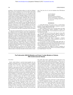
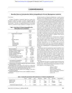
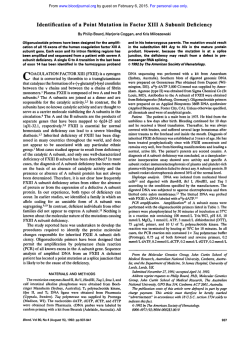
X-Linked Sideroblastic Anemia: Identification of the Mutation
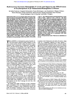
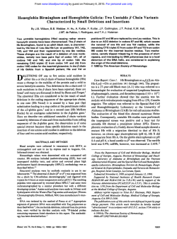
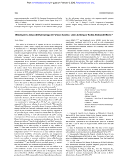
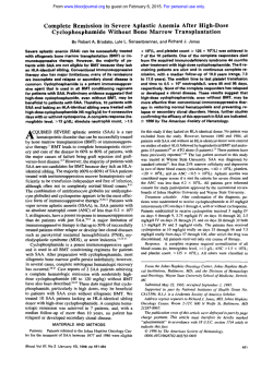
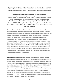
From www.bloodjournal.org by guest on February 6, 2015. For personal use only.
X-Linked Sideroblastic Anemia: Identification of the Mutation in the
Erythroid-Specific S-Aminolevulinate Synthase Gene (ALAS2) in the
Original Family Described by Cooley
By Philip D. Cotter, Donald
L. Rucknagel, and David F. Bishop
In 1945, Thomas Cooley described the firstcases ofX-linked
sideroblastic anemia (XLSA) in two brothers from a large
family in which the inheritance of the disease was documented through six generations. Almost 40 years later the
enzymatic defect in XLSA was identified as the deficient
activity of the erythroid-specific form of S-aminolevulinate
synthase (ALAS2). the firstenzyme in the heme biosynthetic
pathway. To determine the nature of the mutation in the
ALAS2 gene causing XLSA in Cooley's original family, genomic DNAs were isolated from two affected hemizygotes, and
each ALAS2exon was PCR amplified and sequenced. A single transversion(A t o C) was identified in exon 5. The mutation predicted the substitution of leucine for phenylalanine
at residue 165 (F165L) in the first highlyconserved domain
of the ALAS2 catalytic core shared by all species. No other
nucleotide changes were found by sequencing each of the
x
-LINKED sideroblastic anemia (XLSA) is the most
common of several inherited forms of sideroblastic
anemia. The first report of this metabolic disorder was by
Thomas Cooley in 1945.' The same family was further described in 1946 by Rundles and Falls' and is the subject of
this report. Since this time, more than 100 cases of XLSA
have been de~cribed,~
demonstrating that XLSA is a clinically heterogeneous disorder of erythropoiesis characterized
by hypochromic, microcytic erythrocytes; anemia; splenomegaly; elevated tissue and serum iron; and ringed sideroblasts in the marrow. Onset is typically in infancy or early
childhood, and X-linked inheritance of the disorder has been
established by pedigree analysis. Affected heterozygotes
have been identified: as expected for lyonization in X-linked
disorders. Carrier detection requires careful determination of
erythrocyte size distribution if lyonization is markedly
skewed toward activation of the normal allele.
Many patients with XLSA respond to oral pyridoxine therapy of 5 to 500 mg/d. Hemoglobin levels increase by varying
degrees but rarely return to normal levels. Even with near
normal hemoglobin concentrations, microcytic erythrocytes
remain. Although transfusions are often given to correct the
severe anemia, the lack of effective erythropoiesis typically
leads to iron overload, requiring chelation therapy and possibly interfering with the benefits of pyridoxine therapy by
disturbing mitochondrial function. Notably, phlebotomy has
proven effective in obviating the iron overload without exacerbating the anemia in several case^.^.^
6-Aminolevulinate synthase (succinyl-coenzyme A
[CoA]:glycine C-succinyltransferase [decarboxylating]; EC
2.3.1.37; [ALAS]) catalyzes the first step of heme biosynthesis and requires the B6 vitamer pyridoxal 5"phosphateto
accomplish the condensation of succinyl CoA and glycine
to form 6-aminolevulinic acid in the mitochondria of animal
cells. Two isozymes of ALAS exist in vertebrates: a
housekeeping isozyme and an erythroid-specific isozyme.8-"
These tissue-specific isozymes are encoded by two separate
In humans, the erythroid gene ALAS2 has been
Blood, Vol 84, No 11 (December l), 1994: pp 3915-3924
11 exons, including intron/exon boundaries, 1 kb of 5"flanking and 350 nucleotides of 3"flanking sequence. The mutation introduced anMse I site and restrictionanalysis of PCRamplified genomic DNA confirmed the presence of thelesion
in the two affected brothers and in three obligate heterozygotes from three generations of this family. Carrier diagnosis
in one
of additional family members identified the mutation
of the proband's sisters. After prokaryotic expression and
affinity purification of both mutantnormal
and ALAS2 fusion
proteins, the specific activity of the F165L mutant enzyme
was about 26% of normal. The cofactor, pyridoxal 5"phosphate, activated and/or stabilized the purified mutant recombinant enzyme in vitro, consistent with the pyridoxineresponsive anemia in affected hemizygotes from thisfamily.
0 1994 b y The American Society of Hematology.
mapped to the distal subregion of chromosome band
Xpl l.21.14 The fact that ALAS2 is on the X chromosome
and that its B,-dependent enzymatic activity is often reduced
in patients with XLSA15316
pointed to ALAS2 as a candidate
gene for this disorder.
Recently, we reported the first mutation in ALAS2 in a
patient with pyridoxine-responsive XLSA.I7 In this patient,
a point mutationin exon 9, which contains the predicted
pyridoxal 5"phosphate attachment site, resulted in a greater
than 95% reduction of the activity of the recombinant mutant
enzyme as compared with the recombinant normal enzyme.
Because responsiveness of ALAS2 to pyridoxal 5"phosphate was demonstrated in vitro, the X-linked defect in
ALAS2 was consistent, both genotypically and phenotypically, with XLSA in the patient. In this report, we identify
and characterize a new ALAS2 mutation in three generations
of the original XLSA family described by Cooley. This exon
5 point mutation results in reduced recombinant ALAS2
activity and shows activation by pyridoxal 5"phosphate in
vitro. These results confirm the conclusion of Cooley and
From the Department of Human Genetics, Mount Sinai School of
Medicine, New York, and the Cincinnati Comprehensive Sickle Cell
Center, Children ' S Hospital Medical Center, Cincinnati, OH.
Submitted February 28, 1994; accepted August 3, 1994.
Supported in part by a grant (R01 DK40895) from the National
Institutes of Health to D.F.B. and a grant (USPHS 5P60-HL 15996)
for the Comprehensive Sickle Cell Center, University of Cincinnati
to D.L.R. P.D.C.is the recipient of a March of Dimes Birth Defects
Foundation Predoctoral Graduate Research Training Fellowship.
Address reprint requests to D.F. Bishop, PhD, Department of
Human Genetics, Mount Sinai School of Medicine, New York, NY
10029.
The publication costs of this article were defrayed in part by page
charge payment. This article must therefore be hereby marked
"advertisement" in accordance with 18 U.S.C. section 1734 solely to
indicate this fact.
0 1994 by The American Society of Hematology.
0006-4971/94/8411-0038$3.00/0
3915
From www.bloodjournal.org by guest on February 6, 2015. For personal use only.
3916
COTTER,RUCKNAGEL,
AND BISHOP
VI1
6
7
8
0
10
l 118 12 1713
19
14
M
I
Fig 1. Pedigree of the family originally reported by Cooley in 1945.' Those family members marked by an asterisk have been scrwned by
PCR and Mse I restriction for the F165L mutation. The number at the upper left of some symbols indicates the age at deeth (inf, died in
infancy).
his associates that the defect was a congenital inability to
synthesize hemoglobin in the face of ample iron stores.
CASE REPORTS
An extensive kindred with XLSA, now documented through eight
generations (Fig l), was the first family with XLSA to be reported,
initially by Cooley in 1945' and subsequently as family I1 of Rundles
and Falls in 1946.' Affected males of this kindred exhibited the
classic features of anemia, peripheral erythrocyte anisocytosis and
poikilocytosis, splenomegaly, and a markedly elevated serum iron
level.' The first female in this large pedigree was from Holland,
whereas most of the American branch of the family now resides in
Michigan. Before 1945, affected males in this family usually died
in their first year of life, but not later than 17 years of age, due to
a severe microcytic, hypochromic anemia. Transfusion therapy was
initiated in patients VI-l6 and VI-l9 (Fig l), which resulted in
clinical improvement.' At 28 years of age, one of these transfusiondependent males (VI-16) was studied in another laboratory'8 and
was found to have hemosiderosis of the marrow with a hemoglobin
level of 6.5 g/dL. Although he had been transfusion dependent for
the previous 15 years, he was subsequently able to maintain a hemoglobin level of 7.8 to 9.6 g/dL on oral pyridoxine therapy (100 mg/
d)."
Patient VII-21. The proband for this report was patient VII-21,
a first cousin once removed of the brothers reported by Cooley' (VI16 and VI-19, Fig 1). He wasborn on March 21, 1961, at the
University of Michigan Medical Center and received subsequent
medical care there. At birth, he weighed 2.18 kg and hada hemoglobin concentration of 7 g/dL. Peripheral blood films showed the classic microcytic, hypochromic erythrocytes with abundant anisocytes
and poikilocytes, including ovalocytes and lacrimatocytes (teardrop
shaped cells) (Fig 2B). His response to vitamin B, administration
and transfusion is summarized in Fig 3. Over the course of his first
21 years, the proband demonstrated a consistent but modest increase
in hemoglobin concentration with vitamin B, therapy. Hemoglobin
levels decreased to values of 6.5 to 7.0 g/dL when B, was withdrawn
and increased to 7.5 to 9.0 g/& when B, therapy was reinstated.
The mean corpuscular volume (MCV) deviated little from a median
value of 56 fL/cell from 14 years of age to hismost recent evaluation
at 32 years of age. His reticulocyte level ranged from 0.5% to 2.2%
with no obvious relationship to therapy. At 2 years of age, results
of a bone marrow examination were normal. At 19 years of age, the
ferritin concentration was 450 pg/L and the serum iron 263 pg/dL,
with 100% transfemn saturation. The bone marrow contained large
numbers of proerythroblasts, and the bone marrow stain for iron
showed abundant classic ringed sideroblasts (Fig 2C). A chromium51 red cell survival time was 21 days (normal 26 to 30 days). Slight
splenomegaly wasnoted for the first time at 19 years of age by
scanning, giving a sp1een:liverratio of 2.2: 1; compatible with hypersplenism. Fetal hemoglobin concentration was 1.8%, and hemoglobin A2 was 1.8% (normal 2.3% to 3.3%). At 21.5 years of age the
liver edge was palpable 6 cm below the costal margin and the span
was 10 cm. The spleen descended 6 cm below the left costal margin.
Because of exertional dyspnea, a transfusion program was begun to
maintain the hemoglobin concentration between 9 and 12 g/dL. At
25 years of age, with a ferritin concentration of 3,700 p g L , desfemoxamine chelation was begun. Three years later, with the development of retinopathy and type I diabetes, the transfusion and chelation
therapy were discontinued. At 32 years of age, increasing the pyridoxine to 500 mg/d produced no hematologic improvement.
Patient VII-22. Patient VII-22, born 15 months after his brother,
VU-21, weighed 2.32 kg and had a hemoglobin concentration of 8
g/dL. He was allegedly placed on pyridoxine intradermally at birth,
but as with his brother, by 1 month of age the hemoglobin concentration had decreased to 5.2 g/dL (hematocrit 21.5%, reticulocyte count
1.9%, MCV 56.5 %/cell). He was transfused once and continued to
From www.bloodjournal.org by guest on February 6, 2015. For personal use only.
INHERITANCE OF ANALAS2
A
MUTATION IN XLSA
L
I
-
B
C
3917
receive intradermal pyridoxine hydrochloride (25 mg/d) until 9 years
of age. He did not showas clear a responseto B6 therapyas did his
brother.Uptotheageof3years,
his hemoglobinconcentration
/
&
with B6 therapy, and from age 3 to 9 years the
averaged 9.3 g
average hemoglobin was 9.0g/&. In the subsequent8 years without
Bstherapy, the average hemoglobin concentration
was 8.5 g/&. The
reticulocyte level varied between 0.6% and2.4%, with no apparent
explanation for the fluctuation. The spleen became palpable at 3.5
years of age. At 4 years of age his height was at the tenth percentile,
his weight at the third percentile. At 5 years of age, when an interatrial septal defect
was repaired, the spleen was palpablecm4 below
the costal margin. At 7 years of age, when an orchiopexy was performed, his height and weight were at the 25th percentile. At age
17, bone marrow examination showed a picture similar to his brother’s witherythroid hyperplasia and large numbers
of proerythroblasts
and ringed sideroblasts.His serum iron level was 275p&&, saturation 100%. His ferritin level was 240 p@. At 18 years of age,
regular transfusions were begun because
of exercise intolerance.
Two years later, liver and spleen were palpable
10 cm below the
costal margins. Because of dramatically increased transfusion
requirements, splenectomy was performed
at 20.5 years of age. Subsequently his hemoglobin decreased from 8.5 to6.7 g/&. He then
experienced repeated episodesof deep venous thrombosis, although
his platelet count
was only 575,000cells/&. He transferred his medical care elsewhere from the Universityof Michigan and continued
to have venous thrombosis and pulmonary emboli, some of which
were life threatening. A bone marrow transplant was performed at
another institution, and hedied from graft-versus-host disease at 23
years of age.
Other relatives. During the past 20 years the blood of over 25
members of this family were studied by one of us (D.L.R.). Eight
obligate heterozygotes had normal hemoglobin concentrations, although their erythrocytes contained a microcytic, hypochromic
subpopulation.Forexample,theproband‘smother(VI-5)showeda
dimorphic film with only a few pale, small cells and an occasional
g
/
&
and an MCV
poikilocyte, with a hemoglobin level around 12.3
of101 %/cell (Fig 2A). By automated red cell
size analyses, the
microcytes comprised approximately 10% of the red cell mass with
an MCV of 50 &/cell.
MATERIALS AND METHODS
Fig 2. Morphology of peripheral erythrocytes and marrow erythroblasts in XLSA. (A) Peripheral blood film from VI-5, the mother
of the proband, showing a dimorphic erythrocyte population. The
majority of erythrocytes are normal with two microcytic, hypochromic cells visible (original magnification x 313). (B) Peripheral blood
film from the probandWI-21. The erythrocytes are characteristically
hypochromic and microcytic.Note the presence of ovalocytes, lacrimatocytes, and anisocytes (original magnification x 313). (C) Bone
marrow film from the proband, Vll-21, stained with Ped‘s. Shown
are characteristic ringed sideroblasts. These are erythroblastswith
red-stained nudei andPrussianblue-stainedirongranulescorresponding to iron-ladenmitochondriain a perinuclear distribution
(originalmagnification x313, panel C isprinted with 1 . 4 ~more
magnification than panelsA and B).
Histochemistry. PeripheralfilmswerepreparedwithWright’s
stain, and the bone marrow film was analyzed
for iron by Perl’s
stain. In the latter, potassium ferrocyanide reacts withfemc iron to
form Prussian blue, and neutral
red dye counterstains the nuclei
red.
The cytoplasm and hemoglobin are not stained. Photography was
on Ektachrome or Fuji
IS0 64tungsten film using a Nikon Labophot2 microscope with a Fluor 100/1.30
oil immersion objective, a2.5X
projection lens and
1.2% tube magnification for 3X magnification
13
before photographic enlargement.
Molecular analysisof the ALAS2 gene. Genomic DNA from the
proband (W-21) and otherfamilymemberswasextractedfrom
peripheral blood, cultured lymphoblasts, and other
tissues according
to standard techniques.lg DNA from the proband’s deceased brother
(VU-22)wasextractedfromhisspleenwhichhadbeenkeptat
-20°Cby one ofus(D.L.R.) since his splenectomy.Subcloning
and sequencing of the polymerase chain reaction (PCR)-amplified
as preALAS2geneexonsandflankingregionswasperformed
viously described.” Each 100-pL PCR reaction mixture contained
1 pg of genomic DNA, 100 pm01 each of the sense and antisense
oligonucleotides containingEcoRI sites, 50 pmol/L each dNTP, 50
mmom KCI, 10 mmol/L Tris-HC1 (pH 9.0), 0.1% Triton X-100,
1.5 mmol/L MgCl,, and 2.5 U Taq polymerase (Promega, Madison,
From www.bloodjournal.org by guest on February 6, 2015. For personal use only.
3918
COTTER, RUCKNAGEL, AND BISHOP
20
1
3
25 4
..
24 5 23 6 22
19
21
20
Age (YO
Fig 3. Hematologic profile of the proband, Vll-21, from 0 to 6 and l 9 to 25 years of age. For vitamin Bs administration the solid bars are 25
mgld intradermal or oral PLP and the grey bars are 25 mgld intradermal pyridoxine. Transfusion of 2 U every 2 to 4 weeks are indicated by
dark grey bars.
WI). The reaction mixture was incubated at 94°C for 4 minutes, then
30 cycles of amplification were performed in a PTC-100 Programmable Thermal Cycler (MJ Research, Watertown, MA) with denaturation at 94°Cfor 30 seconds, primer annealing at the melting temperature (t,) of the oligonucleotide pairs for 30 seconds, and extension
at 72°C for I minute. All PCR products were restricted and subcloned
into pGEM-4Z (Promega). ALAS2 exon 5 was amplified (annealing
temperature 62°C) with the following primers: sense 5' AGACTAGCCAGGGAGAGACT 3' and antisense 5' GCCGCCGAATTCTTTCCATGTGTGGTT"C 3'. Sequences were confirmed in
multiple subclones if differences were found from our published
sequence." The numbering of the complementary DNA (cDNA)
previously usedt7has been corrected by adding five nucleotides (nt)
to include an upstream in-frame methionine identified in the genomic
sequence (Bishop, unprlhlished observations) and not present in the
reported cDNA clone." Primer extension studies (Bishop, unpublished observations) identified this as the first methionine after the
CAP site, confirming the result of Cox et al.'3
Products from PCR amplification of ALAS2 exon 5 were restricted with 4 U Mse I (New England Biolabs, Beverly, MA). All
restriction digests were electrophoresed in 2% agarose (Ultra Pure
grade, GIBCO BRL, Gaithersburg, MD) gels containing 0.1 pg/ml
ethidium bromide and photographed on a Fotodyne UV Transilluminator with Polaroid Type 57 film.
Protein homology comparisons and secondary structure predictions were performed withthe PILEUP program of the Genetics
Computer Group (GCG) Sequence Analysis Software Package"'and
with the Predictprotein program,*' respectively.
Expression ofthe nomulandF165LAUS2 enzymes. Recombinantnormal and mutant ALAS2 were generated using the pMAL
fusion protein expression system (New England Biolabs). resulting
in maltose binding protein (MBP) attached tothe N-terminus of
ALAS2 with a Factor Xa-cleavable linkage. The EcoRl fragment
from pGEM4Z-AE2" was initially subcloned into EcoRI-restricted
pMAL-c2 (New England Biolabs) to yield the clone pMALc2-AEI.
The 5' endof the ALAS2 portion of the fusion protein was engi-
neered by PCR to start at Asp 79 as determined by homology to the
predicted mouse erythroid ALAS N-terminus.",'3 PCR amplification
was accomplished as previously described, using I O ng of template
DNA (pGEM4Z-AE2) and oligonucleotides: sense 5' GATGGGAAGAGCAAGATTGTG 3' and antisense 5' GCCGCCAAGCTTCTCGAGGGGACAGATGGC 3' (annealing temperature 56°C). PCR
products were blunt-ended using T4 DNA polymerase by incubating
for 15 minutes at15°Cin 50 mmol/L NaCI, 10 mmol/L Tris-HCI,
10 mmol/L MgClz, 1 mmol/L DTT, 125 pmol/L of each dNTP and
3 U T4 DNA polymerase (New England Biolabs). The enzyme was
inactivated by incubating at 100°C for 5 minutes. The blunt-ended
PCR products were restricted with Xho 1, gel-purified, and cloned
into Xmn IIXho I-restricted andgel-purified pMALc2-AEI. The
F165L mutation was initially introduced into the pET5a-AE2I7clone
by site-directed mutagenesis using the PCR ovcrlap mcthodZ4to
yield the clone pET5a-AE7. The Xtnn YXho 1 fragment from pET5aAE7was cloned into Xmn IIXho I-restricted and gel-purified
pMALc2-AE2. The resulting clones, pMALc2-AE2 and pMALc2AE7, were sequence confirmed.
The pMALc2-AE2 and pMALc2-AE7 expression constructs were
transfected into Escherichiu coli BL2 I , pLysS (Novagen, Madison,
WI) and overnight cultures grown in LB (GIBCO BRL) media with
50 pglmL ampicillin (Sigma, St Louis, MO) and 34 pglml chloramphenicol (Sigma). The next day, 100-mL cultures in LBlampicillin
media were initiated with a 1:40 dilution of the overnight cultures
and grown to 0.4 A, units. Inductionwith 0.4 mmol/L isopropyl
0-D-thiogalactopyranosidewas performed in LBIampicillinmedia
for 4 hours at 37°C. Cells were pelleted at 4 , 0 0 0 ~for 15 minutes,
resuspended in 5 mL of SO mmol/L HEPESlpH7.5, 1 mmol/L
EDTA, 5 mmol/L DTT,and frozen at -7OT for at least 1 hour.
MgCIZ,RNase A (Sigma), andDNase I (Sigma) were added to the
thawed lysate to final concentrations of 1 mmol/L, 200 pgImL, and
20 pglmL, respectively. The lysate was incubated for 20 minutesat
room temperature, then centrifuged at 4°C for 30 minutes at 10,000~.
The supernatant was retained, filtered through a 0.45-pm filter (Gelman, Ann Arbor, MI) toyield the crude E coli extract. This was
From www.bloodjournal.org by guest on February 6, 2015. For personal use only.
INHERITANCE OF AN ALAS2 MUTATION IN XLSA
3919
then diluted with 3 v01 of 50 mmol/L HEPES/pH 7.5, 1 m m o l n
EDTA, and 5 mmol/L DTT and applied to an amylose resin affinity
column (New England Biolabs). MRP-ALAS2 fusion proteins were
eluted from the column with 10 m m o l n maltose, SO mmol/L
HEPESlpH 7.5, 1 mmol/L EDTA, and S mmol/L DTT and stored
at -20°C.
ALAS activity was assayed as previously described" with detection by Ehrlich's reagent after reaction with ethyl acetoacetate."
One unit of activity is that amount of enzyme required to catalyze
the production of I nmol of 6-aminolevulinate per hour under the
conditions of the assay. Protein concentration was determined by a
modification of the Ruorescamine method." Both the normal and
F16SLfusion proteins were assayed at various concentrations of
added pyridoxal S'-phosphate (PLP).
RESULTS
Characterization the
of
ALAS2 mutation. Genomic
DNA from peripheral blood was isolated from patient VII2 1. Each exon of the ALAS2 gene, including 50 to 150 nt
of flanking intron sequence, 1 kb of 5', and 350 nt of 3'
flanking sequence, was PCR-amplifiedand sequenced. A
single base change in exon 5 was the only difference from
the normal sequence. This C-to-A transversion at nt 547 (Fig
4) predicted the substitution of leucine for phenylalanine at
residue 165 (F165L) and introduced an Mse I site. PCRamplified exon 5 from 50 unrelated white females was restricted with Mse I and analyzed by agarose gel electrophoresis for detection of the predicted normal 325-bp or mutant
208-bp and 117-bp fragments. The absence of restriction in
1 0 0 alleles demonstrated that the mutation was not a common polymorphism (data not shown).
Restriction analysis of exon 5 amplified genomic DNA
from family members demonstrated 100% concordance between the presence of the Mse I site and a microcytic, hypochromic red blood cell population in affected hemizygotes
and obligate heterozygotes. X-linked inheritance of the
F165L mutation was demonstrated in three generations. The
proband, his grandmother, mother, aunt, sister, and brother
all had beenpreviously diagnosed as hemizygotes or carriers,
and all had the mutation as evidenced by 208 bp and 1 17
bp Mse I restriction fragments (Fig 5). Two phenotypically
normal sisters and an unaffected nephew all lacked the mutation as demonstrated by uncut 325-bp fragments (Fig 5 ) .
Expression and characterization of recombinant normal
and
mutant
fusion proteins. Recombinant normal
and
F165L ALAS2 proteins were expressed in E coli as fusion
proteins with the maltose-binding protein (MBP) using the
pMAL expression system. Previous expression of recombinant ALAS2 in the pET5a system" resulted in only a small
fraction (less than 0.1%) of soluble recombinant ALAS2,the
remainder being localized in inclusion bodies. Expression of
recombinant ALAS2 as a fusion with the 43-kD MBP resulted in greater than 10% of the predicted 90-kD ALAS2
fusion protein remaining soluble in the supernatant. The
MBP moiety also allowed partial purification by affinity
chromatography using an amylose resin (Fig 6). Crude extract and affinity-purified normal and mutant ALAS2 fusion
proteins were analyzed by sodium dodecyl sulfate-polyacrylamide gel electrophoresis in 10% gels with a 3% stacking gel according to the method of Laemmli and Favre."
Approximately 1 0 0 pg of crude extract and 4 pg of affinitypurified fusion protein were applied to each well. The 14to 94-kD protein molecular weight standards (Pharmacia,
Piscataway, NJ) were in the firstandlast
wells (Fig 6).
Although similar amounts of both recombinant normal and
mutant 90-kD fusion proteins were visible in the gel for the
crude extracts, the amount of F165L mutant fusion protein
was reduced relative to normal for the affinity-purified material. Extracts from cells lacking the pMAL fusion construct
lacked the 90-kD band (data not shown). The specific activity
of the purified mutant ALAS2 was about 26% of the purified
normal enzyme. Because the 90-kD mutant band was also
reduced in intensity to about one third of the normal band
in the affinity-purified lanes (Fig 6), it is possible that the
mutation affects stability. The presence of the fusion protein
on the aminoterminus of the ALAS2 protein apparently does
not inhibit activity because the specific activity was unchanged after complete cleavage by factor Xa (data not
shown).
3'
3'
A T C
i4rA
G
Fig4.Sequenceanalysis
of
ALAS2 exon 5of the proband
(Vll-21) demonstrating the C-toA transversion atnt 547,predicting the substitution of leucine forphenylalanineat residue
165 (F165L).
G
5'
/g
/%
A
G
$f
C
G A T C
Normal
'\%
C
5'
From www.bloodjournal.org by guest on February 6, 2015. For personal use only.
3920
COTER, RUCKNAGEL, AND BISHOP
v Q
“I
b8
VI11
U
Std.
bp
1
1
A
2
3
4
5
6
7
8
Effect of PLP on normal and mutantALAS2 activity. The
affinity-purified recombinant normal and F165L fusion proteins were assayed with 0 or 5 pmol/L PLP after two and
three freeze-thaw cycles (Table 1). The residual specific activity of the F165L enzyme at 0 pmol/L added PLP was
13% that of the normal enzyme and at 5 pmoVL PLP averaged 26%. The specific activity of the normal MBP-ALAS2
protein increased about 9% from 0 to 5 pmoVL added PLP.
In contrast, the specific activity of the F165L MBP-ALAS2
fusion protein increased over twofold in the presence of 5
pmol/L PLP. The addition of protease inhibitors in the assay
had no effect, suggesting that PLP activated or stabilized the
enzymes rather than protected them against proteolysis.
Homology comparison and secondary structure prediction. Homology comparisons of human ALAS2 exon 5
with 14 other ALAS sequences, using the PILEUP program
of the GCG package,*’ demonstrated that the mutation occurred in a region that was well conserved (Fig 7) and that
the mutated phenylalanine residue was invariant in all but
the chick erythroid sequence. The beginning of exon 5 corresponds to the aminoterminus of prokaryotic ALAS sequences, suggesting that exons 5 through 1 1 constitute the
essential catalytic region of the enzyme. The F165L mutation
occurs within 22 residues of the beginning of exon 5 and is
the most aminoterminal mutation found to date in patients
with XLSA.
Secondary structure predictions for the region of exon 5
surrounding the mutation were obtained with version 11.93
of the PHD program of Rost and Sander.” The expected
accuracy was about 64% because the program was run without comparison to ALAS homologues. Although the normal
sequence predicted a transition from &sheet to a-helix structure in the region of F165 (data not shown), the F165L
mutation resulted in a prediction of all a-helix structure in
this region. Although this predicted structural change was
consistent with the altered phenotype, accurate correlation
awaits tertiary structure determination.
9
10
Fig 5. Mse I digestionof PCR products from exon
5 of the ALAS2 gene from the proband (Vll-21) and
members of his family. After Mse I digestion of PCR
products from normal individuals a 325-bp band is
visible. The presence ofthe F165L mutation resulted
in two fragments of 208 and 117 bp. Lane 1 is the
size standard (Hae 111 digestion of /X174). Lanes 2
to 4 and 7 are carrier females. Lanes 5 and 6 are the
hemizygous males (Vll-22 and Vll-21). Lanes 8 to 10
are normal individuals.Pedigreenumberscorresponding to Fig 1 are above each symbol.
DISCUSSION
In 1945, Cooley described “a severe type of hereditary
anemia with elliptocytosis.”’ This first reported case of what
is now known as XLSA has been shown here to be caused
by a point mutation in the ALAS2 gene. An F165L mutation
was identified in the proband (VII-21) and his affected
brother (VII-22). descendants of the family members originally studied by Cooley.’ In this family, the Mse I site created
by the mutation provided definitive heterozygote diagnosis.
All tested female family members with dimorphic erythrocyte populations were confirmed heterozygous for the F165L
mutation, andall individuals tested in this pedigree with
normal red cell populations lacked the F165L mutation. The
X-linked inheritance of this specific ALAS2 mutation was
established through three generations of this large kindred.
Normaland mutant ALAS2 were expressed from the
pMAL-c2 vector in E coli as maltose binding protein (MBP)ALAS2 fusion proteins and affinity purified for enzymatic
characterization. Perhaps due to the fact that a substantial
part of the aminoterminal region may be unnecessary for
catalytic activity (absent in prokaryotes), the presence of the
maltose binding protein did not significantly reduce ALAS2
catalysis: it could be cleaved with Factor Xa with no change
in ALAS2 activity (data not shown). The fusion protein
helped maintain the enzyme in a soluble form and facilitated
affinity purification with an amylose resin column. The final
specific activity of the normal enzyme, which was estimated
to be about 70% pure (Fig 6), was about 20,000 U h g . This
was over 100-fold more active than the homogeneous murine
erythroid ALAS22’ and may reflect loss of murine ALAS2
activity during purification involving denaturation and renaturation from inclusion bodies.
The effect of PLP on ALAS2 enzymatic activity wascharacterized for the normal and mutant recombinant enzymes.
Assayed inthe presence of 5 p m o l n PLP, the F165L enzyme
had about 26% residual specific activity relative to the normal enzyme after affinity purification.The presence of muta-
From www.bloodjournal.org by guest on February 6, 2015. For personal use only.
3921
INHERITANCE OF AN ALAS2 MUTATION IN XLSA
Crude
Extract
Affinity
Purified
kD
94 -
67 43 Fig 6. SDS-PAGE gel of recombinant normal and
F165LMBP-ALAS2 fusion proteins. Lanes 1 and 6 are
molecular weight standards. Lanes 2 and 3 are total
crude lysate supernatants after induction of E coli
transformed with the normal and F165L expression
constructs, respectively. Lanes 4 and 5 are the purified normal and Fl65L fusion proteins after amylose
affinity chromatography.
!
30
tion-specific lower molecular weight species in this preparation suggested that the mutant enzyme may be more
susceptible to degradation during purification. It also may
be more susceptible to inactivation by loss of the PLP moiety
because addition of PLP resulted in over 110% activation
as opposed to a 9% activation for the normal enzyme. Our
working hypothesis is that the mutation alters the local secondary structure and possibly perturbs the overall conformation decreasing stability and/or reducing the affinity for PLP.
The activating and/or stabilizing effect of PLP may be the
explanation for the modest increase in hemoglobin seen in
hemizygotes after pyridoxine supplementation. Additional
PLP binding studies and stability experiments with homogeneous normal and mutant ALAS2 will be needed to test this
hypothesis.
Comparison of all reported ALAS sequences demonstrated that the phenylalanine at residue 165 was highly conserved. When the 15 published amino acid sequences for the
ALAS2 and ALAS 1 isozymes were aligned in the region of
the F165L mutation, phenylalanine 165 was invariant in all
sequences except that for the chicken erythroid isozyme (Fig
7). The immediate context was also highly conserved as
compared with flanking sequences, suggesting that this domain is important for enzyme structure and/or catalytic activity. Although there are 139 and 193 amino acids in human
erythroid and hepatic ALAS I , respectively, before the beginning of the residues encoded by exon 5 , the beginning of all
six of the bacterial peptides corresponds to the fourth residue
of this exon (Fig 7). Thus, it is expected that phenylalanine
143 defines the minimal aminoterminal boundary of the region of the ALAS2 peptide required for catalytic activity.
The F165L mutation is the most aminoterminal ALAS2 mutation identified to date and occurs in the first highly conserved domain of this catalytically competent core. The secondary structure prediction program of Rost and Sander?'
predicted that the F165L mutation would introduce a local
change from &sheet to a-helix in the region of the mutation.
This could provide a mechanism for the observed reduced
stability of the F165L recombinant ALAS2 fusion protein.
Our case studies confirm and extend the original report
From www.bloodjournal.org by guest on February 6, 2015. For personal use only.
COTTER, RUCKNAGEL, AND BISHOP
3922
Table 1. Effect of PLP on Normal and F165L Mutant
ALAS2 Enzyme Activity
ALAS2 Specific Activity (U/
mg)t
Experiment*
0 pmollL
PLP
5.0 prnol/L
PLP
Increase
(%l*
1
Normal ALAS2
F165L ALAS2
F165L (% of normal)
17,300
2,170
(13)
19,100
4,620
(24)
10
113
Normal ALAS2
F165L ALAS2
F165L (% of normal)
13,700
1,810
(13)
14,600
4,120
(28)
8
128
2
Experiment 1 was conducted after the enzyme samples had been
frozen and thawed twice and experiment 2 after a third freeze thaw
cycle.
t Normal and F165L recombinant ALAS2 fusion proteins were affinity purified.
Percentage increase of activity
at 5 p n o l / L PLP over 0 pmol/L PLP.
*
by Cooley'anddemonstrate thatthisfamily
is aclassic
example of XLSA. Hemizygotes presented atbirth with predominantly microcytic, hypochromic erythrocytes of variable shapes and sizes, and their anemia showed a moderate
response to pyridoxine therapy, as was previously demonstrated in this family." Iron overloadbecame significant with
time as evidenced by abundant ringedsideroblastsin
the
marrow, elevated plasma ferritin, and saturated plasma transferrin. The obligate heterozygotes were
uniformly dimorphic
in peripheral blood cell size, although this was often a small
population. This result could be due to skewed lyonization,
Mutation F165L
impaired hematopoiesis, or diminished survival of the erythrocytes derived from the mutant allele. Diagnosis of XLSA
was aided by careful determination of MCV and/or red cell
distribution width in mothers and femalerelatives of affected
individuals.
The moderate in vitro response of the recombinant F16SL
enzyme to PLP was mirrored in the partial clinical response
of affected males in this family to pyridoxine supplementation and appeared to be compromised by iron overload. For
example, patient VII-21 showed repeated response to PLP
or pyridoxine therapy and wasable tomaintain a hemoglobin
of 8 to 9 g/dL for 16 years on 25 to 50 mg pyridoxineld and
even in the absence of pyridoxine for a decade, until he was
placed on a transfusion program due to exercise intolerance
(Fig 3 ) . The patient's 16-year period with tolerable anemia
wassignificant because previouslymales in this family, if
untreated by pyridoxine or transfusion, typically died in the
first year of life(Fig l). It isnotclearwhy
the brothers
became so anemic in the first month of life, even while
receiving pyridoxine. The fluctuations in hemoglobin concentration,with or withoutpyridoxineadministration,
are
unexplained, as is the seemingly greater response to pyridoxine of VII-21than his brother. Their splenic involvement
also differed, the spleen becoming palpableat least 3.5 years
earlier in VI-22and requiringremovalaftertransfusions
were initiated.
Although transfusions immediately alleviated the anemia,
thesubsequent increaseinmitochondrial
iron stores may
have compromised what limited heme synthetic capacity was
available. The cause of death in XLSA patients, other than
of severeanemia in infancy, is often dueto progressive
iron overload.' Several reports have highlighted the value of
phlebotomy to remove excess iron in XLSA patient^.'"^^^ In
7
L
0
.
.
.
0
0
0
.
.
0
0
0
H-ry-X56352
Musery-M15268
Opsery-L02632
Humhep-X56351
Rathep-J04044
Chkhep"24366
Aspnid-X64170
Saccer"26329
Chkery-M24367
Brajap-M16751
Agrrad-P26505
Rhosph-LO7489
Rhimel-X02853
Rhocap-X53309
Rhosph-LO7490
Exon W5 Boundary
Exon 5/6 Boundary
3
Fig 7. Homology between the human ALASZ'*,35 residues encoded by exon 5 and the corresponding residues in all the published ALAS
sequences. From top to bottom are human erythroid
mouse ALAS2,= op?anus tau erythroid ALAS"(GenBankAccession No L026321,
human housekeeping ALASl'2,'3,36rat ALASl,"" chicken ALAS1,'O Aspergillusnidulans,"Saccharomycesceravisiae,u
chicken erythroid
ALAS2," Bradyrhizobium japonicum," Agrobscterium radiobacter/" Rhodobacter sphaeroides," Rhizobium meliloti," Rhodobacter capsula?us,"' and a secondALAS isozyme from Rhodobacter sphaeroides." The shaded region indicates identity to thenormal human ALAS2 sequence.
The solid circles indicate that the amino acid is invariant and the open circles indicate one or two differences in the 15 sequences.
From www.bloodjournal.org by guest on February 6, 2015. For personal use only.
INHERITANCE OF AN ALAS2 MUTATION IN XLSA
these cases, repeated phlebotomy resulted in reduced iron
levels without adversely affecting hemoglobin concentration,
indicating that stored iron was capable of being mobilized
for bone marrow hemoglobin production and that the bone
marrow was capable of responding to blood loss despite the
limitation to e r y t h r o p ~ i e s i s . ~Significantly,
”~~~
patients who
were previously refractory to pyridoxine therapy became
responsive after the iron stores were d e ~ l e t e d . ~ . ~
In this and our previous study” we have demonstrated an
in vitro response to PLP of the mutant recombinant enzymes
consistent with the clinical response to pyridoxine seen in
the patients. In addition, we have reported in abstracts the
existence of four additional unrelated XLSA families with
unique pyridoxine-responsive ALAS2 mutation^,^^.^' and
others have reported in abstracts four mutations in five families, one of which was suggested not to be pyridoxine respon~ i v e . ~ Thus,
‘ . ~ ~ mutations causing this disorder seem to be
largely private.
If X-linkage is not demonstrated, hereditary sideroblastic
anemia refractory to pyridoxine is most likely due to some
other cause, and we recently reported exclusion of X-linkage
for pyridoxine-refractory hereditary sideroblastic anemia,
with evidence for autosomal i n h e r i t a n ~ e . ~ ~
In conclusion, we have reported a newmutationin the
erythroid-specific ALAS2 gene in a large kindred with moderately pyridoxine-responsive XLSA and have demonstrated
an in vitro activation of the F165L recombinant mutant enzyme by PLP. This was the first XLSA family to be described
in the literature,’.’ and their mutation has now been identified
and confirms the involvement of the X-chromosomal ALAS2
gene in the etiology of XLSA.
ACKNOWLEDGMENT
We thank Drs Ron Bishop, Robert Cody, Ruth Heyn, and Mark
Roth at the University of Michigan for excellent care of the patients
reported herein. The authors appreciate the gift of pyridoxal 5’phosphate from Dr John D. Hines.
REFERENCES
1. Cooley TB: A severe type of hereditary anemia with elliptocytosis. Interesting sequence of splenectomy. Am J Med Sci 209561,
1945
2. Rundles RW, Falls H F Hereditary (?sex linked) anemia. Am
J Med Sci 211:641, 1946
3. Bottomley SS: Sideroblastic anaemia. Clin Haematol 11:389,
1982
4. Lee GR, MacDiamid WD, Cartwright GE, Wintrobe MM:
Hereditary, X-linked, sideroachrestic anemia. The isolation of two
erythrocyte populations differing in Xg” blood type and porphyrin
content. Blood 3259, 1968
5. Vogler WR, Mingioli ES: Porphyrin synthesis and heme synthetase activity in pyridoxine-responsive anemia. Blood 32:979,
1968
6. Verloop MC, Bierenga M, Diezeraad-Njoo A: Primary or essential sideroachrestic anaemias. Pathogenesis and therapy. Acta
Haematol 27: 129, 1962
7. Weintraub LR, Conrad ME, Crosby WH: Iron-loading anemia.
Treatment with repeated phlebotomies and pyridoxine. N Engl J
Med 275:169, 1966
8. Bishop DF, Kitchen H, Wood WA: Evidence for erythroid and
3923
nonerythroid forms of 6-aminolevulinate synthetase. Arch Biochem
Biophys 206:380, 1981
9. Watanabe N. Hayashi N, Kikuchi G: 6-Aminolevulinate synthase isozymes in the liver and erythroid cells of chicken. Biochem
Biophys Res Commun 113:377, 1983
10. Yamamoto M, Fujita H, Watanabe N, Hayashi N, Kikuchi
G: An immunochemical study of 6-aminolevulinate synthase and 6aminolevulinate dehydratase in liver and erythroid cells of rat. Arch
Biochem Biophys 245:76, 1986
11. Fernindez J, Gonzilez 0, Martin M, Ruiz Ami1 M: Erythroid
5-aminolevulinate synthetase from trout (Salmo gairdneri R.). Comp
Biochem Physiol [B] 85675, 1986
12. Bishop D F Two different genes encode 6-aminolevulinate
synthase in humans: Nucleotide sequences of cDNAs for the
housekeeping and erythroid genes. Nucleic Acids Res 18:7187, 1990
13. Cox TC, Bawden MJ, Martin A, May B K Human erythroid
5-aminolevulinate synthase: Promoter analysis and identification of
an iron-responsive element in the mRNA. EMBO J 1 0 1891, 1991
14. Cotter PD, Willard HF, Gorski JL, Bishop DF: Assignment
of human erythroid 6-aminolevulinate synthase (ALAS2) to a distal
subregion of band Xp11.21 by PCR analysis of somatic cell hybrids
containing X; autosome translocations. Genomics 13:211, 1992
15. Aoki Y, Urata G , Takaku F: 6-Aminolevulinic acid synthtase
activity in erythroblasts of patients with primary sideroblastic anemia. Acta Haematol Jpn 36:74, 1973
16. Aoki Y, Urata G , Wada 0,Takaku F: Measurement of 6aminolevulinic acid synthetase activity inhuman erythroblasts. J
Clin Invest 53:1326, 1974
17. Cotter PD, Baumann M, Bishop DF: Enzymatic defect in “Xlinked” sideroblastic anemia: Molecular evidence for erythroid 6aminolevulinate synthase deficiency. Proc Natl Acad Sci USA
89:4028, 1992
18. Bishop RC, Bethell W:Hereditary hypochromic anemia with
transfusion hemosiderosis treated with pyridoxine. Report of a case.
N Engl J Med 261:486, 1959
19. Sambrook J, Fritsch EF, Maniatis T: Molecular Cloning: A
Laboratory Manual. 2nd ed, v01 1-3. Cold Spring Harbor, NY, Cold
Spring Harbor Laboratory, 1989
20. Devereux J, Haeberli P, Smithies 0: A comprehensive set of
sequence analysis programs for the VAX. Nucleic Acids Res 12:387,
1984
21. Rost B, Sander C: A prediction of protein secondary structure
at better than 70% accuracy. J Mol Biol 232584, 1993
22. Dierks P Molecular biology of eukaryotic 5-aminolevulinate
synthase, in Dailey HA (ed): Biosynthesis of Heme and Chlorophylls. New York, McGraw-Hill, 1990, p 201
23. Ferreira GC, Dailey HA: Expression of mammalian 5-aminolevulinate synthase in Escherichia coli. Overproduction, purification,
and characterization. J Bioi Chem 268:584, 1993
24. Ho SN, Hunt HD, Horton RM, Pullen JK, Pease LR: Sitedirected mutagenesis by overlap extension using the polymerase
chain reaction. Gene 7751, 1989
25. Bishop DF, Wood WA: An assay for 6-aminolevulinic acid
synthase based on a specific, semiautomatic determination of picomole quantities of 6-[’4]aminolevulinate. Anal Biochem 80:466,
1977
26. Mauzerall D, Granick S: The occurrence and determination
of 6-aminolevulinic acid and porphobilinogen in urine. J Bioi Chem
219:435, 1956
27. Bishop DF, Wampler DE, Sgouris JT, Bonefeld RI, Anderson
DK, Hawley MC, Sweeley CC: Pilot scale purification of u-galactosidase A from Cohn fraction IV-l of human plasma. Biochim Biophys
Acta 524:109, 1978
From www.bloodjournal.org by guest on February 6, 2015. For personal use only.
3924
28. Laemmli UK, Favre M: Maturation of the head of bacteriophage T4 I. DNA packing events. J Mol Biol 80:575, 1973
29.Bottomley SS, Healy HM, Brandenburg MA, May BK: 5Aminolevulinate synthase in sideroblastic anemias: mRNA and enzyme activity levels in bonemarrow cells. Am J Hematol 41:76,
I992
30. Cotter PD, Baumann M, Rucknagel DL, Fitzsimons EJ, May
A, Bishop DF: Heterogeneity in X-linked sideroblastic anemia due
to unique mutations in the erythroid 6-aminolevulinic acid synthase
gene. Am J Hum Genet 51:A45, 1992
3 1. Cotter PD, Bishop DF: Congenital sideroblastic anemia: Correlation of the microcytic, pyridoxine-responsive phenotype with
coding region mutations in the erythroid 6-aminolevulinate synthase
gene. Am J Hum Genet 53:145A, l993
32. Cox TC, Bottomley SS, Wiley JS, MayBK: Erythroid 5aminolevulinate synthase deficiency due to a point mutation in a
kindred with X-linked sideroblastic anemia. Blood 80:341A, 1992
33. Bottomley SS, Wise PD, Whetsell LH, Schaefer FV: Heterogeneity of molecular defects of erythroid 5-aminolevulinate synthase
in X-linked sideroblastic anemia. Blood 82:433A, 1993
34. Jardine PE, Cotter PD, Johnson SA, Fitzsimons EJ, Tyfield L,
Lunt PW, Bishop D F Pyridoxine refractory congenital sideroblastic
anemia with evidence for autosomal inheritance: Exclusion of linkage to ALAS2 at Xp11.21 by polymorphism analysis. J Med Genet
31:213, 1994
35. May BK, Bhasker CR, Bawden MJ, Cox TC: Molecular regulation of 5-aminolevulinate synthase. Diseases related to heme biosynthesis. Mol Biol Med 7:405, 1990
36. Schoenhaut DS, Curtis PJ: Nucleotide sequence of mouse 5aminolevulinic acid synthase cDNA and expression of its gene in
hepatic and erythroid tissues. Gene 48:55, 1986
37. Bruning G, Ferkowicz M, Cornell N: Heme biosynthesis in
fish and land vertebrates: Enzyme and cDNA comparisons. Biol Bull
185:327, 1993
38. Yamamoto M, Kure S, Engel JD, Hiraga K: Structure, turnover, and heme-mediated suppression of the level of mRNA encod-
COTTER, RUCKNAGEL, AND BISHOP
ing rat liver 6-aminolevulinate synthase. J Biol Chem 263: 15973,
1988
39. Srivastava G, Borthwick IA, Maguire DJ, Elferink CJ, Bawden MJ, Mercer JF, May BK: Regulation of 5-aminolevulinate synthase mRNA in different rat tissues. J Biol Chem 263:5202, 1988
40. Borthwick IA, Srivastava G, Day AR, Pirola BA, Snoswell
MA, May BK, Elliott WH: Complete nucleotide sequence of hepatic
5-aminolaevulinate synthase precursor. Eur J Biochem 150:48 I ,
1985
41. Bradshaw RE, Dixon SWC, RaittDC, Pillar TM: Isolation
and nucleotide sequence of the 5-aminolevulinate synthase gene
from Aspergillus nidulans. Curr Genet 23:501, 1993
42. Urban-Grimal D, Volland C, Gamier T, DehouxP, LabbeBois R: The nucleotide sequence of the HEM1 gene and evidence
for a precursor form of the mitochondrial 5-aminolevulinate synthase
in Succharomyces cerevisiae. Eur J Biochem 156:511. 1986
43. Riddle RD, Yamamoto M, Engel JD: Expression of b-aminolevulinate synthase in avian cells: Separate genes encode erythroidspecific and nonspecific isozymes. Proc Natl Acad Sci USA 86:792,
1989
44. McClung CR, Somerville JE, Guerinot ML, Chelm BK:
Structure of the Bradyrhizobium japonicum gene hemA encoding 5aminolevulinic acid synthase. Gene 54: 133, 1987
45. Drolet M, Sasarman A: Cloning and nucleotide sequence of
MolGen Genet
the hernA gene of Agrobucteriumradiobucter.
226:250, 1991
46. Neidle EL, Kaplan S: Expression of the Rhodobacter sphaeroides hemA and hemT genes, encoding two 5-aminolevulinate acid
synthase isozymes. J Bacteriol 175:2292, 1993
47. Leong SA, Williams PH, Ditta GS: Analysis of the 5’ regulatory region of the gene for 6-aminolevulinic acid synthetase of Rhizobium melilofi. Nucleic Acids Res 13:5965, 1985
48. Hornberger U, Liebetanz R, Tichy HV, Drews G: Cloning
and sequencing of the hemA gene of Rhodobucter cupsulatus and
isolation of a 6-aminolevulinic acid-dependent mutant strain. Mol
Gen Genet 221:371, 1990
From www.bloodjournal.org by guest on February 6, 2015. For personal use only.
1994 84: 3915-3924
X-linked sideroblastic anemia: identification of the mutation in the
erythroid-specific delta-aminolevulinate synthase gene (ALAS2) in
the original family described by Cooley
PD Cotter, DL Rucknagel and DF Bishop
Updated information and services can be found at:
http://www.bloodjournal.org/content/84/11/3915.full.html
Articles on similar topics can be found in the following Blood collections
Information about reproducing this article in parts or in its entirety may be found online at:
http://www.bloodjournal.org/site/misc/rights.xhtml#repub_requests
Information about ordering reprints may be found online at:
http://www.bloodjournal.org/site/misc/rights.xhtml#reprints
Information about subscriptions and ASH membership may be found online at:
http://www.bloodjournal.org/site/subscriptions/index.xhtml
Blood (print ISSN 0006-4971, online ISSN 1528-0020), is published weekly by the American
Society of Hematology, 2021 L St, NW, Suite 900, Washington DC 20036.
Copyright 2011 by The American Society of Hematology; all rights reserved.


© Copyright 2026