
Hemoglobin Stimulates Mononuclear Leukocytes to Release
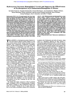
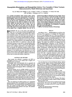
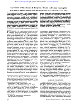
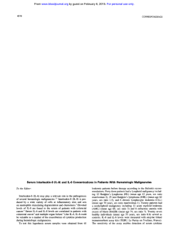
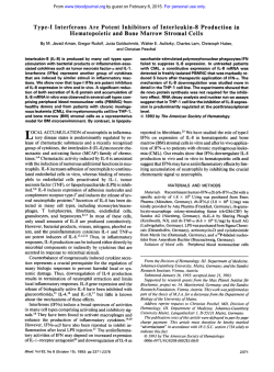
From www.bloodjournal.org by guest on February 6, 2015. For personal use only.
Hemoglobin Stimulates Mononuclear Leukocytes to Release Interleukin-S
and Tumor Necrosis Factor Q!
By Steve J. McFaul, Phil D. Bowman, Vilmar M. Villa, Marcelina J. Gutierrez-lbanez, Maria Johnson, and Dan Smith
Incubation of human mononuclear leukocytes (MNL) with
human stroma-free hemolysate (SFH), purified adult hemoglobin Ao (HbAo), andoxidized HbAo (METHb) causedMNL
t o release compoundsinto the
supernate that mediated neutrophil (polymorphonuclear leukocytes, PMN) chemotaxis
and PMN adherence t o human umbilical vein endothelial
cells (HUVEC). Chemotaxis and PMN adherence t o HUVEC
were reduced significantly when supernates were preincubated with neutralizing antibodies t o interleukin-8 (IL-8) and
tumor necrosis factor (U (TNF-m), respectively, suggesting
that 11-8 and TNF-a played significant roles in mediating
these activities, Greatest chemotactic activity was observed
in supernates of MNL treated with HbAo; while greatest
PMN/endothelial cell (EC) adherence activity was observed
in supernates of MNL treated with METHb. Furthermore,
PMN/EC adherence activity was a function of METHb con-
tent in each hemoglobin solution. PMN chemotaxis, PMN
adherence t o HUVEC, and cytokine release increased as a
function of increasing incubation time. Chemotactic activity
was detected in HbAo-treatedand METHb-treated MNL supernates after incubation for 6 hours and was maximal by
10 hours. 11-8 was detected in both HbAo and METHb-MNL
supernates by 4 hours. PMN/EC adherence activity was detected in HbAo-MNL supernatesat 10 hours and in METHbMNL supernates at 4 hours. TNF-(Uwas detected in METHb
and HbAo-MNL supernatesat 4 and 12 hours, respectively.
These results suggest that hemoglobin solutions stimulate
MNL t o release IL-8 and TNF-a in quantities sufficient to
induce PMN chemotaxis and PMN adherencet o HUVEC.
This is a US government work. There are no restrictions on
its use.
C
produce a variety of side effects in vivo. Two types of cells
that hemoglobin solutions contact immediately following infusion are leukocytes and endothelial cells. Both of these
cell types play major roles in modulating a number of hematologic events including blood clotting, antibody production,
and sequestration of monocytes and neutrophils from the
circulation into the tissues, which is characteristic of the
inflammatory response.I3 Furthermore, mononuclear leukocytes (MNL, monocytes and lymphocytes), polymorphonuclear leukocytes (PMN, neutrophils), and endothelial cells
(EC) respond to various stimuli by releasing cytokines that
alter the behavior of target cells.
The data from earlier studies indicate also that hemoglobin
solutions must be extremely low in endotoxin and phospholipid impurities for results to be interpreted without ambiguity. This is particularly true when looking at the interaction
of hemoglobin solutions and leukocytes since monocytes are
stimulated to release cytokines and prostaglandins by low
levels of endotoxin. Therefore, we developed a protocol for
preparing solutions of SFH and HbAo that do not contain
measurable quantities of endotoxin and phospholipid^.'^
Using these solutions, we examined whether hemoglobin
ELL-FREE HEMOGLOBIN is a potentially useful oxygen-carrying blood substitute that obviates the need
for blood typing, could be prepared free of human immunodeficiency and hepatitis viruses, and would have a considerably longer storage life than whole blood. Early in the development of a hemoglobin-based blood substitute, however,
hemoglobin solutions were found to be toxic when administered to animals.’.’ Subsequently, Rabiner et a13 showed that
residual fragments of the erythrocyte membrane (“stroma”)
present in the earlier hemoglobin preparations were toxic.
Removal of the stroma, however, did not render hemoglobin
preparations free of side effects. When infused into animals,
preparations of stroma-free hemolysate (SFH) have demonstrated a variety of toxic effects including disseminated intravascular coagulation,“ liver necrosis,5 vasoconstriction,6and
effects on renal hemodynamic^.^ Furthermore, when a very
low dose of SFH was administered to eight men in a clinical
trial, they exhibited transient bradycardia, mild hypertension,
decreased urine output and endogenous creatinine clearance,
and a mild prolongation of the activated partial thromboplastin time during the SFH infusion and several hours thereafter.8
These results prompted several investigations into the
source of the toxicities. Feola et al9*I0 showed that bovine
hemoglobin itself was not toxic to rabbits and did not stimulate endothelial cells to release prostaglandins. Rather, it
was endotoxin and phospholipid impurities that gave rise to
toxicity and cell activation. In contrast, however, Macdonald
et a l l ’ showed that high pressure liquid chromatography
(HPLC) purified human adult hemoglobin Ao (HbAo)
evoked a slow rise in coronary resistance (vasoconstriction)
in isolated perfused rabbit hearts. These solutions were below detectable limits of endotoxin and phospholipids. Furthermore, Smith et a l l 2 showed that preparations of HbAo
with endotoxin levels less than 0.12 endotoxin units (EU)/
mL activated complement from human plasma and stimulated human monocytes to express procoagulent factors on
their surface.
Taken as a whole, these data suggest that purified HbAo
can exert direct effects on various cell types, which can
Blood, Vol84, No 9 (November l),
1994 pp 3175-3181
From theLetterman Army Institute of Research, Division of Blood
Research, Presidio of San Francisco, CA; USAlSR, SGRD-ULM-T,
Ft. Sam Houston, TX; and Alliance Pharmaceutical, San Diego, CA.
Submitted April 4, 1994; accepted July 12, 1994.
The opinions and assertions contained herein are the private views
of the authors and are not to be construed as ofJicial nor do they
reflect the views of the Department of the Army or the Department
of Defense (AR360-5).
Address reprint requests to Steve J. McFaul, PhD, Walter Reed
Army Institute of Research, Blood Research Detachment, Washington, DC 20307-5100.
The publication costs of this article were defrayed in part by page
charge payment. This article must therefore be hereby marked
“advertisement” in accordance with 18 U.S.C. section I734 solely to
indicate this fact.
This is a US government work. There are 110 resm’ctions on its use.
00W-497I/94/8409-0033$0.00/0
3175
From www.bloodjournal.org by guest on February 6, 2015. For personal use only.
McFAUL ET AL
3176
stimulated MNL to release substances that mediate two critical events in an inflammatory response: PMN adherence
to endothelium and PMN chemotaxis. PMN adherence is
considered to be the primary step of an inflammatory re~ponse"*'~
because cells must attach to the endothelium before they can enter into the tissues. The presence of a chemotactic substance in the tissues then promotes the movement
of PMNs through the endothelial lining. We found that when
exposed to hemoglobin solutions that contained less than
0.06 EU/mL of endotoxin, MNL released cytokines that promoted both PMN adherence to endothelial cells and PMN
chemotaxis.
MATERIALS AND METHODS
Reagents
All reagents used in the preparation
of hemoglobin solutions were
of the highest grade available. RPM1 1640 medium without phenol
red, glutamine, antibiotic-antimycotic mixture (loox), and 1 m o m
HEPES were purchased from Gibco BRL (Grand Island, NY). Human serum albumin (HSA, 25%) was purchased
from Miles, Inc
(Elkart, IN). Quantikineenzyme-linkedimmunoassay kits for interleukin-8 (IL-8) and tumor necrosis factor a (TNF-a),and neutralizing antibodies toward IL-8 and TNF-a! were purchased from
R & D Systems (Minneapolis, MN).
Hemoglobin Preparation
Stroma-free hemolysate and purified HbAo were prepared from
outdated human packed red blood cells in a pilot plant production
facility as described by Winslow and Cha~rnan.'~
The final concentration of SFH was 60 mg/mL,buttheHbAowasconcentrated
further to 250 mg/mL using a Clirans C081 hollow fiber type dialyzer, 0.8' (TerumoCorp,Tokyo,Japan)attached
to a vacuum.
Solutions of SFH and HbAo were made isotonic by the addition of
lox Hanks' balanced salt solution(HBSS). Penicillin G, streptomycin sulfate, amphotericin B, and HEPES were added
to final concentrations of 100 U/mL, 100 pg/mL, 0.25 pg/mL (1X antibiotic-antimycotic solution), and 25 mmoVL, respectively. Solutions of SFH
and HbAo were stored frozen at -20°C and -8O"C, respectively as
10-mL aliquots. The aliquots were thawed in a 37°C water bath as
needed. Stroma-free hemolysate and HbAo were partially oxidized
to 2% and 7% methemoglobin, respectively, when
frozen. Methemoglobin levels rose to 7% and 14% for SFH and HbAo, respectively,
duringthe%monthperiod
of research. This gradualincreasein
oxidation did not
alter results from those seen when both hemoglobin
solutions were freshly prepared.
Methemoglobin. HbAo
was
oxidized
to methemoglobin
(METHb) by the slow additionof potassium ferricyanide K3Fe(CN)6
at a ratioof 1.2 mol of K,Fe(CN), to 1 mol of heme.The K,Fe(CN),
was dissolved in 100 pL of HBSS, and four 25-pL aliquots were
added to the HbAo solution at 10-minute intervals. The HbAo was
rocked gently and maintained4°C
at during the process. The METHb
wasthensterile-filtered,and excess K,Fe(CN)6wasremovedby
bed resin
passing the METHb through an analytical grade mixed
(AG 501-X8 (D); Bio Rad, Richmond, CA). The resin was poured
in a sterile syringe barrel in a laminar flow hood, and was washed
with several column volumes of sterile pyrogen-free water just before addition of the METHb. These precautions were takento minimize endotoxin contamination. The METHb effluent was collected,
made isotonic with the addition of1OX HBSS, supplemented with
25 mmoVLHEPES and antibiotic-antimycotic solution, sterile filtered, and stored at 4°C. All METHb analyses were performed on
a COBAS automatic analyzer usingthe method of Evelyn-Malloy."
Final endotoxin levelsof SFH, HbAo, and METHb solutions were
<O.M EU/mL using the gel-clot limulus amebocyte
lysate assay
(BioWhittaker, Inc). The sensitivity of this assay was not affected
by the presenceof hemoglobin. Phospholipids were undetectable
by
thin layer chromatography and phosphorous analysis.
Leukocyte Isolation
MNL were isolated from the blood of healthy volunteers. Blood
was drawninto EDTA Vacutainer tubes and layered over Histopaque
1077 (Sigma, St Louis, MO) at a blood-to-Histopaque ratio of 2:l.
The mixture was centrifuged for 20 minutes at 300 X g at room
temperature in a swinging bucket rotor.The MNL band at the interface wasremovedwith a pipet,washedtwicein
HBSS without
calcium and magnesium(HBSS-), then resuspended in 2mL HBSSand maintained on a rocker until used.MNL contained 20% monocytes and 80% lymphocytes.
PMNs were isolated by the method of McFaul." Human
blood
was collected into EDTA Vacutainer tubes and 30 mL were layered
onto 15 mL of Mono-Poly Resolving Medium (MPRM) (ICN Biomedicals, Inc, Irvine, CA) in a sterile conical polypropylene tube.
The blood was centrifugedfor 50 minutes at 750 x g and the PMN
band (band 2 from top) was removed using a syringe and 18 gauge
needle. Contaminating erythrocytes were removed from the PMN
suspension by underlayering the suspension with 5 mL of MPRM
and centrifuging it for 15 minutes at 750 X g and the PMNs were
collected from the upper phase and were washed twice with HBSS-,
then resuspended in HBSS. Cells were maintained on a rocker at
room temperature until used. The PMN band contained greater than
96% neutrophils. Viability of both cell types was >98% by trypan
blue exclusion.
MNL Incubation
Unlessotherwisenotedinthelegend,
8 X l@ MNUmL were
suspended in multiwell plates with RPM1 1640 medium containing
25 mmol/L HEPES buffer,100 U/mL penicillin G, 100p@mLstreptomycin sulfate, 2.5 pg/mL amphotericin B (RPM complete), and
differenttreatments as indicatedinthelegends.
The suspensions
were incubated with gentle shaking for 1 to 14 hours at 37°C and
5% CO2. The plates were centrifuged and the supernates were transferred to sterile multiwell plates and kept frozen ateither -20°C or
-80°C until used.
PMN Chemotaxis
Chemotaxis was assayed according to a modification
of the procedure of Megyerietwhichisbased
on the migration of PMNs
through 3.0 pm pores inp~ly~arbonate
membranes of Transwell cell
culture inserts (Costar Inc. Cambridge, MA).MNL supernates (300
pL) were placed into the inner 12 wells of 24-well plates, and 300
pL of RPM complete media containing 0.25% HSA (RPMUHSA)
were added. Then 50 pL ofRPMUHSA media were placed into
each Transwell, andthe plates were incubated at37°C and 5% CO2
for 20 minutes to equilibrate the solutions. After equilibration, 1 X
l@ PMNs in 50 pL of RPM complete media were added to the
Transwells, andthe Transwells were placedinto the wells of the 24well plates that contained the MNL supernates. The plateswere
incubated for 40 minutes at 37°C and 5% COz, and the bottom side
of each Transwell membrane was then rinsed with 2
mL of cold
HBSS- containing 0.2% EDTA into its respective well. The plates
were incubated at room temperature for 30 to 60 minutes to allow
PMNstodetachfrom
the plastic,and the detached PMNs were
countedusinganElzone282PCsizingparticlecounter(Particle
Chemotaxis was expressed as the percent
Data, Inc, Elmhurst, L).
of the number of PMNs added initially to the Transwell that had
From www.bloodjournal.org by guest on February 6, 2015. For personal use only.
HEMOGLOBINSTIMULATESMONONUCLEAR
3177
CELLS
migrated through the membrane: (no. of PMNs in welVl
100.
X
*
T
IO5) X
60
PMN Adherence to Endothelial Cells
Endorhelial cell cultures. Second passage human umbilical vein
endothelial cells (HUVEC) (Clonetics Corp, San Diego, CA) were
cultured in endothelial cell growth medium supplied with the cells.
Endothelial cells were subcultured in HBSS- containing 0.25% trypsin and 0.01% EDTA, then seeded into 48-well multi-plates at 5,000
cells/well and grown to confluence.
PMN adherence assay. PMN adherence was quantitated by the
method of McFaul and Bowman.” HUVEC were incubated at 37°C
and 5% CO, for 4 hours with 250 pL of MNL supernate mixed with
250 pL of media. The supernates were then removed and the HUVEC were washed 3 times with RPMI 1640 medium containing 25
mmol/L HEPES. Then 250 pL of RPM1 1640 containing 5 X lo5
PMNs was added to each well, and the cells were incubated for 20
minutes at 37°C and 5% CO?.
Nonadherent PMNs were removed by washing the HUVEC 3 to
5 times with fresh RPMI 1640 medium. The results of the washings
were monitored by microscopic examination to assure complete removal of nonadherent PMNs. After the last wash, HUVECand
PMNs were dislodged by the addition of 250 pL of HBSS- containing 0.25% trypsin and 0.01% EDTA at room temperature. When
the HUVEC rounded up (about 5 minutes), a single-cell suspension
was obtained by gentle up and down dispersal with a pipette. The
cell suspensions in each well were fixed by the addition of 250 pL
of 1% glutaraldehyde in HBSS-, and the multiwell plates were stored
in the refrigerator until the cells were counted.
The number of PMNs that adhered to the HUVEC wasdetermined
by counting both PMNs and HUVEC simultaneously with the Elzone
282PC sizing particle counter used for quantitating chemotaxis.
PMNs were counted between 7.02 and 11.63 pm and HUVEC were
counted between 11.63 and 25.66 pm. Size limits varied slightly
among experiments. PMN adherence was calculated as:
PMNs
-=
EC
Counts Between 7.02-11.63 pm
Counts Between 11.63-25.66 pm
The PMNEC ratios were adjusted for spurious counts induced by
trypsin treatment of the cells by subtracting the PMNEC ratios of
untreated endothelial cells that were incubated without PMNs.
Statistical Analyses
The data in this study were tested for significance by analysis of
variance followed by Dunnett’s test for multiple comparisons against
a single control group. The levels of significance are indicated in
the figure legends.
RESULTS
Figure 1 shows the effects of different hemoglobin solutions on the release of chemotactic and PMNEC adherence
factors from human MNL. Supernates from MNL treated
with 1% SFH, 1% HbAo, and 1% METHb caused significantly greater PMN chemotaxis and PMN adherence to HUVEC than did supemates from MNL treated with 1% HSA.
HbAo supemates exhibited the greatest level of chemotactic
activity, equal to that of the LPS supernates, while there was
no significant difference in chemotactic activities between
SFH and METHb supernates. Adherence of PMNs to HUVEC was greatest when HUVEC were treated with supernates of MNL previously incubated with METHb and least
with supernates of MNL previously incubated with SFH.
50
40
30
20
10
0
-5
SFH
HSA
A
Ao
METAo
LPS
TREATMENTS
l5
B
lr
*
+MNL
HSA
SFH
HbAo
METHb
*
LPS
TREATMENTS
Fig 1. Effect of hemoglobinsolutions on the release of (A) chemotactic and (B) PMNlEC adherence factorsfrom MNL. Human MNL (8
x 10‘ cells/mL) were incubated with 10 mg/mL solutions of HSA,
SFH, HbAo, METHb,and 500 ng LPSlmL for 14 hours at 37°C and 5%
CO2.All solutions contained
25 mmol/L HEPES, 2 mmol/L glutamine,
100 U/mL penicillin G, 100 pg/mL streptomycin sulfate, and 2.5 pg/
mL amphotericin B in RPM1 1640 medium. After incubation, the MNL
were removed by centrifugation and the cell-free supernates were
tested for chemotacticand PMN/EC adherence activitiesas described
in Materials and Methods. Values represent the means 2 SEM from
eight replicates. PMN/EC values indicate the averagenumber of
PMNs stuck to each endothelialcell. Significantdifferencesfrom HSA
data are indicated by * ( P < .001) and + ( P < .07).
METHb supemates were as active in promoting PMN adherence to HUVEC as were LPS supernates, and both HbAo
and METHb supernates were more active than the positive
control, 10 ng IL-lflIrnL (PMNEC = 6.33 ? 0.37). Adherence of PMNs to HUVEC treated with supernate from MNL
incubated with HSA was not significantly different from the
adherence of PMNs to untreated HUVEC (PMNEC = 0.09
? 0.04). Hemoglobin solutions incubated in the absence of
MNL did not exert either chemotactic or PMNEC adherence
activity.
Figure 2 shows the time course of the release of mediators
of chemotaxis and PMN adherence to HUVEC from MNL.
MNL incubated with HbAo and METHb released increasing
amounts of chemotactic factor(s) and PMNEC adherence
From www.bloodjournal.org by guest on February 6, 2015. For personal use only.
3178
McFAUL ET AL
*
*
loo
80
T
*
-
+
-+
Tt
/
i
I
’ -
~~
2
0
A
4
6
8
l 0 1 2 1 4
INCUBATION TIME (HOURS)
l5
rI
*
T
,i
either HbAo or METHb released significant amounts of IL8 and TNF-a into the supernates, whereas, MNL incubated
with HSA did not. Significant levels of IL-8 were detected
in supernates from HbAo and METHb-treated MNLby 4
hours, and IL-8 levels continued to increase with incubation
time. METHb-treated MNL released significantly higher levels of TNF-a than didHbAo at alltimes beginning at 4 hours.
TNF-a release was detected in HbAo-MNL supernates by
12 hours.
To determine whether IL-8 and TNF-a were responsible
for the chemotactic and PMN/EC adherence activities, respectively, we performed the chemotactic and PMN/EC adherence assays with HSA andHbAo-treated MNL supernates
in the presence of neutralizing antibodies to IL-8 and TNFa. Figure 4 shows that neutralizing antibodies to IL-8 and
TNF-a reduced chemotaxis and PMN/EC adherence activities significantly in supernates of MNL treated with HbAo.
Chemotactic activity mediated by HSA-treated MNL supernates was also reduced. Chemotactic activity in the presence
*
*
.’-
*
I*
.’
.A’
_.
- .
-.....m
0
2
4
6
8
l 0 1 2 1 4
B
INCUBATION TIME (HRS.)
Fig 2. Effect of MNL incubation time on the
release of (A) chemotactic and(B) PMN/EC adherence factors fromMNL. MNL wereincubated with IO
0 )HSA, l.-----.)
HbAo, and (A - -- A) METHb;
incubation conditions were the same as in Fig 1. At various times,
aliquots of each suspension were removed andcentrifuged, and the
cell-free supernates were tested for
chemotactic and PMN/EC adherence activities as described in Materials andMethods. Values represent the means f SEM from six replicates. PMN/EC values indicate
the average number of PMNs stuck t o each endothelial cell. Significant differences from HSA data are indicated by * ( P < .0021 and +
( P < .05).
--
-.
-
factors into the supernates as a function of incubation time.
The earliest times when a significant increase in chemotactic
activity could be detected in MNL supernates were 6 and
8 hours for HbAo and METHb, respectively. Chemotactic
activity by HbAo-MNL supernates was higher than that of
METHb-MNL supernates at all times.
PMNEC adherence activity in HbAo-MNL supernates
was not detectable until after 10 hours of incubation. However, METHb stimulated MNL to release adherence factors
within 4 hours. Adherence activity was significantly greater
in METHb-MNL supernates than in the HbAo-MNL supernates at all times.
PMN chemotaxis and adherence to EC can be promoted
by IL-8 and TNF-a, respectively. Therefore, we measured
the release of these cytokines into the supernate as a function
of incubation time. Figure 3 shows that MNL incubated with
--.-3
0
A
n
I
2
0
i
~
6
4
8
1 0 1 2 1 4
INCUBATION TIME (HOURS)
250
300
*
1
*
U
z
100
c
1
50
3
.
i
t
Y
+
T
.
*
*
- -,v
4.- -
-
,A
,.
~
..
.
L
~
- +
-
L
4
8
m
~
..
-
-
-
0
B
0
2
8
1 0 1 2 1 4
INCUBATION TIME (HRS)
Fig 3. Release of (A) IL-8 and (B) TNF-a from MNL as a function
--
of incubationtime.MNLwereincubated
with IO
0 ) HSA,
(U-----.) HbAo, and (A
A) METHb; incubation conditions were
the same as in Fig l.At various times, aliquots of each suspension
- -- -
were removed and centrifuged, and the cell-free supernates were
assayed for cytokines using Quantikine
ELISA kits. Values represent
the mean f SEM of four replicates. Significant dMerences from HSA
data are indicated by * ( P < .03) and + ( P < .08).
From www.bloodjournal.org by guest on February 6, 2015. For personal use only.
HEMOGLOBIN STIMULATES MONONUCLEAR CELLS
*
.Anti-IL-8
+
-
Anti-IL-8
H SA
Ao
TREATMENTS
. AnlkTNF
*
+ Anll-TNF
T
Ao
HSA
TREATMENTS
Fig 4. Effect of neutralizingantibody to IL-8 and TNF-a on (A)
chemotaxis and (B) PMN adherence t o HUVEC, respectively. MNL
supernates were incubated with 30p g anti-IL-S/rnL and 4 p g of antiTNF-dmL for 60 minutes before performing the chemotactic and
PMN/EC adherence assays as described in Materials and Methods.
Incubations were performed at 37°C and 5% CO2 in a humidified
chamber. Values represent the means ? SEM from tworeplicates. A
significant difference from HSA data in the absence of antibody is
indicated by * ( P <: .01). A significant difference from Ao data in the
absence of antibody is indicated by * * ( P < ,031.
of anti-IL-8 was the same for HSA and HbAo-treated MNL
supernates; while P M N E C adherence activity in the presence of anti-TNF-a was higher for HbAo-treated MNL supernates than for HSA-treated MNL supernates.
DISCUSSION
We have shownin vitro that hemoglobin solutions stimulate MNL to release compoundsthatmediatechemotaxis
and PMN adherence to HUVEC, activities associated with
inflammation. In the absence of MNL, none of the hemoglobinsolutions exhibited theseactivities demonstratingthat
the source of the mediators of these activities was the MNL.
Furthermore, MNL incubated
in the presenceof HSA instead
of hemoglobin did not release mediatorsof chemotaxis and
PMNIECadherence.Thus,hemoglobinwasalso
required
for MNL to release these compounds.
We showed also that 1L-8 and TNF-a played significant
3179
roles in mediating the chemotactic and P M N E C adherence
activities, respectively. In the presence of neutralizing antibodies to IL-S and TNF-a, chemotaxis and PMNECadherence, respectively, were reduced significantly (Fig 4). Furthermore, the releaseof IL-8 and TNF-a increased with time
as did chemotaxis and P M N E C adherence (Figs 2 and 3).
Our data suggest thatpartially oxidized andfully oxidized
hemoglobin play different roles in the release of these cytokines from MNL. P M N E C adherence activity in hemoglobin-MNLsupernateswasdetected
at 4 and 12 hoursfor
METHb and HbAo, respectively. At 12 hours the level of
METHb in the HbAo-MNL supernate was close to 50%.
Supemates of METHb-treated MNL exhibited the highest
level of P M N E C adherence activity (Fig lB), and a correlation between METHb content andP M N E C adherence activity was shown when P M N E C ratios were plotted as a function of the METHb contentof hemoglobin-MNL supernates
(Fig 5). Furthermore, MNL incubatedwith METHb released
greater amounts of TNF-a than did MNL
incubatedwith
HbAo (Fig 3B). These results suggest that the ferric form
of hemoglobin played a primary role in stimulating MNL to
release TNF-a.
With respect to chemotactic
activity, however, HbAoelicited a higher response from MNL than did either SFH or
METHb (Fig 1A). However, there was no
correlation between chemotaxis and the amountof reduced hemoglobin in
the hemoglobin treatments. SFH-MNL supernates contained
greateramountsof
reducedhemoglobin than didHbAoMNL supernates throughout theincubation,yetexhibited
lower chemotacticactivity. The data suggest that stimulation
of MNL to release chemotactic factorsmay have resulted at
least in part from the processof hemoglobin oxidation since
HbAo oxidized to a greater extent than SFH during the 14hour incubation. METHblevels in the SFH-MNL and HbAoMNL supernates increased from 7% to 30% and from 14%
to 54%, respectively, over the 14-hourincubationperiod.
Autooxidation of hemoglobinproduces 0; and H202,Z1.22
which may oxidize cellular components thus leading to the
10
5
0
0
20
40
60
80
100
% METHEMOGLOBIN
Fig 5. Correlation of PMN adherence to HUVEC and methemoglobin content in each treatment. Methemoglobin contents were measured for each of the treatmentsused. PMN/EC values are taken from
Fig 1B.
From www.bloodjournal.org by guest on February 6, 2015. For personal use only.
3180
McFAUL ET AL
release of chemotactic factors such as IL-8. In this regard,
release of the chemotactic and PMN/EC adherence factors
DeForge et ai23,24showed that LPS-induced release of IL-8
is currently under investigation.
from whole blood was inhibited by oxygen radical scavenIt is difficult to speculate on whatthese results mean with
gers implying that reactive oxygen species were involved.
respect to infusing hemoglobin solutions into humans as a
The decreased degreeof oxidation observed with SFH superresuscitation fluid because this is a relatively simple in vitro
nates presumably was attributed to the presenceof the H2O2
system. The concentration of hemoglobin used in this study
and 0 ; degrading enzymes, catalase and superoxide dismu(10 mg/mL) was significantly less than the concentrations
tase (SOD). Accordingly, the breakdown of these reactive
proposed to be infused as aresuscitation fluid (70 to 140
oxygen species would inhibit the release of IL-8.
mg/mL). Furthermore, it is not yet known whether acellular
Although partially oxidized hemoglobin elicited the highsolutions of hemoglobin behave similarly in vivo. However,
est response from MNL with respect to chemotacticactivity,
if they do, then our results suggest that acellular solutions
fully oxidized hemoglobin also elicitedasignificantreof hemoglobin are likely to invokean inflammatory response
sponse. Thus, oxidation of the iron is not essential to stimuin the recipient after infusion. Within 6 hours of infusion,
late MNL torelease IL-8. Another mechanismof stimulation
MNL could release cytokines that would activate PMNs and
may involve binding of the globin chains to receptors on the endothelial cells resulting in increased movement of PMNs
surface of MNL. Binding may eitherinitiate transmembrane
through the endothelial layer into thetissues where activated
signaling or be followed
by internalization of the hemoglobin
0; and proteoPMNs couldrelease toxic compounds such as
(phagocytosis). Both processes may trigger the synthesis and
lyticenzymes. In addition,activated PMNsmaydamage
release of cytokines. Opioid receptors in rat brain homogeendothelium following their adherence, thus making the ennates have been shown to bind short polypeptide fragments
dothelium lining leaky.
(4-9 amino acids) of the ,&chain of human hem~globin.’~
Hemoglobin is metabolized in the spleen, liver, and kidThese polypeptides are called hemorphins.” Such receptors
ne^.^' Thespleenand livercontainresident macrophages
may also exist on leukocytes, but when bound with hemoglo- and,
therefore,
these
organs may becomeconcentrated
bin may elicit the release of cytokines instead of the release
sources of inflammatory mediators as they take up the hemoof opioids. Weare currentlyinvestigating if hemoglobin
globin. Chemotactic gradients could be established fromthe
binds to MNL membranes and is internalized.
tissue macrophages to thevascular bed, thus drawing PMNs
The stimulating activity of METHb could also be associintothesetissues.
Therefore,over the course of 10 to 12
ated with either its peroxidase activity or the instability of
hoursafterinfusion,asignificantinflammatoryresponse
could develop resultingintissue
damage. Indeed, hepatic
its heme-globin linkage. In the presence of H202, METHb
centrolobular necrosis has been reported in
pigs that were
has been showntooxidize
hydroxylated aromaticcomexchange-transfusedwithhemoglobinsolutions,andthis
pounds such as tyrosine residues of proteins.’’ Normal cells
was accompanied by a significant movement of PMNs into
produce oxygen radicals (eg, 0 ; ) as by-products of mitochondrialrespiration andother biochemical p r ~ c e s s e s . ’ ~ ~ the
~ ~ liver tissue.3h
Currently, we are studying the mechanismof cellular actiSuperoxide spontaneously dismutates into H202 and02,and
vation, and are determining if other cytokines such as ILH2O2 moves freely across the
cell membrane into the sul a and I L - l p are released. Thesecytokinesalsoactivate
pernate. Thus, H 2 0 2would be available to react with METHb
endothelial cells to promote PMN/EC adheren~e,’~
and may
without additional stimulation of the MNL.
precede the release of IL-8 and TNF-a. We are also examinSeveral investigators have shown that the heme-globin
ing the effect of hemoglobinsolutions on leukocytes in
linkagein METHb is unstable and thatthe heme moiety
whole blood to determine towhat extent these inflammatory
readily exchanges between globin moleculesand fromglobin
reactions may occur in a more in vivo-like environment. The
to human serum albumin.3”,3’Furthermore, heme readily asgoal of this research is to understand the interactions that
sociates with thehydrophobic environment of cell memoccur between hemoglobin and leukocytes at the molecular
branes and oxidizes sulfhydryl compounds.” Thus, the exlevelsuchthat
wecan find effective ways of preventing
change of heme between METHb and cell membranes is
deleterious responses to hemoglobin infusion.
likely. In the presence of H202.heme catalyzesthe peroxidation of unsaturated lipids present in cellular and organelle
Thus, perturbation of proteins and lipids in
ACKNOWLEDGMENT
thecell membranes of MNL maystimulatetherelease
of
We thank Mike Cross and Rob Jesse for preparing the HbAo used
cytokines.
in these studies.
That there was no difference
in the amount of IL-8 released from MNL incubated with either HbAo or METHb
REFERENCES
(Fig 3A) even though supernatesof HbAo-treated MNL ex1.
Hamilton
PB,
Hiller
A,
Van Slyke DD: Renal effects of hemohibited greaterchemotactic
activity
than
supematesof
globin infusions in dogs in hemorrhagic shock. J Exp Med 86:477,
METHb-treated MNL suggests that other chemotactic sub1948
stances werereleased from MNLwhen incubated with HbAo
2 . Baker SB, deC, Dawes RLF: Experimental haemoglobinuric
that were not releasedwhen MNL wereincubatedwith
nephrosis. J Pathol Bacteriol 87:49, 1964
METHb. The possibility that other inflammatory mediators
3. Rabiner SF, Helbert JR, Lopas H, Friedman LH: Evaluation
are releasedin MNLsupematesas wellas theroles that
of a stroma-free hemoglobin solution for use as a plasma expander.
J Exp Med 126:1127, 1967
METHb and the oxidation of HbAo to METHb play in the
From www.bloodjournal.org by guest on February 6, 2015. For personal use only.
HEMOGLOBINSTIMULATESMONONUCLEAR
CELLS
4. White CT, Murray AJ, Greene JR. Smith DJ, Medina F, Makovec GT, Martin EJ, Bolin RB: Toxicity of human hemoglobin solution infused into rabbits. J Lab Clin Med 108:121, 1986
5. Friedman HI, DeVenuto F, Lollini L, Mellick P, Zuck TF:
Morphologic effects following massive exchange transfusions with
a stroma-free hemoglobin solution: I. Liver Lab Invest 39:167, 1978
6. Lieberthal W, Wolf EF, Merrill EW, Levinsky NG, Valeri
C R Hemodynamic effects of different preparations of stroma free
hemolysates in the isolated perfused rat kidney. Life Sci 41:2525,
1987
7. Vogel W M , Dennis RC, Cassidy G, Apstein CS, Valeri CR:
Coronary constrictor effect of stroma-free hemoglobin solutions. Am
J Physiol 251:H413, 1986
8. Savitsky JP, Doczi J, Black J, Arnold JD: A clinical safety
trial of stroma-free hemoglobin. Clin Pharmacol Ther 23:73, 1978
9. Feola M, Simoni J, Fishman D, Tran R, Canizaro PC: Biocompatibility of hemoglobin solutions: I. Reactions of vascular endothelial cells to pure and impure hemoglobins. Artif Organs 13:209, 1989
10. Feola M, Simoni J, Canizaro PC, Tran R, Raschblaum G,
Behal FJ: Toxicity of polymerized hemoglobin solutions. Surg Gynecol Obstet 166:211, 1988
1 l . MacDonald VM, Winslow RM, Marini MA, Klinker MT:
Coronary vasoconstrictor activity of purified and modified human
hemoglobin. Biomat Art Cells Art Org 18:263, 1990
12. Smith DJ, Winslow RM: Effects of extraerythrocytic hemoglobin and its components on mononuclear cell procoagulent activity. J Lab Clin Med 119:176, 1992
13. Movat HZ: Leukocyte emigration and its sequelae. Karger,
New York, 1981, p 1
14. Winslow RM, Chapman KW: Pilot-scale preparation of hemoglobin solutions in Everse J, Vandegriff, KD, Winslow RM (eds):
Methods in Enzymology, v01 231. Academic, San Diego, 1994, p 3
15. Lawrence MB, Springer TA: Leukocytes roll on a selectin at
physiologic flow rates: Distinction from and prerequisite for adhesion through integrins. Cell 5:859, 1991
16. Harlan JM: Leukocyte-endothelial interactions. Blood 65:513,
1985
17. Evelyn KA, Malloy HT: Microdetermination of oxyhemoglobin, methemoglobin, and sulfhemoglobin in a single sample of blood.
J Biol Chem 126:655, 1938
18. McFaul SJ: A method for isolating neutrophils from moderate
volumes of human blood. J Immunol Methods 130:15, 1990
19. Megyeri P, Issekutz TB, Issekutz AC: An endotoxin-induced
factor distinct from interleukin-la and tumor necrosis factor a produced by the THP-l human macrophage line stimulates polymorphonuclear leukocyte infiltration in vivo. J Leuk Biol 47:70, 1990
20. McFaul SJ, Bowman PD: Quantitation of polymorphonuclear
leukocyte adherence to endothelial cells by electronic particle size
discrimination. J Immunol Methods 130:171, 1990
21. Winterbourn CC: Free-radical production and oxidative reactions of hemoglobin. Environ Health Perspect 64:321, 1985
3181
22. Wallace WJ, Houtchens RA, Maxwell JC, Caughey WS:
Mechanism of autooxidation for hemoglobins and myoglobins: Promotion of superoxide production by protons and anions. J Biol Chem
257:4966, 1982
23. DeForge LE, Fantone JC, Kenney JS, Remick DG: Oxygen
radical scavengers selectively inhibit interleukin-8 production in human whole blood. J Clin Invest 90:2123, 1990
24. DeForge LE, Preston AM, Takeuchi E, Kenney J, Boxer LA,
Remick DG: Regulation of interleukin 8 gene expression by oxidant
stress. J Biol Chem 268:25568, 1993
25. Giamsta EL, Marklund A, Hellman U, Wernstedt C, Terenius
L, Nyberg F: Isolation and characterization of a hemoglobin-derived
opioid peptide from human pituitary gland. Regul Pep 34:169, 1991
26. Brant1 V, Gramsch C, Lottspeich F, Mertz R, Jaeger KH, Herz
A: Novel opioid peptides derived from hemoglobin: hemorphins. Eur
J Pharmacol 125:309, 1986
27. Shiga T, Imaizumi K. Electron spin resonance study on peroxidase- and oxidase-reactions of horse radish peroxidase and methemoglobin. Arch Biochem Biophys 167:469, 1975
28. M m JL: Oxygen free radicals linked to many diseases. Science 235:529, 1987
29. Van den Berg JJM, Kuypers FA, Qu JH, Chiu D, Lubin B,
Roelofsen B, Op den Kamp JAF: The use of cis-parinaric acid to
determine lipid peroxidation inhuman erythrocyte membranes.
Comparison of normal and sickle erythrocyte membranes. Bioch
Biophys Acta 994:29, 1988
30. Bunn HF, Jandl JH: Exchange of heme among hemoglobins
and between hemoglobin and albumin. J Biol Chem 243:465, 1968
31. Benesch RE, Kwong S : The stability of the heme-globin linkage in some normal, mutant, and chemically modified hemoglobins.
J Biol Chem 265:14881, 1990
32. Chiu D, Lubin B: Oxidative hemoglobin denaturation and
RBC destruction: The effect of heme on red cell membranes. Semin
Hematol 26: 128, 1989
33. Tappel AL: Unsaturated lipid oxidation catalyzed by hematin
compounds. J Biol Chem 217:721, 1955
34. Vincent SH: Oxidative effects of heme and porphyrins on
proteins and lipids. Semin Hematol 26:105, 1989
35. Keipert PE, Triner L: Catabolism and excretion of crosslinked
hemoglobin, in Brewer GJ (ed): The Red Cell: Seventh Ann Arbor
Conference, v01 319. Liss, New York, NY, 1989, p 383
36. Smith CD, Shuschereba ST, Hess JR, McKinney L, Bunch
D, Bowman PD: Liver and kidney injury after administration of
hemoglobin cross-linked with bis(3,5-dibromosalicyl) fumarate. Biomater Art Cells Artif Organs 18:251, 1990
37. Bevilacqua MP, Pober JS, Wheeler ME, Cotran RS, Gimbrone MA Jr: Interleukin-l acts on cultured human vascular endothelium to increase the adhesion of polymorphonuclear leukocytes,
monocytes, and related leukocyte cell lines. J Clin Invest 76:2003,
1985
From www.bloodjournal.org by guest on February 6, 2015. For personal use only.
1994 84: 3175-3181
Hemoglobin stimulates mononuclear leukocytes to release
interleukin-8 and tumor necrosis factor alpha
SJ McFaul, PD Bowman, VM Villa, MJ Gutierrez-Ibanez, M Johnson and D Smith
Updated information and services can be found at:
http://www.bloodjournal.org/content/84/9/3175.full.html
Articles on similar topics can be found in the following Blood collections
Information about reproducing this article in parts or in its entirety may be found online at:
http://www.bloodjournal.org/site/misc/rights.xhtml#repub_requests
Information about ordering reprints may be found online at:
http://www.bloodjournal.org/site/misc/rights.xhtml#reprints
Information about subscriptions and ASH membership may be found online at:
http://www.bloodjournal.org/site/subscriptions/index.xhtml
Blood (print ISSN 0006-4971, online ISSN 1528-0020), is published weekly by the American
Society of Hematology, 2021 L St, NW, Suite 900, Washington DC 20036.
Copyright 2011 by The American Society of Hematology; all rights reserved.

© Copyright 2026