
Hemoglobin Birmingham and Hemoglobin Galicia: Two
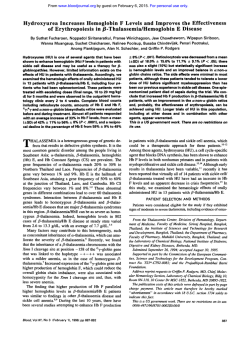
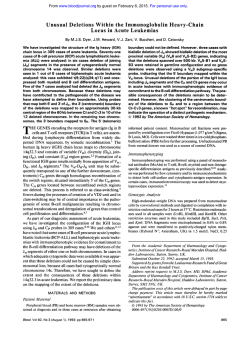
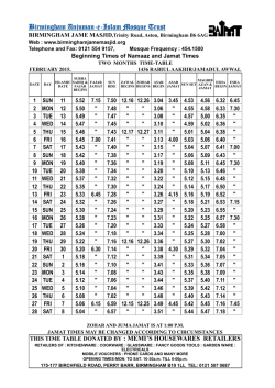
From www.bloodjournal.org by guest on February 6, 2015. For personal use only.
Hemoglobin Birmingham and Hemoglobin Galicia: Two Unstable B Chain Variants
Characterized by Small Deletions and Insertions
By J.B. Wilson, B.B. Webber, H. Hu, A. Kutlar, F. Kutlar, J.F. Codrington, J.T. Prchal, K.M. Hall,
J.M!. de Pablos, I. Rodriguez, and T.M.J. Huisman
Two unstable hemoglobins (Hbs) causing rather severe
hemolytic anemia have been characterized. The B chain of
Hb Birmingham, found in an adult black man, is characterized by the loss of -Leu-Ala-His-Lys- at positions 141,142,
143, and 144 and their replacement by one Gln residue.
These changes are the result of a deletion of nine nucleotides, namely two base pairs (bp) of codon 141, all of
codons 142 and 143. and one bp of codon 144; the
remaining CAG triplet (C from codon 141 and AG from
codon 144) codas for the inserted glutamine. In the B chain
of Hb Galicia from a Spanish patient, His and Val at
D
ELETIONS OF one to five amino acid residues in
either the a or the j3 chain of human hemoglobin (Hb)
cause a change in the stability of the protein and a mild to
severe hemolytic anemia. Two deletions in the a chain and 12
such mutations in the @ chain have been reported; these are
listed' and many are discussed in detail by Bunn and Forget.'
The abnormal Hbs are considered to be the result of small
deletions in the coding sequences of the appropriate chains;
in one case ( H b Natal) it is caused by a base pair (bp)
substitution leading to a stop codon at the penultimate codon
of the a2-globin gene,3 and in a second case (Hb McKees
Rocks) by an analogous substitution in the @-globin gene.'
Here we describe two additional unstable @ chain variants
caused by deletions of nine and three nucleotides from coding
sequences of the @-globin gene; the observation is of some
special interest because these specific deletions result in the
insertion of one amino acid residue in addition to the deletion
of four and two amino acid residues, respectively.
MATERIALS AND METHODS
Blood samples were collected in vacutainers with EDTA as
anticoagulant and sent in ice by express mail to Augusta, GA.
Informed consent was obtained.
Hematologic values were determined with an automated cell
counter. Hb analyses included isoelectrofocusing (IEF), heat and
isopropanol stability tests, and cation and reversed phase high
performance liquid chromatography (HPLC); methodology was as
described before?.'
Structural analyses were by methods routinely in use in our
laborat~ries.~,~
The abnormal ,9 chain (,9' or fly) was separated from
the (Y chain by CM-cellulose chromatography' and digested with
trypsin for 6 hours at room temperature and at pH 8.5. The resulting
peptides were separated by reversed phase HPLC,8 and some were
rechromatographed by a similar procedure but with a different
developing system.' Amino acid analyses were made in 24-hour acid
hydrolysates with the Waters Pico Tag amino acid analyzer (Waters
Chromatography Division, Milford, MA) (see manufacturer's manual).
DNA was isolated by the method of Poncz et a1."' Appropriate
segments of genomic DNA were amplified with Taq polymerase as
described before"; the nucleotide deletions,inferred from the protein
data, were verified by dot-blot analysis with 32P-labeledprobes
containing sequences listed elsewhere in this report. The methodology has been detailed before."
Blood, Vol 75, No 9 (May 11, 1990: pp 1883-1 887
positions97 and 98 are replaced by one Leu residue. This is
due to an ACG deletion in codons 97 and 98, which causes
the removal of one His and one Val residue, while the
remaining CTG triplet (C from codon 97 and TG from codon
98) codes for the inserted leucine residue. Two mechanisms, namely slipped mispairing in the presence of short
repeats, and misreading by DNA polymerase due to a local
distortion of the DNA helix, are considered in explaining
the origin of the small deletions.
0 1990 b y The American S o c i e t y of Hematology.
RESULTS
Cuse Report. Cuse 1. H b Birmingham or a2&( [Leu-AlaHis-Lys]+Gln at positions 141 through 144). The propositus is a 27-year-old Black man (A.J.) who was referred to a
hematologist for evaluation of suspected lymphoma because
of splenomegaly, anemia, and thrombocytopenia (1 10 x 10I2/
L). Further evaluation showed that the anemia was hemolytic with reticulocytosis ranging from 6% to 12%, Coombs
negative. The subject was referred to the Special Red Cell
and Hemoglobinopathy Laboratory at the University of
Alabama at Birmingham (UAB) for red cell enzyme studies.
The evaluation of the blood film showed numerous Heinz
bodies. Consequently, unstable H b studies were performed;
the isopropanol screen was positive and a heat test for
unstable H b showed a precipitate (about 20%). Electrophoretic evaluation of a freshly drawn blood sample showed a
mutant H b with a migration identical to that of H b S;
however, on citrate agar electrophoresis (pH 6), H b X did
not separate from H b A. On the globin electrophoresis at pH
8.4 and pH 6, a band anodic to PAwas observed. The metHb
level was 0.9%; sulfH'b, however, was increased a t 2.45%.12
From the Department of Cell and Molecular Biology. Medical
College of Georgia, Augusta; Division of Hematology and Oncology, University of Alabama at Birmingham and the Veterans
Administration Hospital, and the Special Red Cell and Hemoglobinopathy Laboratory, Birmingham: Servicio de Hematologia, Hospital General de Especialidades, Granada; and Servicio de Hematologia, Hospital Jeran Canalejo, La Coruna, Spain.
Submitted November 6,1989; accepted January 12.1990.
Supported by National Institutes of Health Research Grants
HLB-05168 and HLB-41544 (to T.H.J.H.)and by support of the
Veterans Administration IFCAP/2237 (to J. T.P.). This is contribution no. 1193 from the Department of Cell and Molecular Biology
at the Medical College of Georgia, Augusta.
Address reprint requests to Titus H.J. Huisman, PhD, Department of Cell and Molecular Biology, Medical College of Georgia,
Augusta, GA 3091 2-21 00.
The publication costs of this article were defrayed in part by page
charge payment. This article must therefore be hereby marked
"advertisement" in accordance with 18 U.S.C.section 1734 solely to
indicate this fact.
6 I990 by The American Society of Hematology.
0OO6-4971/90/7509-0012$3.OO/0
1883
From www.bloodjournal.org by guest on February 6, 2015. For personal use only.
WILSON ET AL
1084
Estimation of the P,, from venous blood gasesi3 showed a
Birmingham after the area of family residence and original
evaluation.
slightly decreased level at 22.0 mm Hg (normal range 22.6 to
29.4). The erythrocyte 2,3-DPG leveli4 was in the normal
Case Report. Case 2. H b Galicia or a,&([His-Val]-Leu
at positions 97 and 98). This variant was present in a young
range of 12,270 5 1,870 nmol/g Hb). A somewhat variable
pattern was noted by reversed phase HPLC (Fig 1); two or
Spanish patient (0.0.)with a hemolytic anemia, hepatothree abnormal '
0 zones eluted ahead of the normal /3* peak. splenomegaly, and a history of jaundice. Hematologic findH b F was slightly elevated at 3.5%, which was confirmed by
ings for a blood sample that had been in transit for 5 days
the presence of small amounts of 'y and Aychain in the HPL
were as follows: H b 7.5 g/dL; PCV 0.290 1/L; RBC 2.22 x
lO"/L; MCV 131 fL;M C H 33.8 pg; MCHC 26.0 g/dL;
chromatogram (Fig 1).
these data indicated a rather severe anemia. The reticulocyte
The unstable H b was isolated by preparative isopropanol
count in a freshly collected sample was 25%; total bilirubin
precipitation (20 mL 17% isopropanol in 0.2 mol/L TrisHCI, pH 7.4, mixed with 2 mL red cell lysate and incubated
1.94 mg/ 100 mL; indirect bilirubin 1.08 mg/ 100 mL. Heinz
bodies were positive (2 hours at 37OC). A slow band was
for 30 minutes at 37OC). The fix chain was isolated by
detectable by IEF, which was slightly faster than Hb A,.
CM-cellulose chromatography and digested with trypsin. Fig
Heat stability and isopropanol tests were positive. Reversed
2 (top) illustrates the separation of the tryptic peptides. Most
peptides eluted at their expected positions and had normal
phase HPLC (Fig 1) detected a distinct zone that eluted
amino acid compositions (data not shown). The peptides
behind the a chain; its quantity (as percent of 6" + BY + y)
T-14 and T-15 were absent, while a combination of these two
was 26.7%. H b F was elevated; its total quantity (as
fragments (T-14',15) eluted together with peptide T-3. The
'y + Ay)was 10.3%.
two peptides were easily separated by rechromatography
The variant was isolated by DEAE-cellulose chromatogra(Fig 2, top insert). The amino acid composition of the
0') was studied by the
phy and the abnormal 0 chain (
methods described above for the 6" of H b Birmingham.
T-14',15 fragment was Asp 0.99(1); Glu 1.12(0); Gly
Figure 2 (bottom) illustrates an HPL chromatogram of the
0.98(1); His 0.92(2); Ala 3.02(4); Tyr 0.82(1); Val 2.15(3);
tryptic peptides of the fly chain. All soluble peptides were
Leu O( 1); Lys O( 1). Expected values are between parentheses;
present and had the expected compositions (data not shown).
the valine value was increased to nearly three residues when a
72-hour acid hydrolysate was analyzed. These results sugAn extra zone eluted in front of T-13; its amino acid
gested a loss of one histidine, one alanine, one leucine, and
composition was Asp 2.08(2); Glu l.Ol(1); His O(1); Arg
one lysine residue and the addition of one glutamic acid or
0.93(1); Pro l.OO(1); Val O(1); Leu 1.91(1); Phe 0.94(1).
This peptide resembled PT-11, which is normally not recovglutamine residue. Sequence analyses gave the following
ered in notable quantities from a digest of a nonaminoethsequence: Val-Val-Ala-Gly-Val-Ala-Asn-Ala-Gln-Tyr-His,
confirming the deletion of Leu-Ala-His-Lys at positions 141,
ylated /3 chain. The sequence of this T-11' peptide, Leu-LeuAsp-Pro-Glu-Asn-Phe-Arg, confirmed a deletion of His-Val
142, 143, and 144 and the substitution of one glutamine
at positions 97 and 98 of the 0 chain and the insertion of an
residue.
extra leucine residue. The variant was named H b Galicia;
The patient has two sons and a living mother; none were
family studies were not conducted.
available for the study. However, electrophoretic studies
were performed on the propositus' parents in routine laboraDISCUSSION
tories in the past; the father was said to have H b A and the
Each of the two H b variants described here is charactermother was told she had sickle cell disease with only H b S
ized by a deletion and an insertion and by an instability that
and Hb F detected; however, she has never had a clinical
causes a rather severe hemolytic anemia. It was not possible
history of sickle cell crisis or other stigmata of sickle cell
to determine whether one or both anomalies were the result
disease. She has been known to be anemic with increased
of de novo genetic events because family studies could not be
reticulocyte counts, The abnormal variant was named H b
E
Heme
Heme
PATIENT 0.0.
t
h
0
Jb
cu
cu
(63.0%) a
-4-
cu
a,
0
C
ea
(26.7%)
8
(6.8%)
a
10
20
30
40
50
60
70
I
10
I
20
Time in Minutes (1 ml/min)
I
30
I
40
I
50
Av
(3.5%)
I
60
I
70
Fig 1. Separation of globin
chains by reversed phase HPLC
with a Vydac C, column (The
SeplAIRations Group. Hesperia, CA)."," (Left) Patient A.J.
with the &Birmingham chain
(@"I. (Right) Patient 0.0. with
the 8-Galicia chain (By). The
gradients used in the two chromatograms were slightly different. which explains the differences in the elution times.
From www.bloodjournal.org by guest on February 6, 2015. For personal use only.
khS BIRMINGHAM AND GALlClA
1886
-
Fb2. 8.pmknd.dubk
p.ptLb..*"dig.md
abnormal B ctuinr by r w u n d
phew W L C with C,,cdumn.'
(Top) Potimt A.J. with tho
8-8irmingh.m c h i n WV. Tho
Inoort illustrotos tho ..p.mtkn
of 8 1 - 3 and BT-14'.16 h,
roporoto orpuimont. IBottom)
P~tkm
0.0.with tho B - a l k h
c h i n (L?').
Time in Minutes (1.5 ml/min)
conducted. However. in the case of Hb Birmingham. it is
almost certain that the mutant was inherited from the
mother whose phenotype was HbS-Hb Birmingham.
Figure 3 provides additional details about the deletions
responsible for the formation of the two unstable 6 chains.
Patient A.J. with Hb Birmingham had the deletion of four
residues (Leu-Ala-His-Lys) and the insertion of a glutamine
residue at positions 141 through 144 of the 6 chain. This
change is explained by the deletion of nine nucleotides as
indicated in Fig 3: the C of codon 141 and the AG of codon
144 combine to code for a glutamine residue. Further proof
-*Q)
cooo(
was obtained from hybridization experiments with specific
synthetic oligonucleotide p r o k , only the amplified DNA of
patient A.J. hybridized with the 9-bpdeletion probe (Fig 4).
The data for patient 0.0. arc comparable (Fig 3). The
replacement of a His-Val dipeptide at positions 97 and 98 by
a leucine residue results from the deletion of three nucle-
139 140 141 142 143 144 1 4 s 146
.ASN.ALA.LEU.ALA.HlS.LYSIYR.nlS.IER
.AAI.OCC.CIO~OCC~CAC.AAO~TAI.CAC.TAA
o(LcIID:-
139 140
14s
IR.mA J ) W rcD .ASN*ALA-OLN
coooN
-*Q)
. A I 1 .OCC-CAO-----TAT
140
TVR.HlS.TER
. C A C * TAA
91 05
00 07 00
89 1 0 0 101
.ASP.LVS.LEU.HlS.VALASP.PRO.OLU.
coooN .OAC.AAO.ClO.CAC.OIO.OAT.CCT.OAO.
Qur(D:-
(Rrr 0 0 )W IcD
coooN
Flg3.
9 1 0 5 0 0
89 loo 101
. A S P . L V S . LEV-LEU-ASP*PRO.OLU~
.01C~AAO~C10-CTO-O~T~CCT.OAO.
pWadth.nornrlmd.bnanr(.mlno#IdmdoNA
~ d ~ h u r w r , B d u lTh.d.(.tkodn(m
n .
nuckocid..
(TQGCCCACAI 0k.rmd in pnkm A.J. k.dr to t h d.kckn
~
d
L ~ - A ) . - H h - L ~ . t p 0 & t i 0 ~ 1 4 1* h r o u g h l U m d t h . m k n o t ~
Qln. t8ottom) Tho d.ktkn of ACQ in potkm 0.0. u
u... tho
d.ktkn of Hh-V.1 .t pooitkm97 and 90. and th.hloutkn o f 0
LW.
Dar-bkr~dmpl(kdoNAwlthPP#.kd
fh. antiro B - u W n 0.m horn ch.
-4.
s"th .-
promotor r o g h 10 tho -ban Jgcw( woo wnp(l(kd.
Hybridtntbn w n w t t h prok.dur.ctwhtk tor th.Brwd.ot(d.
d.kckn (oxpukrrmt I) 0. tor tho 3-nud.otld.d.kt&n (0rp.r)mw 11). Probow
Expt.1 "
k 6'
mvunt:C
~rpt.w"k
6'
w o n t : 6'
CCTaacccAcMoTATCAC Y
QQCTMTQCCC
AOTATCAC Y
AAQCTQCACOTOOATCCTO S'
OACAAQCTOC-TOQATCCTO S'
oNA from pmhta A.J. nd 0.0. md from lour
nu)y..d.
--
From www.bloodjournal.org by guest on February 6, 2015. For personal use only.
WILSON ET AL
1886
otides (ACG); the remaining nucleotides form codon CTG,
which codes for Leu. Data from hybridization experiments
with specific probes indicate that amplified D N A from
patient 0.0.hybridizes to the probe containing this deletion
(Fig 4).
As many as 11 different H b types have been described that
are characterized by the deletion of 1 (6 variants), 2 (2
variants), 3 (1 variant), 4 (1 variant), or 5 (1 variant) amino
acid residues in the (3 chain.’ None of these seems to have an
insertion’af an amino acid residue as do the two (3 chain
variants described here.
There are at least two different mechanisms that may
account for the origin of variants with deleted residues. One
is based on data by Farabaugh et al” and Albertini et all8
who studied spontaneously occurring small deletions in the
lac I gene of Escherichia coli, and on considerations by
Efstratiadis et all9 concerning deletions occurring in the
human globin gene family. Small deletions apparently can
result from slippage and mispairing of two homologous D N A
sequences during DNA replication. This removes the DNA
between these sequences as well as one copy of the repeated
sequence. This slipped mispairing mechanism may readily
account for the deletion of Leu-His-Cys-Asp-Lys at (391-95
in the (3 chain of H b Gun Hill,” of Phe-Glu-Ser a t (343-45 in
the B chain of Hb Niteroi,2’of the seven nucleotides causing a
p-thalassemia in a Turkish family,22and of four nucleotides
in the Ay-globin gene p r ~ m o t e r . ~When
’
the same rules are
applied to the deletions causing the variant (3 chains described here, only the one present in patient A.J. can be
partially explained by this mechanism. As indicated in Fig 3,
a repeat of GCCC is observed, and slipped mispairing would
account for the loss of six nucleotides; ie, one GCCC
sequence and the T G dinucleotide located between the
repeated sequences. The additional loss of the ACA trinucleotide is difficult to explain; it is to be noted that loss of ACA
has also been observed a t codon (387 in H b Tours, which
results in the deletion of a Thr residue in the (3 chain of this
variant.24
The ACG deletion present in the (3 gene of patient 0.0.
can also not readily be explained. This small deletion is at the
edge of the eight nucleotide sequence (AGCTGCAC; Fig 3)
that is repeated a t codons 90,91, and 92, and may be used to
account for the 15-nucleotide H b Gun Hill deletion. Perhaps
the ACG deletion is the result of a local distortion of the
D N A helix and misreading by D N A polymerase leading to
the deletion of the three nucleotides. This mechanism has
been considered as an explanation for the occurrence of small
deletions leading to frameshifts. It is also possible that the
deletion of ACG is the direct result of mispairing of these
closely related sequences. For instance, the 11-nucleotide
sequence AG-CTG-CAC-GTC- in codons 95 (two nucleotides), 96, 97, 98 is similar to AG-CTG-CAC-TGT-G in
codons 90 (2 nucleotides), 91, 92, 93, 94 (1 nucleotide)
except for the first T in codon 93. When these sequences are
mispaired, this T might remain unpaired and interfere with
the pairing between the ACG sequence in codons 97 and 98
and the ACG in codons 92 and 93; this could result in the
deletion of ACG from codons 97 and 98 and in the observed
abnormal (3 chain sequence.
ACKNOWLEDGMENT
We thank Dr T.A. Stoming for providing the primers and probes.
REFERENCES
1. The International Hemoglobin Information Center Variant
List. Hemoglobin 13:223, 1989
2. Bunn HF, Forget BG: Hemoglobin: Molecular, Genetic and
Clinical Aspects. Philadelphia, PA, Saunders, 1986
3. Jogessar VB, Westermeyer K, Webber BB, Wilson JB, Hu H,
Gonzalez-Redondo JM, Kutlar A, Huisman THJ: Hb Natal or a2
(minus Tyr-Arg)& a high oxygen affinity a chain variant with a
deleted carboxy-terminus resulting from a TAC-TAA (Tyrterminating codon) mutation in codon a140. Biochim Biophys Acta
hemoglobins chains by high-performanceliquid chromatography. J
Chromatogr 179:271,1979
9. Schroeder WA: High performance liquid chromatography
used in structural analyses of hemoglobin variants, in Huisman THJ
(ed): The Hemoglobinopathies, Methods in Hematology Series, vol
15. Edinburgh, UK, Churchill Livingstone, 1986, p 142
10. Poncz M, Solowiejczyk D, Harpel B, Mory Y,Schwartz E,
Surrey S: Constructionof human gene libraries from small amounts
of peripheral blood: Analysis of @-likeglobin genes. Hemoglobin
951:36, 1988
4. Huisman THJ: Introduction and review of standard methodol-
6:27, 1982
11. Gonzalez-Redondo JM, Stoming TA, Lanclos KD, Gu YC,
ogy for the detection of hemoglobin abnormalities,in Huisman THJ
(ed): The Hemoglobinopathies, Methods in Hematology Series, vol
15. Edinburgh, UK, Churchill Livingstone, 1986, p 32
5. Huisman THJ: Separation of hemoglobins and hemoglobin
chains by high-performanceliquid chromatography. J Chromatogr
Kutlar A, Kutlar F, Nakatsuji T, Deng B, Han IS, McKie VC,
Huisman THJ: Clinical and genetic heterogeneity in black patients
with homozygous &thalassemia from the Southeastern United
States. Blood 72:1007,1988
12. Evelyn KA, Malloy HT: Microdeterminationof oxyhemoglobin, methemoglobin, and sulfhemoglobin in a single sample of blood.
J Biol Chem 126:655,1938
13. Lichtman MA, Murphy MS, Adamson JW: Detection of
mutant hemoglobins with altered affinity for oxygen. Ann Intern
Med 84517.1976
14. Beutler E: Metabolism: A Manual of Biochemical Methods
(ed 2). Orlando, FL, Grune and Stratton, 1975
15. Shelton JB, Shelton JR, Schroeder W A High performance
liquid chromatographic separation of globin chains on a large-pore
C, column. J Liquid Chromatogr 7:1969, 1984
16. Kutlar F, Kutlar A, Huisman THJ: Separation of normal and
418:277, 1987
6. Wilson JB, Chen SS, Webber BB, Kutlar A, Kutlar F, Villegas
A, Huisman THJ: The identification of five rare 6-chain abnormal
hemoglobins by high performance liquid chromatographic procedures. Hemoglobin 10:49, 1986
7. Clegg JB, Naughton MA, Weatherall DJ: Abnormal human
haemoglobins. Separation and characterization of the a and 6 chains
by chromatography,and the determination of two new variants, Hb
Chesapeakeand Hb J (Bangkok).J Mol Biol 19:91,1966
8. Wilson JB, Lam H, Pravatmuang P, Huisman THJ: Separation of tryptic peptides of normal and abnormal a, 0, y, and 6
From www.bloodjournal.org by guest on February 6, 2015. For personal use only.
Hes BIRMINGHAM AND GALlClA
abnormal hemoglobin chains by reversed-phase high-performance
liquid chromatography. J Chromatogr 357:147,1986
17. Farabaugh PJ, Schmeissner U,Hofer M, Miller JH: Genetic
studies of the lac repressor. VII. On the molecular nature of
spontaneous hotspots of the lac1 gene of Eschericia coli. J Mol Biol
126:847, 1978
18. Albertini AM, Hofer M, Calm MP, Miller JH: On the
formation of spontaneous deletions: The importance of short sequence homologies in the generation of large deletions. Cell 29:3 19,
1982
19. Efstratiadis A, Posakony JW, Maniatis T, Lawn RM,
OConneil C, Spritz RA, DeRiel JK, Forget BG, Weissman SM,
Slightom JL, Blechl AE, Smithies 0, Baralle FE, Shoulders CC,
Proudfoot NJ: The structure and evolution of the human &globin
gene family. Cell 21:653, 1980
20. Bradley TB, Wohl RC, Rieder RF: Hemoglobin Gun Hill:
1887
Deletion of five amino acid residues and impaired heme-globin
binding. Science 157:1581, 1967
21. Praxedes H, Lehmann H: Haemoglobin Niteroi-A new
unstable variant. Proc 14th Int Congr Hematol, Sau Paulo, Brazil,
1972
22. Schnee J, Griese E-U, Eigel A, Horst J: @-Thalassemiagene
analysis in a Turkish family reveals a 7 bp deletion in the coding
region. Blood 73:2224, 1989
23. Gilman JG, Johnson ME, Mishima N: Four base-pair DNA
deletion in human Ay-globingene promoter associated with low Ay
expression in adults. Br J Haematol68:455, 1988
24. Wajcman H, Labie D, Schapira G: Two new hemoglobin
variants with deletion. Hemoglobin tours: Thr @87(F3) deleted and
Hemoglobin St Antoine: Gly-Leu 074-75 (E18-19) deleted. Consequences for oxygen affinity and protein stability. Biochim Biophys
Acta 295:495, 1973
From www.bloodjournal.org by guest on February 6, 2015. For personal use only.
1990 75: 1883-1887
Hemoglobin Birmingham and hemoglobin Galicia: two unstable beta
chain variants characterized by small deletions and insertions
JB Wilson, BB Webber, H Hu, A Kutlar, F Kutlar, JF Codrington, JT Prchal, KM Hall, JM de Pablos
and I Rodriguez
Updated information and services can be found at:
http://www.bloodjournal.org/content/75/9/1883.full.html
Articles on similar topics can be found in the following Blood collections
Information about reproducing this article in parts or in its entirety may be found online at:
http://www.bloodjournal.org/site/misc/rights.xhtml#repub_requests
Information about ordering reprints may be found online at:
http://www.bloodjournal.org/site/misc/rights.xhtml#reprints
Information about subscriptions and ASH membership may be found online at:
http://www.bloodjournal.org/site/subscriptions/index.xhtml
Blood (print ISSN 0006-4971, online ISSN 1528-0020), is published weekly by the American
Society of Hematology, 2021 L St, NW, Suite 900, Washington DC 20036.
Copyright 2011 by The American Society of Hematology; all rights reserved.


© Copyright 2026