
Identification of a Point Mutation in Factor XI11 A Subunit
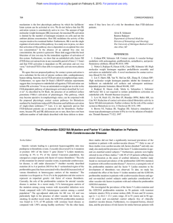
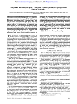
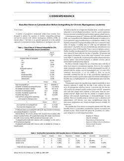
From www.bloodjournal.org by guest on February 6, 2015. For personal use only.
Identification of a Point Mutation in Factor XI11 A Subunit Deficiency
By Philip Board, Marjorie Coggan, and Kris Miloszewski
Oligonucleotideprimers have been designed for the amplification of all 15 exons of the human coagulationfactor Xlll A
subunit gene. Each exon and its intron flanking regions has
been amplified and sequenced from a patient with severe A
subunit deficiency. A single G to A transition in the last base
of exon 14 has been identified in the homozygous proband
and in his heterozygous parents. The mutation would result
in the substitution 681 Arg to His in the mature protein
product. However, because the mutation is at a splice
junction, the deficiency may result from a defect in premessenger RNA splicing.
8 1992by The American Society of Hematology.
C
DNA sequencing was performed with a kit from Amersham
(Sydney, Australia). Southern blots of digested genomic DNA
were prepared on Genescreen Plus obtained from Dupont (Wilmington, DE). ct3$ dATP 3,000Ci/mmol was supplied by Amersham. Agarose (type 11) was obtained from Sigma Chemical Co (St
Louis, MO). Antibodies to the A subunit of FXIII were obtained
from Behringwerke (Marburg, Germany). Oligonucleotide primers
were prepared on an Applied Biosystems 380B DNA synthesizer
(Applied Biosystems, Foster City, CA). Unless otherwise specified,
all chemicals used were of analytical grade.
Patient. The patient is a male born in 1975. He bled from the
umbilicus a few days after birth. Bleeding continued for 10 days
and he received a blood transfusion. Thereafter, he was always
covered with bruises, and suffered several large hematomas after
minor trauma to the forehead and inside the mouth. Diagnosis of
inherited FXIII deficiency was established at 3 years of age. He has
been treated prophylactically since with FXIII concentrate and
remains very well, free from bleeding manifestations and leading a
normal, active life. The patient’s parents are second cousins. A
diagnosis of A subunit deficiency was concluded, as a dansylcadaverine incorporation assay showed zero activity and specific A
subunit rocket immunoelectrophoresis of plasma and platelet-rich
plasma with lysed platelets failed to detect any A subunit protein. B
subunit rocket electrophoresis showed 36% of the normal level.
Haplotype anaZysis. DNA was isolated from nucleated blood
cells12 and digested with BamHI, Bcl I, HindIII, and Taq I
according to the conditions specified by the manufacturers. The
digested DNA was subjected to agarose electrophoresis and then
blotted onto nylon membranes.13The blotted DNA was probed
with FXIII A cDNA labeled with ct32pdATP.14
of A subunit exons were
PCR amplification. Amplificati~n’~
performed with the oligonucleotide primers listed in Table 1. The
primers were phosphorylated by 30 minutes of incubation at 37°C
in a reaction mix containing 100 mmol/L Tris/HCl, pH 8.0, 10
mmol/L MgC12, 1 mmol/L ATP, 3 mmol/L dithiothreitol (DTT),
75 pg/mL primer, and 10 U of T4 polynucleotide kinase. The
reaction was terminated by heating at 70°C for 10 minutes. In all
cases, the PCR reaction mix contained 1x Tag polymerase buffer
(Promega), 0.75 pg of both forward and reverse primers, 0.2
mmol/L dATP, 0.2 mmol/L dCTP, 0.2 mmol/L dGTP, 0.2 mmol/L
OAGULATION FACTOR XI11 (FXIII) is a zymogen
that is converted by thrombin to a transglutaminase
that catalyzes the formation of E-(y-glutamy1)-lysyl crosslinks
between the y chains and between the a chains of fibrin
monomers.’ Plasma FXIII is composed of two A and two B
subunits2 The A subunits are joined as a dimer and are
responsible for the catalytic a c t i ~ i t y . In
~ ? contrast,
~
the B
subunits have no known catalytic activity and are thought to
serve as a carrier molecule stabilizing the A subunits in the
cir~ulation.~
The A and the B subunits are the products of
separate genes that have been mapped to 6~24-25and
1q31-32.1, respectively.5S6 FXIII is essential for normal
hemostasis and deficiency can lead to a severe bleeding
diathesis.’~~
Inherited deficiency of FXIII has been diagnosed in many countries throughout the world, and does
not appear to be associated with any particular ethnic
gr0up.l Most cases studied appear to result from deficiency
of the catalytic A subunit, and only one well-characterized
deficiency of FXIII B subunit has been described.8 In most
cases, the diagnosis of A subunit deficiency has been made
on the basis of an absence of catalytic activity and the
presence or absence of A subunit protein has not always
been determined. Therefore, it is not clear how frequently
FXIII A subunit deficiency arises from either the absence
of protein or from the expression of a defective A subunit
protein. In our experience, both types of deficiency can
occur. In earlier studies, we identified a family in which an
allele coding for an unstable form of A subunit was
~egregating.~,’~
In contrast, deficient individuals from other
families did not appear to express A subunit.ll Nothing is
known about the molecular nature of the mutations causing
FXIII A subunit deficiency.
The study reported here was undertaken to develop the
procedures required to identify the precise molecular
changes responsible for inherited FXIII A subunit deficiency. Oligonucleotide primers have been designed that
permit the amplification by polymerase chain reaction
(PCR) of all known exons in the A subunit gene. Sequence
analysis of amplified DNA from an FXIII A deficient
patient has located a point mutation at a splice junction that
is likely to be the cause of the deficiency.
MATERIALS AND METHODS
The restriction enzymesBamH1,Bcl I, HindIII, Tag I, Sma I, and
calf intestinal alkaline phosphatase were obtained from Boehringer Mannheim (Sydney, Australia); T4 polynucleotide kinase,
Hpa 11, and T4 DNA ligase were obtained from Pharmacia
(Uppsala, Sweden). Tag polymerase was supplied by Promega
(Madison, WI). The nucleotides dATP, dGTP, dCTP, and dTTP
were obtained from Pharmacia. cDNA probes were labeled by
random priming with a kit from Besatek (Adelaide, Australia). All
Blood, Vol80, No 4 (August 15), 1992: pp 937-941
From the Molecular Genetics Group, John Curtin School of
Medical Research, Australian National University, Canberra, Australia; and the Department of Medicine, St James Hospital, University of
Leeds, Leeds, UK.
Submitted November 27,1991; accepted April 14, 1992.
Address reprint requests to Philip Board, PhD, Molecular Genetics
Group, John Curtin School of Medical Research, The Australian
National University, GPO Box 334, CanberraACT 2601, Australia.
The publication costs of this article were defrayed in pari by page
charge payment. This article must therefore be hereby marked
“advertisement” in accordance with 18 U.S.C. section I734 solely to
indicate this fact.
8 1992 by The American Society of Hematology.
0006-497119218004-0002$3.00/0
937
From www.bloodjournal.org by guest on February 6, 2015. For personal use only.
BOARD, COGGAN, AND MILOSZEWSKI
938
Table 1. Primers and Annealing Conditions for the Amplification of
FXlll A Subunit Exons
Exon
1 Forward
1 Reverse
2 Forward
2 Reverse
3 Forward
3 Reverse
4 Forward
4 Reverse
5 Forward
5 Reverse
6 Forward
6 Reverse
7 Forward
7 Reverse
8 Forward
8 Reverse
9 Forward
9 Reverse
10 Forward
10 Reverse
11 Forward
11 Reverse
12 Forward
12 Reverse
13 Forward
13 Reverse
14 Forward
14 Reverse
15 Forward
15 Reverse
Primer 5’
-
3’
TACTCCCAGCAACTGGTTGC
CTGGCTCATAGGGTGCAGG
CATGCCTTTTCTGTTGTCTTC
CTGGACCCAGAGTGGTGG
GATTATITCTTCAACCCTTG
TCTACAATGCAACCCATGG
AATGGCTTGTGAAATCAACC
GAAAACTAAATGTCTGCCTC
GTTTGGTAATAGTCACTATG
CAATAACAAATTTTAAGTGGC
GCTTGCAGAGTGAACACTAG
GCAAATGACAGGTGTAACAG
CCTTCTCACTTCTCACGGAC
AATGTCTTAGAGTGAAGTTTCC
GCTGGTGATGTGmAGCTG
CATCAGCCAATGCCATTGTC
ATTACAGGCATGAGCCACTG
TCAGCAATGAAGCAAGTTCC
TGGACAGAATTGGAGATGAC
AAACAGCACTTTCCTCCAGC
ATGATGGCTAATGCTCTCC
GAACTCATCTCTGAGTGAC
TTGCCTGTCATTATCTCTGG
GACAGCGAGTCTCACAAAG
AAGTCTTGATGCCAGGCCTG
TCTGTTCCAGGATGAGACGC
GCTGCTAATGACCTGCATTC
ACAGCTCTGCACTGCCTG
GATCTTCCGAACCTCTCCTC
ATGCCAGGGTTCATCTCAGC
382*
55
230
56
277
52
326
53
184
50
preparations were sequenced with an Amersham kit. All exons
were sequenced in both strands. On average, five single-stranded
clones were sequenced from each exon. Because of the possible
infidelity of Taq polymerase, differences from the published
may be due to amplification errors. To confirm
differences from the published sequence, appropriate exons were
recloned and sequenced from a separate PCR experiment.
Restriction analysis of PCR products. The amplified exon 14
DNA was digested with Hpa I1 under the conditions suggested by
the supplier (Pharmacia). The digested DNA was subjected to
electrophoresis on a 12% acrylamide gel in a buffer comprising 90
mmol/L Tris, 90 mmol/L boric acid, and 2.5 mmol/L EDTA. The
gel was stained with ethidium bromide and photographed under
UV irradiation.
205
52
RESULTS
262
52
21 1
56
246
56
172
56
266
53
360
52
231
49t
211
52
226*
52
Length of
Annealing
Product Temperature
(bp)
(“C)
‘The amplified region extends from nucleotide -268 in the 5’
flanking region.
tThis reaction contained 5 pL of dimethyl sulphoxide in total volume
of 50 pL.
*The amplified region extends to 11 bp beyond the translation stop
codon.
dTTP, 0.5 to 1 pg template DNA, and 0.5 to 1 U of Taq DNA
polymerase (Promega). Denaturation was performed at 96°C for 1
minute, annealing was performed for 1minute at the temperatures
indicated for each exon in Table 1, and the extension reaction was
performed at 73°C for 1minute. All PCR reactions were performed
in an Innovonic Gene Machine (Bartelt Instruments, Melbourne,
Australia) and the amplification continued for 30 to 35 cycles.
Sequence analysis. The exon DNA amplified by PCR with
phosphorylated primers was cloned into M13 phage mp18.16 The
mp18 vector DNA was digested with Sma I and treated with
alkaline phosphatase to remove 5’ phosphates. The amplified
DNA was purified by electrophoresis in 2% agarose in TBE buffer
(90 mmol/L Tris, 90 mmol/L boric acid, 2.5 mmol/L EDTA). The
DNA was recovered from the gel electrophoretically on an LKB
(Stockholm, Sweden) Extractaphore. The amplified exon DNA
was ligated to the Sma I-digested mp18 with T4 DNA ligase in
“BRL,” blunt end ligation buffer (5x contains 250 mmol/L
Tris/HCI, pH 7.6, 50 mmol/L MgCIZ, 5 mmol/L ATP, 5 mmol/L
DTT, 25% wtlvol polyethylene glycol 8000 [PEG]) and transformed into competent Escherichia coli (TGI strain) by standard
techniques. M13 clones containing inserts in different orientations
were identified electr~phoretically~~
and single-stranded DNA
Amplification of FXIII A exons. The gene encoding the
A subunit of FXIII is composed of 15 exons.*9 Pairs of
oligonucleotide primers suitable for the amplification of
each exon and its intron splice junctions have been designed (Table l).The amplified exon l fragment contained
5’ flanking nucleotides that extended to -268. Because the
3’ end of exon 15 is not defined, the reverse primer
sequence was positioned 11 bp 3’ of the translation stop
codon. Under the conditions described in Materials and
Methods, each exon was amplified from genomic DNA with
little nonspecific contamination (Fig 1). The validity of the
amplification of each exon was confirmed by sequence
analysis. In normal subjects, the amplified sequences did
not differ from the published
Haplotype analysis. To determine if the patient we
chose to study was potentially a true homozygote, or a
compound heterozygote having inherited different FXIIIA
mutations from each parent, we studied the restriction
fragment length polymorphism (RFLP) haplotypes of the
patient and his parents. Two allele RFLPs detected by
BamHI, HindIII, and Taq I digestionsI4 and a multiple
allele polymorphism detected by Bcl IZ1 digestion were
evaluated. As shown in Fig 2, the proband was homozygous
for each RFLP and has apparently inherited an identical
haplotype from each parent. This suggests that the proband
has inherited the same deficiency causing mutation from
each parent and is a true homozygote.
Sequence analysis. The A subunit exons were amplified
from genomic DNA obtained from the proband and sequenced on both strands. A single base transition G + A
was detected in the last base of exon 14 (Fig 3). This change
alters codon 681 from CGT to CAT and would change the
amino acid sequence of the A subunit product from 681Arg
to 681 His.
The deduced amino acid sequences of all other exons
agreed exactly with that derived from the gene,I9 except for
residue 651 Glu, which was in agreement with the protein
sequence.Z0This difference results from a C to G change in
the first base of the codon encoding residue 651. No
changes were detected in intron sequences flanking the
splice junctions and the GT-AG rule was adhered to in each
case. Furthermore, no changes in the 5’ flanking sequence
as far as -268 bp were detected.
To confirm that this substitution was a property of the
From www.bloodjournal.org by guest on February 6, 2015. For personal use only.
939
MUTATION IN FACTOR Xlll DEFICIENCY
thcrc was no cvidencc of thc 165-bp fragment. In contrast,
both thc 165-bp and the 189-bp fragments could bc detected in thc parental samples. Thcsc rcsults indicate that
the proband is homozygous for the cxon 14 G + A transition and his parents arc hctcrozygous for thc samc mutation.
It is possiblc that the mutation wc obscrved in the
proband and his family is a common polymorphism without
dclcterious cffccts. Therefore, we studied the DNA from 20
normal blood donors by this RFLP technique and in each
casc we detected the small 165-bp fragment, suggesting that
they did not havc the G + A transition and that this
mutation is not common and is rclatcd to the deficiency in
this family.
154
220
#!
506
I632
DISCUSSION
1
2
3
4
5
6
7
9
10 11 12 13 14 15
8
bP
154
220
298
344
396
506
1632
Fig 1. The PCR reaction product for each pair of primers has been
fractionated on 2% agarose gel. The number beneath each track
indicates the pair of primers used and the exon amplified. Standard
molecular size markers have been run at the side of each gel and their
lengths are indicated in base pairs (bp).
patient's genomic scqucncc and not dcrivcd from Ta9
polymcrasc infidelity, scvcral MI3 cloncs from two scparatc
PCR amplifications of cxon 14 wcrc scqucnccd. In each
case. the G + A transition was obscrved. The normal
scqucncc (G) was not dctcctcd in any MI3 clonc dcrivcd
from the proband.
The G + A mutation obscrvcd in thc proband results in
thc loss of a Hpa 11 sitc in the PCR amplification product.
As shown in Fig 4A, digestion of thc 211-bp cxon 14
amplification product from a normal control with Hpa I1
should give risc to fragments of 165, 24, and 22 bp. In
contrast, digestion of thc samc PCR product from thc
proband should givc rise to fragments of 189 and 22 bp. To
dctcrminc if thc G + A substitution is inherited and could
bc dctcctcd in thc proband and his parents, amplified cxon
14 fragments wcrc digested with Hpa I I and fractionated by
electrophoresis on acrylamidc gcls. As shown in Fig 4B, thc
normal control showcd thc prcscncc of thc cxpcctcd 165-bp
fragment. The proband showed a 189-bp fragment and
In this study, we have designed oligonucleotidc primers
and defined reaction conditions for thc amplification of the
15 FXIll A subunit gene exons. The primers for exon 1
encompass 5' flanking scqucnces extending to nucleotide
-268. This rcgion contains sevcral potential regulatory
clcments'" and may contain a promotcr, although there is
no experimental evidence demonstrating that thcsc clcmcnts contributc to the regulation of FXffU transcription.
Except for the 3' cnd of exon 15, the amplified fragments all
contain thc cxon coding scquences with their 5' and 3'
splicc junctions and immcdiatc flanking regions. Sequencing thc fragments amplified from deficient patients under
the conditions dcscribcd in the present experiments should
show thc vast majority of mutations giving rise to deficiency
of FXIll A subunit.
Thc A subunit genc FXIII A is autosomal and has been
mappcd to the distal region of the short arm of chromosome 6.s In many autosomal recessive disorders, such as
FXlll A deficiency, affectcd individuals are compound
hctcrozygotes with two mutations affecting A subunit synthesis or function. Thus, at the site of each mutation, the
deficient patient also has a normal allele on the alternate
chromosome. If fragments containing particular mutations
arc amplificd by PCR, the DNA from the normal and
BamHI B
BamHI C
Hind I11
Taq I
Bcl I
-++
++-
++ -+
++-
aa
b a
a a
Fig 2. The 0 family showing the inheritance of Fxlll A subunit
deficiency (darkenedsections) and RFLP haplotypes. The RFLPs with
two alleles are indicated as
or and the Ecl I polymorphism that
has multiple alleles has been identified alphabetically. The nomenclature for the 8cl I RFLPs relates only to this family.
+
-
From www.bloodjournal.org by guest on February 6, 2015. For personal use only.
BOARD, COGGAN, AND MILOSZEWSKI
940
G
Normal
G
A
T
C
G
Exon14
Patient
T
C
C
C
--G
V
G
T
A
A
A
Intron N
C
A
G
T
A
A
A
G
A
T
C
Fig 3. Part of the nucleotide sequence of exon 14
and intron N of the human FXlll A subunit gene. The
normal sequence is on the left and the dotted line
shows the normalsplice junction. The mutation in the
patient is shown by an arrow.
mut d chromosomes will be amplified equally and, when
cloned in M I 3 for sequencing, will give rise to equal
numbers of clones with the normal or mutant sequence. In
such cases, many M13 clones from each amplified fragmcnt
need to be sequenced to identify the mutation. Altcrnatively, direct sequencing can be used, but these methods
have other associated drawbacks. In the present case, the
sequence analysis was simplified by the fact that the
proband appeared to be a true homozygote because he was
homozygous for five closely linked RFLPs and showed the
inheritance of an identical RFLP haplotype from each
parent. Furthermore, it was known that the parents were
second cousins, increasing the probability that the mutation
had an identical familial origin.
Sequence analysis of amplified exons from the patient
identified a G + A transition in the last base of exon 14.
Two additional changes were detected in individual clones;
however, they could not be confirmed by sequencing other
clones from the original amplification or after rcamplification and cloning. We concluded that these changes were
amplification artifacts. The mutation we identified was
confirmed by sequencing multiple MI3 clones in both
orientations and M 13 cloncs dcrivcd from two independent
PCR experiments. The G 4 A transition was idcntificd in
cvcry clone of amplified exon 14 from the proband. The
absence of a normal sequence confirmed his predicted
homozygosity.
It is not immediately clear what effcct the obscrved
mutation has on A subunit function and expression. The
point mutation occurs in the coding sequcncc and would
result in the amino acid substitution 681 A% to 681 His.
Because littlc is known about the active site of the A
subunit and the residues required for substrate binding and
catalysis, it is not clear whether the change would substantially altcr the mutant subunit's catalytic function, stability,
structure, or interaction with B subunits. The lack of any
immunologically detectable A subunit in plasma and platelets from the proband suggests that the defect primarily
influences its expression or stability. It may be significant
that the mutation is located at a splice junction. Introns in
higher eukaryotic prc-messenger RNAs (mRNAs) are
bounded at the 5' end by the consensus sequence (C/A)AG/
A
Normal
Patient
165
c
CCG
Fig 4. (A) The amplified exon 14 fragment is 211
bp and contains two Hp.9 II sites indicated by arrows.
The mutation in the patient deletes one site. The
expected sizes of the DNA fragments after Hpe II
digestion are indicated. (6) Electrophoretic fractionation of amplified exon 14 digested with Hps II. The
normal control sample is indicated N and the different members of the 0 family are indicated by the
family tree.
bP
189
165
CGG
1
189
C CAC
B
c
22
C CGG
From www.bloodjournal.org by guest on February 6, 2015. For personal use only.
MUTATION IN FACTOR Xlll DEFICIENCY
941
GU(G/A)AGU.zz,z3Although the GU at the start of the
intron (underlined) is largely invariant, variations in other
~ , survey
~
of 5’ splice sitesz4
positions are known to O C C U ~ ? A
has shown that, while G is the most common base in the -1
position, A occurred in 8 of 99 sites studied. Although the
G --* A transition detected at position -1 in exon 14 does
not absolutely exclude normal splicing, there is evidence
that components of the flanking exon sequence can influence the site specificity of ~ p l i c i n g . ~ ~ - ~ ~
A number of studiesz3-z5have implicated the hybridization of U1 small nuclear (sn) RNA in the specific recognition and cleavage of 5’ splice junctions. Mutations in the
exon sequence adjacent to the splice junction have been
shown to directly influence U p R N A hybridization and to
modify the efficiency of cleavage. Because the G + A
transition observed in these experiments is in the exon
immediately adjacent to the normal cleavage site, it is clear
that it will substantially decrease potential hybridization of
the UlsnRNA in the correct position, which may lead to
either a decrease in the efficiency of cleavage, the elimination of cleavage, or even cleavage at an adjacent cryptic site.
Thus, the cause of the A subunit deficiency in the present
case may be due to a splicing defect.
At this stage, it is not possible to conclude if the observed
point mutation alters pre-mRNA splicing or if the subunit
with the 681Arg +His mutation is unstable. To resolve this
question, in future studies it may be possible to express a
cDNA clone containing the same mutation and to directly
evaluate the properties of the recombinant protein?
In conclusion, we have defined oligonucleotide primers
that allow amplification of all the coding regions of the
FXIII A subunit gene and their intron/exon splice junctions. We have amplified and sequenced all of these regions
from a homozygous deficient patient and identified a point
mutation that could cause the deficiency. In accordance
with the convention established with other coagulation
factors, we propose that this FXIII A subunit defect be
described as the Canberra I mutation.
REFERENCES
1. Lorand L, Losowsky MS, Miloszewski KJM: Human factor
XI11 fibrin-stabilizingfactor. Prog Hemost Thromb 5:245,1980
2. Schwartz ML, Pizzo SV, Hill RL, McKee P A Human factor
XI11 from plasma and platelets. J Biol Chem 248:1395,1973
3. Board PG: Genetic polymorphism of the A subunit of human
coagulation factor XIII. Am J Hum Genet 31:116,1979
4. Mary A, Achyuthan KE, Greenberg CS: B-chains prevent
proteolytic inactivation of the a-chains of plasma factor XIII.
Biochem Biophys Acta 966:328,1988
5. Board PG, Webb GC, McKee J, Ichinose A Localization of
the coagulation factor XIIIA subunit gene (F13A) to chromosome
bands 61324 + p25. Cytogenet Cell Genet 48:25,1988
6. Webb GC, Coggan M, Ichinose A, Board PG: Localization of
human coagulation factor XIIIB subunit gene (F13B) to chromosome bands 1q31-32.1 and restriction fragment polymorphism of
the locus. Hum Genet 81:157,1989
7. Duckert F, Jung E, Schmerling DH: A hitherto undescribed
congenital haemorrhagic diathesis probably due to fibrin stabilizing factor deficiency. Thromb Diath Haemorrh 5:179,1960
8. Saito M, Asakura H, Yoshida T, Ito K, Okafugi K, Yoshida T,
Matsuda T: A familial factor XI11 subunit B deficiency. Br J
Haematol74:290, 1990
9. Castle S, Board PG, Anderson RAM: Genetic heterogeneity
of factor XI11 deficiency: First description of unstable A subunits.
Br J Haematol48:337,1981
10. Castle S, Board PG: Biochemical characterization of genetically variant and abnormal blood coagulation factor XI11 A
subunits. Clin Chim Acta 133:141,1983
11. Board PG, Coggan M, Hamer JW: An electrophoretic and
quantitative analysis of coagulation factor XI11 in normal and
deficient subjects. Br J Haematol45:633,1980
12. Grunebaum L, Cazenave J-P, Camerino G, Kloepfer C,
Mandel J-L, Tolstoshev P, Jaye M, De La Salle H, Lecocq J-P:
Carrier detection of haemophilia B using restriction site polymorphism associated with the coagulation factor IX gene. J Clin Invest
73:1491,1984
13. Reed KC, Man D A Rapid transfer of DNA from agarose
gels to nylon membranes. Nucleic Acids Res 13:7207, 1985
14. Board PG, Chapple R, Coggan M: Haplotypes of the
coagulation factor XIIIA subunit locus in normal and deficient
subjects. Am J Hum Genet 42:712,1988
15. Saiki RK, Scharf SJ, Faloona F, Mullis KB, Horn GT, Erlich
HA, Arnheim N Enzymatic amplification of P-globin genomic
sequences and restriction site analysis for diagnosis of sickle cell
anaemia. Science 230:1350,1985
16. Norrander J, Kempt T, Messing J: Construction of improved
M13 vectors using oligodeoxy nucleotide-directed mutagenesis.
Gene 26:101,1983
17. Messing J: New M13 vectors for MI3 cloning. Methods
Enzymol101:20,1983
18. Ichinose A, Hendrickson LE, Fujikawa K, Davie E W The
amino acid sequence of the A subunit of human factor XIII.
Biochemistry 25:6900,1986
19. Ichinose A, Davie EW: Characterization of the gene for the
A subunit of human factor XI11 (plasma transglutaminase), a blood
coagulation factor. Proc Natl Acad Sci USA 85:5829,1988
20. Takahashi N, Takahashi Y, Putnam FW: Primary structure
of blood coagulation factor XIIIa (fibrinoligase, transglutaminase)
from human placenta. Proc Natl Acad Sci USA 83:8019,1986
21. Zoghbi HY, Daiger SP, McCall A, OBrien WE, Beaudet
AL: Extensive DNA polymorphism at the factor XIIIa (F13A)
locus and linkage to HLA. Am J Hum Genet 42:877,1988
22. Mount SM: A catalogue of splice junction sequences. Nucleic Acids Res 10:459,1982
23. Aebi M, Hornig H, Weissmann C: 5’ Cleavage site in
eukaryotic pre-mRNA splicing is determined by the overall 5’
splice region, not by the conserved 5’ GU. Cell 50:237,1987
24. Oshima Y, Gotoh Y: Signals for the selection of a splice site
in pre-mRNA computer analysis of splice site junction sequences
and like sequences. J Mol Biol195:247,1987
25. Smith CWJ, Patton JG, Nadal-Ginard B: Alternative splicing in the control of gene expression. Ann Rev Genet 23:527,1989
26. Board PG, Pierce K, Coggan M: Expression of functional
coagulation factor XI11 in Escherichia coli. Thromb Haemost
63:235,1990
From www.bloodjournal.org by guest on February 6, 2015. For personal use only.
1992 80: 937-941
Identification of a point mutation in factor XIII A subunit deficiency
P Board, M Coggan and K Miloszewski
Updated information and services can be found at:
http://www.bloodjournal.org/content/80/4/937.full.html
Articles on similar topics can be found in the following Blood collections
Information about reproducing this article in parts or in its entirety may be found online at:
http://www.bloodjournal.org/site/misc/rights.xhtml#repub_requests
Information about ordering reprints may be found online at:
http://www.bloodjournal.org/site/misc/rights.xhtml#reprints
Information about subscriptions and ASH membership may be found online at:
http://www.bloodjournal.org/site/subscriptions/index.xhtml
Blood (print ISSN 0006-4971, online ISSN 1528-0020), is published weekly by the American
Society of Hematology, 2021 L St, NW, Suite 900, Washington DC 20036.
Copyright 2011 by The American Society of Hematology; all rights reserved.


© Copyright 2026