
Endothelial Cells Express the Interleukin-1 Receptor Type I
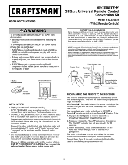
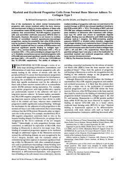
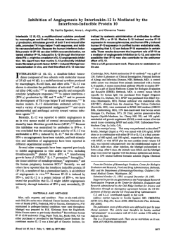
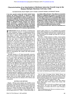
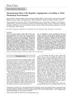
Endothelial Cells Express the Interleukin-1 Receptor Type I
By Diana Boraschi, Alessandro Rambaldi, Antonio Sica, Paolo Ghiara, Francesco Colotta, Ji Ming Wang, Marco de Rossi,
Carla Zoia, Giuseppe Remuzzi, Federico Bussolino, Giuseppe Scapigliati, Antonella Stoppacciaro, Luigi Ruco,
Aldo Tagliabue, and Alberto Mantovani
Interleukin-1 (11-1) profoundly affects a number of functions
of vascular cells. Two distinct IL-1 receptors (IL-IR) are
expressed on different cell types: the 80 Kd IL-IR, on T cells
and fibroblasts, and the 68 Kd IL-IR,, on B cells and myelomonocytic cells. The presence and functionality of IL-1R
on vascular cells has been investigated by using polyomatransformed mouse endothelial cell (EC) lines (sEnd.1 and
tEnd.1). These cells expressed specific and saturable binding
sitesfor IL-I (1,273 sites per cell with kd 9.5 x lo-" mol/Lfor
sEnd.1, and 771 sites per cell with kd 8.5 x lo-" mol/L for
tEnd.1, with radioiodinated IL-la as ligand). Binding of IL-1
was also evident at single cell level by autoradiography. By
cross-linking studies, the molecular weight of the IL-I binding protein on EC was approximately 80 Kd. This was
confirmed by the presence in EC of mRNAfor the 80 Kd IL-IR,.
The IL-lR, on EC was apparently functional, since EC responded to IL-1 with IL-6 mRNA expression and IL-6 bioactivity production. These results were extended to human EC
and vascular smooth muscle cells, which were also found to
express mRNAfor IL-lR,.
0 1991 by The American Society of Hematology.
I
marrow granulocytes.21 These cells do not express the
IL-lR,.
Little is known concerning the IL-lR present on EC.
Thieme et al' characterized the binding of IL-la to human
umbilical vein ECs. By immunoprecipitation and chemical
cross-linking to the ligand they identified a 78 Kd IL-1binding protein on human EC.24
Here we report that vascular cells (mouse EC; human
EC, and smooth muscle cells [SMC]) express the 80 Kd
IL-lR,.
NTERLEUKIN-1 polypeptides are prototypic pleiotropic cytokines whose action encompasses various organs
and tissues.' Endothelial cells (EC) have emerged as one
important target for IL-l.2s3IL-1 and the functionally
related cytokine tumor necrosis factor (TNF) activate EC
functions mainly related to inflammation and thrombosis.
These include production of procoagulant activity: prostaglandins,5s6platelet-activating factor (PAF),' and plasminogen activator inhibitor'; inhibition of the synthesis of
plasminogen activator* and alterations in the thrombomodulidprotein C anticoagulation pathway'; expression of adhesion molecules'o; and production of cytokines."
The action of IL-1 on cells is mediated via specific
receptors. An 80 Kd IL-1 binding protein (IL-1RJ has been
purified from a murine thymoma cell line" and a cDNA
that encodes this protein has been c10ned.l~This 80 Kd
IL-1R is expressed in mouse and human T cells, fibroblasts,
keratinocytes, and epithelial cell^.'^''^ Affinity cross-linking
suggested that the IL-1R on B cells would be a 60 to 68 Kd
pr~tein.",'~
Further evidence for the existence of a second
distinct IL-1R (IL-lR,J has been provided recently.20-22
This
receptor, identified using Epstein-Barr virus (EBV) transformed human B cell lines and the murine 702/3 pre-B cell
line, is also expressed on mouse macrophages and bone
From Sclavo Research Center, Siena; Istituto di Ricerche Farmacologiche Mario Negri; Milano and Bergamo; the Departments of
Medical Genetics, Biology, and Ckmisby, University of Torino; the
Department of Human Biopathology, University of Roma; and Domp2
S.p.A., Milano, Italy.
Submitted January 28,1991; accepted May 3,1991.
Supported by a fellowship of Consocio Mario Negri Sud, S. Maria
Imbaro, Chieti, Italy. Also supported by Minister0 della SanitdIstituto Superiore della Sanita (National AIDS Project). The generous
contribution of the Italian Association for Cancer Research is gratefully acknowledged.
Address reprint requests to Alberto Mantovani, MD, Laboratoly of
Immunology, Istituto di Ricerche Farmacologiche Mario Negri; via
Eritrea 62, I-20157Milan0, Italy.
The publication costs of this article were defrayed in part by page
charge payment. This article must therefore be hereby marked
"advertisement" in accordance with 18 U.S.C.section 1734 solely to
indicate this fact.
0 1991 by TheAmerican Society of Hematologv.
0006-4971I9117805-0027$3.00/0
1262
MATERIALS AND METHODS
Cells. The mouse endothelioma cell lines sEnd.1 and tEnd.1
were obtained through the courtesy of Dr E.F. Wagner (European
Molecular Biology Laboratory, Heidelberg, FRG). These cell lines,
derived from a subcutaneous (s) and thymic (t) hemangioma,
express the polyoma middle T antigen, have cobblestone endothelial-like morphology, express von Willebrand factor, and cause
hemangiomas in vivo.25**6
Expression of von Willebrand factor was
checked during this study. The cells were maintained in Dulbecco's
modified Eagle's medium (DMEM) containing 15% fetal bovine
serum (FBS) and 750 pg/mL G418. Human ECs were isolated from
umbilical veins and cultured in medium 199 (GIBCO, Paisley,
Scotland) supplemented with 20% FBS in the presence of an EC
cell growth supplement (50 @mL; Sigma Chemical Co, St Louis,
MO) and porcine intestinal heparin (100 pg/mL; Sigma). EC were
used within the eighth passage.
Human vascular SMCs derived from the femoral artery were
kindly donated by Dr G. Gabbiani (University of Geneva School of
Medicine, Switzerland). SMC were cultured in DMEM containing
10% FBS. When cells were confluent, human recombinant IL-1p
(Sclavo, Siena, Italy) was added at 10 ng/mL for 4 or 20 hours
before extraction of mRNA. IL-lp had a specific activity of 1 X l@
U/p,g on D10.G4.1 cells when compared with the interim IL-lp
reference reagent 86/552 (arbitrary titer 1 X l@Ulpg; NBSB,
NIBSC, Potters Bar, South Mimms, UK) and was free of endotoxin
( I 0.08 pg LPS/p,g IL-1).
Autoradiographic determination of IL-1 binding. Human recombinant IL-la, radioiodinated with the chloramine T method, was
purchased from Du Pont-NEN (Boston, MA) and had a specific
activity of 2.2 x lo6 dpdpmol. Endothelioma cells were grown to
confluency on 8-chamber tissue culture chambedslides (Lab-Tek
Division, Miles Laboratories, Naperville, IL). Washed monolayers
were incubated with approximately 0.3 nmol/L '"I IL-la in 0.1 mL
of DMEM with 15% FBS and 0.02% NaN, (binding medium) for 3
hours at room temperature with gentle agitation. At the end of the
incubation, gaskets were removed, slides were extensively washed
Blood, Vol78, No 5 (September l), 1991:pp 1262-1267
1263
IL-1 RECEPTOR IN ENDOTHELIUM
by dipping in PBS and k e d for 30 seconds in acetone at room
temperature. Slides were stored at -20°C until autoradiography.
Slides were postfured for 1minute in cold acetone, then covered by
dipping with NTB3 Kodak photographic emulsion. After 2 days of
exposure, slides were developed with Kodak D19 developer and
Rapid Fixer and counterstained with hematoxylin. Results were
evaluated by the method of Schlessinger et al?’ The cut off for
positivity of IG1 binding was calculated as the median number of
silver grains on cells incubated with cold IG1 plus three times the
standard deviation (SD), multiplied for a field factor (ie, the
number of grains per field without cells in the slides incubated with
labeled IL-lP divided by that in slides incubated with cold IL-lB).
ZL-1binding assay. Replicate confluent monolayers of 1to 2 X
106 cells per well of Cluster6plates (Costar, Cambridge, MA) were
incubated with increasing doses of ‘=I IL-1 in 0.5 mL of binding
medium for 3 hours at room temperature under gentle agitation on
a rocking platform. Nonspecific binding was assessed in the
presence of a 500-fold molar excess of unlabeled IL-1. Wells were
then extensively washed in PBS, monolayers were dissolved in 0.5
mL 9.5 m o m urea, and radioactivity was counted. Control monolayers were trypsinized for exact determination of cell numbers.
Calculations and Scatchard analysis were performed according to
Munson and Rodbard?’
Affinity cross-linking. ‘=I IL-la was cross-linked to intact cells
with disuccinimidyl suberate (DSS) or with dithiobis (disuccinimidyl propionate) (DSP) (Pierce, Rockville, MD) as previously
described.16 Briefly, cell monolayers (3.0 x lo7 cells) in binding
medium were incubated with
IL-la (0.5 nmoUL) for 3 hours at
room temperature in gentle agitation with or without a 500-fold
molar excess of unlabeled IL-1. After washing the unbound IL-1,
DSS or DSP (50 mg/mL in DMSO) were added to a final
concentration of 2 mg/mL, and the reaction was allowed to proceed
for 45 minutes at room temperature with vigorous shaking, then
centrifuged in Eppendorf for 5 minutes. The supernatant was
denatured with Laemmli sample buffer?’ Sodium dodecyl sulfatepolyacrylamidegel electrophoresis (SDS-PAGE) was run on 10%
gel using colored molecular weight markers (Amersham, Little
Chalfont, UK). Dried gels were exposed at -40°C using Kodak
XAR-5 autoradiographic film.
Northern blot analysis. Northern blot analysis was performed
according to standard procedures.30Total RNA was isolated from
confluent cell cultures by guanidine isothiocyanate method.” In
some instances, polyadenylated RNA was prepared by chromatography on oligo dT cellulose columns (Pharmacia, Uppsala, Sweden). Aliquots of 10 pg total RNA were analyzed by electrophoresis through 1% agarose formaldehyde gels followed by Northern
blot transfer to Gene Screen Plus membrane (Du Pont-NEN). A
human IL-1 receptor cDNA (HindIII-EcoRI fragment of 477 bp)”
was obtained through the courtesy of Dr S.K.Dower (Immunex
Corp, Seattle, WA). The mouse IL-1 receptor cDNA (SmaI
fragment of 1,800 bp) was cloned in this laboratory by polymerase
chain reaction according to the sequence cloned by Sims et al.”
The mouse IL-6 cDNA probe (EcoRI-BglII fragment of 650 bp)
was a kind gift of Dr J. Van Snick (Ludwig Institute, Bruxelles,
Belgium). These fragments were labeled to a specific activity of lo9
cpm/pg by using hexanucleotide primers and a3*P-CTP.Membranes were pretreated and hybridized in 50% formamide (Merck,
Rahaway, NJ) with 10% dextran sulfate (Sigma) and washed twice
with 2x SSC (lx SSC: 0.15 moUL sodium chloride, 0.015 m o m
sodium citrate) then twice with 2x SSC plus 1% SDS (Merck) at
60°C for 30 minutes and finally twice with 0 . 1 ~SSC at room
temperature for 30 minutes.
The membranes were exposed for 12 to 24 hours at -80°C with
intensifying screens. RNA loading and transfer to membrane was
checked by examination under UV light and hybridization of the
blot with an a-actin probe.
ZL-6bioactiviry assay. IL-6 was measured as hybridoma growth
factor (HGF) activity using the 7TD1 cell line, obtained through
the courtesy of Dr J. Van Snick. Briefly, 2 x 10’ cells in 200 pL
(four replicates per experimental point) were cultured for 72 hours
with different dilutions of the supernatants to be tested or of the
appropriate control medium. Routinely, cell proliferation was
assessed by the (3-4,5-dimethylthiazol-2-yl)-2,5-diphenyl-tetrazolium bromide colorimetrictest as described.” Alternatively, cells
were pulsed with 0.5 kCi/well [’HI dThd (sp act 185 GBq/mmol;
Amersham) for 6 hours. HGF activity resulting in half maximal
stimulation of target cell growth was arbitrarily defined as 1 U.
Reference standards used in these experimentsconsisted of human
recombinant IG6 or IL-brich supernatant of a mixed lymphocyte
reaction.
RESULTS AND DISCUSSION
Vascular cells are one important target for IL-1. On
interaction with IL-1, ECs undergo a complex reprogramming of function, which favors thrombosis, leukocyte recruitment, and inflammation? A limitation to the detailed
analysis of modulation of EC functions has been the
unavailability of continuous EC lines. Recently, murine
endothelioma cells lines have been established from hemangiomas developed in mice on infection with a retroviral
vector carrying the polyoma virus middle T oncogene.”.26
These lines have endothelial morphology and express EC
markers,” and might thus be suitable for study of E C
modulation in vitro, if they also maintain EC functional
characteristics. In order to evaluate the ability of polyomatransformed cells to respond to IL-1 modulation, two of
these lines, sEnd.1 and tEnd.1, have been analyzed for
expression of IL-lR and functional response to IL-1 stimulation. We focused on mouse EC initially because most
available information concerning the existence of differential distribution of distinct IL-1R has been obtained in the
Specific binding of radioiodinated IL-1 to sEnd.1 cells
was first evaluated at the single cell level by autoradiography on cells cultured on glass slides. As shown in Fig 1,
sEnd.1 cells could efficiently bind radioiodinated IGla.
Binding capacity was, however, not homogeneous, with
approximately 30% of cells showing significant binding of
IL-la (8% had > 100 grains per cell; 9% between 50 and
100 grains per cell; and 13% between 30 and 50 grains per
cell), and the other 70% showing only few associated silver
grains ( < 30 per cell). Cells showing the highest density of
associated grains were highly adherent and with a large
cytoplasm (Fig 1). These data indicate that endothelioma
cells possess receptors for IL-1.
Two structurally distinct IL-1R have been identified, with
differential cellular d i s t r i b ~ t i o n . ’ ~Only
~ ~ *the
~ ~ 80 Kd ILlR,, expressed on T cells and fibroblasts, has been molecularly cloned and ~equenced.’~’~
A second type of IL-1R has
been found on B cells and on myelomonocytic cell^.'^-^^^
The IL-lR,, has a molecular weight of approximately 68 Kd
and, at variance with IL-lR,, apparently binds IL-1p more
abundantly than IL-hZO>’The role of the two types of
IL-lR in the mechanisms of cellular activation by IL-1 is
1264
BORASCHI ET AL
Fig 1. Binding of IL-1 to murine endothelioma
cells. Monolayersof sEnd.1 cells on glass slides were
incubated with '"I IL-lufor 3 hoursthen examined for
IL-1 binding by autoradiography. Original magnification: x 100 (A), x500 (e).
not clear. Presumably, the two IL-1R types are coupled to
distinct intracellular signal transduction pathways" and are
associated with different rates of ligand internalization and
intracellular degradati~n?~
The characteristics of the IL-1R present on endothelioma lines have been assessed by binding studies. As shown
in Fig 2 (A,B), the two endothelioma lines tEnd.1 and
sEnd.1 showed specific and saturable binding of radioiodinated human recombinant IL-la. The number of IL-1
binding sites per cell and the affinity of binding were similar
between the two lines (1,273 sites per cell with kd 9.5 X lo-"
mol/L for sEnd.1, and 771 sites per cell with kd 8.5 X lo-''
mol/L for tEnd.1, respectively). IL-1 receptors on human
EC showed similar characteristics (ref. 23 and 35; 2 experiments not shown), with a somewhat lower number of
binding sites (100 to 500 sites per cell). By chemical
cross-linking of radiolabeled IL-la to sEnd.1 membrane
proteins with DSS, a single cross-linking product could be
seen, of molecular weight of approximately 98 Kd (Fig 2,
C). Accordingly, the presence of a 98 Kd product was
evident also when DSP was used as a cross-linking agent
(data not shown). By subtracting the molecular weight of
bound IL-la (17.5 Kd), this corresponded to an IL-1
binding protein of approximately 80 Kd. This is in agreement with the molecular weight of the IL-lR described on
human EC (ie, 78 Kd)" and suggests that the IL-1R
expressed by EC is the IL-lR,. To clarify this point, the
expression of mRNA encoding the IL-lR, has been examined. As shown in Fig 3, both sEnd.1 and tEnd.1 had
appreciable levels of IL-lR, transcripts, though these were
substantially lower than those detected in 3T3 murine
fibroblasts used as positive controls (these express over
5,000 IL-lR per cell).21s36
Having established that the mouse endothelial lines
express IL-lR,, it was important to evaluate whether these
cells indeed respond to IL-1 stimulation, ie, whether their
IL-1R was functional. As shown in Fig 4,IL-1 was able to
induce high levels of IL-6 mRNA in both the sEnd.1 and
the tEnd.1 lines. In parallel to detection of message
transcription for IL-6, an increase of IL-6 biologic activity
could be detected in the culture supernatant. In fact,
IL-1-stimulated sEnd.1 and tEnd.1 cells produced 568
U/mL and 1,250 U/mL of IL-6, respectively, whereas only
82 U/mL and 83 U/mL of IL-6 bioactivity could be detected
11-1 RECEPTOR IN ENDOTHELIUM
1265
s End. 1
t End. 1
1,273 sites/cell
Kd = 95 pM
0
0.05
0.10
0.15
/
/p
Bound
771 sites/cell
Kd = 85 pM
"."
CROSS-LINKING
0.20
0
0.05
0.10
0.15
Cold IL-1
0.20
Free pmoledwell
Fig 2. Equilibrium binding and cross-linking of "1 IL-lat o murine endothelioma cells. For equilibrium binding analysis, specific binding of
increasing amounts of 161 IL-la (free) was determined in the presence of 500-fold molar excess of unlabeled IL-1. Scatchard analyses are reported
in the insets: B, bound; F, free; in abscissa are bound fmol/well. (A) tEnd.1 cells. (B) sEnd.1 cells. (D) Cross-linking of '=I IL-la on sEnd.1 cells,
performed with DSS in the absence (-) or in the presence (+) of unlabeled IL-1.
in the absence of IL-1. The ability of IL-1 to induce IL-6
synthesis in murine endothelioma cells is in full agreement
with previous observations in human EC." It is of interest
that the recently identified IL-1 receptor antagonist, reportedly interacting with type I receptors, inhibits the action of
IL-1 on human and murine endothelium (data not shown)?'
Thus, murine endothelioma cell lines sEnd.1 and tEnd.1
express functional IL-lR, similar to that observed on
human EC. However, that the IL-lR present on human
1
2
3
vascular cells is in fact the IL-lR, was inferred only by the
size of the IL-1 binding protein observed in cross-linking
studies, ie, approximately 78 Kd.24To ascertain that human
vascular cells possess the IL-lR,, we have examined human
1
2
3
4
5
6
7
..
-
285
-
185
- 185
Fig 3. IL-lR, expression in murine endothelioma cells. Lane 1,
positive control (10 p g of total RNA from 3T3 murine fibroblasts); lane
2, sEnd.1 cells (5 p g poly A+ RNA); lane 3, tEnd.1 cells (2 p g poly A+
RNA).
Fig 4. Kinetics of IL-6 mRNA expression induced by IL-lp in murine
endothelioma cells. sEnd.1 cells (lanes 1,3,5) and tEnd.1 cells (lanes 2,
4, 6) were either left untreated (lanes 1 and 2) or incubated with 10
ng/mL of human recombinant 11-1p f o r 4 hours (lanes 3 and 4) or for 20
hours (lanes 5 and 6). Murine N11 microglioma cells" were used as
positive control (lane 7). On each lane, 10 p g of total RNA were
loaded.
BORASCHI ET AL
1266
SMC
EC
1
2
I --
IL-lR
285
:.,
. .._, ,..
EC and SMC for IL-lR, mRNA expression. As shown in
Fig 5, both EC and SMC express mRNA for IL-lR,.
Exposure of human vascular cells, as well as murine
endothelioma cells, to IG1 did not appreciably affect
IL-lR, mRNA expression (Fig 5, data not shown).
The results reported here demonstrate that murine EC
and human EC and SMC express IL-lR,, though they do
Fig 5. IL-lR, expression in human vascular cells.
(Left) HumanEC. (Right) HumanSMC, preincubated 4
hourswithout (lane 1) or with 10 ng/mL IL-1p (lane 2).
On each lane, 10 pg of total RNA were loaded.
not formally exclude that these cells may at the same time
have other IL-1R types on their membrane.
The availability of the recently cloned soluble form of
IL-lR:9 and that of a neutralizing monoclonal antibodies
for the murine IL-lR,Z' will help to finely evaluate in vitro
and in vivo the precise role of IL-1 in vascular cell activation
in pathologic conditions.
REFERENCES
Damme J, Dejana E, Mantovani A IL-1 stimulates IL-6 produc1. Dinarello C A Interleukin-1 and its biologically related cytotion in endothelial cells. J Immunol142549,1989
kines. Adv Immunol 44:153,1989
12. Urdal DL, Call SM, Jackson JL, Dower S K Affinity purifica2. Pober JS: Qtokine-mediated activation of vascular endothetion and chemical analysis of the interleukin-1 receptor. J Biol
lium. Physiology and pathology. Am J Pathol133:426,1988
Chem 263:2870,1988
3. Mantovani A, Dejana E: Cytokines as communication signals
13. Sims JE, March CJ, Cosman D, Widmer MB, Mac Donald
between leukocytes and endothelial cells. Immunol Today 10370,
HR, Mc Mahan CJ, Grubin CE, Wignall JM, Jackson JL, Call SM,
1989
Friend D, Alpert AR, Gillis S, Urdal DL, Dower SK cDNA
4. Bevilacqua MP, Pober JS, Majeau GR, Cotran RS, Gimbrone
expression cloning of the IL-1 receptor, a member of the immunoMA Jr: Interleukin-1 (IL-1) induces biosynthesis and cell surface
globulin superfamily. Science 241:585,1988
expression of procoagulant activity in human vascular endothelial
14. Chua AO, Gubler U: Sequence of the cDNA for the human
cells. J Exp Med 160:618,1984
fibroblast type interleukin-1 receptor. Nucleic Acids Res 17:10114,
5. Dejana E, Breviario F, Erroi A, Bussolino F, Mussoni L,
1989
Gramse M, Pintucci G, Casali B, Dinarello CA, Van Damme J,
15. Sims JE, Acres RB, Grubin CE, Mc Mahan CJ, Wignall JM,
Mantovani A Modulation of endothelial cell functions by different
March CJ, Dower SK: Cloning the interleukin-1 receptor from
molecular species of interleukin 1. Blood 69:695,1987
human T cells. Proc Natl Acad Sci USA 8623946,1989
6. Rossi V, Breviario F, Ghezzi P, Dejana E, Mantovani A
16. Chin J, Cameron PM, Rupp E, Schmidt J A Identificationof
Prostacyclin synthesis induced in vascular cells by interleukin-1.
a high affinity receptor for native human interleukin 1p and
Science 229:174,1985
interleukin la on normal human lung fibroblasts. J Exp Med
7. Bussolino F, Breviario F, Tetta C, Aglietta M, Mantovani A,
165:70,1987
Dejana E Interleukin-1stimulatesplatelet-activatingfactor produc17. Kupper TS, Lee F, Birchall N, Clark S , Dower S: Interleution in cultured human endothelial cells. J Clin Invest 77:2027,
kin-1 binds to specific receptors on human keratinocytes and
1986
induces granulocyte macrophage colony-stimulatingfactor mRNA
8. Schleef RS, Bevilacqua MP, Sawdey M, Gimbrone MA Jr,
and protein. J Clin Invest 82:1787,1988
Loskutoff DJ: Cytokine activation of vascular endothelium. Effects
18. Matsushima K, Akahoshi T, Yamada M, Furutani Y, Oppenon tissue-type plasminogen activator and type 1 plasminogen
heim JJ: Properties of a specific interleukin-1 (IL-1) receptor on
activator inhibitor. J Biol Chem 263:5797,1988
human Epstein Barr virus-transformed B lymphocytes: Identity of
9. Nawroth PP, Handley CT,Esmon C, Stem D M Interleukin-1
the receptor for IL-1-a and IL-1-p. J Immunol136:4496,1986
induced endothelial cell procoagulant while suppressing cell19. Horuk R, Huang JJ, Covington M, Newton R C A biochemisurface anticoagulant activity. Proc Natl Acad Sci USA 83:3460,
cal and kinetic analysis of the interleukin-1 receptor. Evidence for
1986
differences in molecular properties of IL-1 receptors. J Biol Chem
10. Carlos TM, Harlan JM: Membrane proteins involved in
26216275,1987
phagocyte adherence to endothelium. Immunol Rev 114:5,1990
20. Scapigliati G, Ghiara P, Bartalini M, Tagliabue A, Boraschi
11. Sironi M, Breviario F, Proserpio P, Biondi A, Vecchi A, Van
IL-1 RECEPTOR IN ENDOTHELIUM
D: Differential binding of IL-la and IL-lP to receptors on B and T
cells. FEBS Lett 243:394,1989
21. Chizzonite R, Truitt T, Kilian PL, Stern AS, Nunes P,
Parker KP, K&a KL, Chua AO, Lugg DK, Gubler U: Two
high-affinity interleukin-1 receptors represent separate gene products. Proc Natl Acad Sci USA 86:8029,1989
22. Bomsztyk K, Sims JE, Stanton TH, Slack J, Mc Maban CY,
Valentine MA, Dower S K Evidence for different interleukin-1
receptors in murine B- and T-cell lines. Proc Natl Acad Sci USA
86:8034,1989
23. Thieme TR, Hefeneider SH, Wagner CR, Burger DR:
Recombinant murine and human IL-la bind to human endothelial
cells with an equal affinity, but have an unequal ability to induce
endothelial cell adherence of lymphocytes. J Immunol 139:1173,
1987
24. Thieme TR, Wagner CR: The molecular weight of the
endothelial cell IL-1 receptor is 78,000. Mol Immunol26:249,1989
25. Williams RL, Courtneidge SA, Wagner E F Embryonic
lethalities and endothelial tumors in chimeric mice expressing
polyoma virus middle T oncogene. Cell 52121,1988
26. Williams RL, Risau W, Zerwes H-G, Drexler H, Aguzzi A,
Wagner EF: Endothelioma cells expressing the polyoma middle T
oncogene induce hemangiomas by host cell recruitment. Cell
57:1053,1989
27. Schlessinger J, Schechter Y, Willingham MC, Pastan I:
Direct visualization of binding, aggregation, and internalization of
insulin and epidermal growth factor on living fibroblastic cells. Proc
Natl Acad Sci USA 752659,1978
28. Munson PJ, Rodbard D: LIGAND: A versatile computerized approach for characterization of ligand-bindingsystems. Anal
Biochem 107:220,1980
29. Laemmli U K Cleavage of structural protein during assembly of the head of bacteriophage T4. Nature 227680,1970
1267
30. Sambrook J, Fritsch EF, Maniatis T Molecular Cloning, 2nd
ed. Cold Spring Harbor, NY,Cold SpringHarbor Laboratory, 1989
31. Chirgwin JM, Przybyla AE, Mac Donald RJ, Rutter WJ:
Isolation of biologically active ribonucleic acid from sources
enriched in ribonuclease. Biochemistry 185294,1979
32. Ghiara P, ScapigliatiG, Tagliabue A, Boraschi D: Characterization of receptors for IL-la and IL-la on lymphoid and non
lymphoid cells. J Immunol Res 1:13, 1989
33. Dinarello CA, Clark BD, Puren AJ, Savage N, Rosoff PM:
The interleukin-1 receptor. Immunol Today 10:49,1989
34. Horuk R: Cellular binding, internalization and intracellular
processing of interleukin-1, in Melli M, Parente L (eds): Cytokines
and Lipocortins in Inflammation and Differentiation. New York,
NY,Wiley-Liss, 1990, p 836
35. Cozzolino F, Torcia M, Aldinucci D, Ziche M, Almerigogna
F, Bani D, Stern DM: Interleukin-1 is an autocrine regulator of
human endothelial cell growth. Proc Natl Acad Sci USA 87:6487,
1990
36. Dower SK, Urdal D L The interleukin-1 receptor. Immunol
Today 8:46,1987
37. Righi M, Mori L, De Libero G, Sironi M, Biondi A,
Mantovani A, Donini SD, Ricciardi-CastagnoliP: Monokine production by microglial cell clones. Eur J Immunol19:1443,1989
38. Carter DB, Deiber MR, Dumm CJ,Tomich CS, Heinrikson
RL, Truesdell SE, Shelly JA, Eessalu TE, Taylor BM, Tracey BM:
Purification, cloning, expression and biological characterization of
an interleukin-1 receptor antagonist protein. Nature 344:633,1990
39. Dower SK, Wignall JM, Schooley K, Mc Mahan CJ,Jackson
JL, Prickett KS, Lupton S, Cosman D, Sims JE: Retention of ligand
binding activity by the extracellular domain of the IL-1 receptor. J
Immunoll424314,1989

© Copyright 2026