
Extracellular Truncations of hpc, the Common Signaling
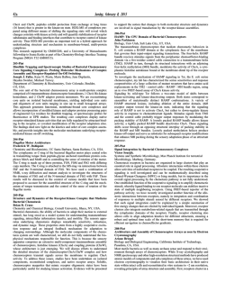
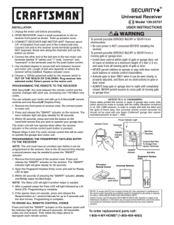
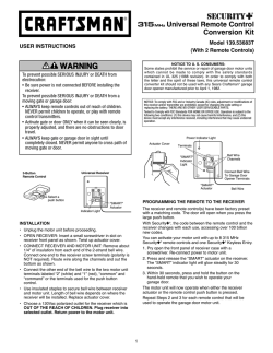
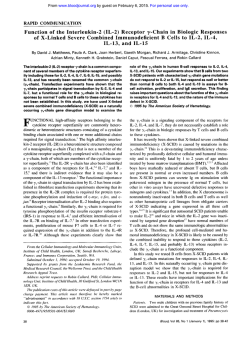
From www.bloodjournal.org by guest on February 6, 2015. For personal use only.
RAPID COMMUNICATION
Extracellular Truncations of hpc, the Common Signaling Subunit for
Interleukin-3 (IL-3), Granulocyte-Macrophage Colony-Stimulating Factor
(GM-CSF), and IL-5, Lead to Ligand-Independent Activation
By Richard J. D‘Andrea, Simon C. Barry, Paul A.B. Moretti, Karen Jones, Sarah Ellis, Mathew A. Vadas,
and Gregory J. Goodall
The hypothesis that extracellular truncation of the
common
receptor subunit for interleukin-3 (IL-3). granulocyte-macrophage colony-stimulating factor, and IL-5 (hpc) can lead t o
ligand-independent activation was tested by infecting factor-dependent hematopoietic celllines with retroviruses encoding truncatedforms ofhpc. A truncation, resembling that
in v-Mpl, and retaining45 hpc-derived extracellular residues,
led t o constitutive activation in the murine myeloid cell line,
FDC-P1. However, infection of cells with retrovirus encoding
a more severely truncated receptor, retaining only 7 hpcderived extracellular residues, did notconfer factor independence on these cells. These experiments show thattruncation activates the receptor and define a 37-amino acid
segment of hpc(H395-A431) which contains two motifs consewed throughout the cytokine receptor superfamily (consensus Y/H XX R/Q VR and WSXWS), asessential for factorindependent signaling. The mechanism of activation
was also investigated in less severetruncations. A receptor
that retains the entire membrane-proximal domain (domain
4) also conferred factor independent growth onFDC-P1 cells;
however, a retrovirus encoding a truncated form of hpc having two intact membrane proximal domains did not have
this ability, suggesting that domain 3 may have an inhibaory
role in hpc. The a b i l i i of these receptors t o confer factor
independence was cell specific as demonstrated by theirinability t o confer factor-independent growth when introduced into the murine IL-3-dependent pro-B cell line BaF603.These results are consistent with a model in which
activation requires unmasking of an interactive receptor surface in domain 4 and association with a myeloid-specific
receptor or accessory component. We suggest that in the
absence of ligand intramolecular interactions prevent inappropriate signaling.
0 1996 by The American Societyof Hematology.
R
gion and the other 184 amino acids are from c-mpl,“ which
encodes the receptor for thrombopoietin (TPO).’2”6The extracellular domain is mostly composed of the F-MuLV env
sequences, but with 43 amino acids derived from c-Mpl (Fig
IA). Thus, in v-Mpl mostof the WO-R extracellular domain
has been deleted, generating a truncated receptor. Although
the mechanism of activation of the v-mpl product has not
been determined it has been shown that Mplcan be activated
by cysteine substitutions in the predicted dimer interface
domain, suggesting one normal mechanism of activation
probably involves ligand-induced homodimerization.’7
Forced receptor homodimerization is involved in aberrant
activation of other cytokine receptors. A constitutively active
form of the erythropoietin receptor (EPO-R) containing an
arginine-to-cysteine substitution at position 129 has been
described. This R129C form of EPO-R forms disulfidelinked homodimers in the absence of EPO, suggesting that
wild-type EPO-R is also activated by ligand-induced homodimerization.IsInfection of mice with recombinant retrovirus
ECEPTORS for hematopoietic growth factors (HGFs)
are members of the expanding cytokine receptor superfamily that is characterized by a 200-amino acid extracellular
receptor module (CRM) composed of two discrete folding
domains, each of which contains seven p strands folded into
antiparallel 0 sandwiches, and bears a structural similarity
to the fibronectin type I11 module and the Ig constant domains.’,’ This structure has been confirmed for two members
of the family, the growth hormone and prolactin receptors,
by x-ray crystallographic
In addition to this similarity in tertiary structure, members of this superfamily share
a number of conserved sequence elements: (1) four conserved cysteine residues located in the N-terminal domain,
(2) a membrane proximal Trp-Ser-Xaa-Trp-Ser (where Xaa
is any amino acid) motif, also known as the “WSXWS
box,” located in the C-terminal domain,’”.6and (3) a prolinerich motif (PRM), which may be involved in signal transduction, located in the membrane proximal region of the cytoplasmic domain of most receptors in this
Members
of this family of receptors do not contain recognizable tyrosine kinase domains; signaling depends on association with
cytoplasmic tyrosine kinases of the JAK family and the subsequent activation of signal transducers and activators of
transcription.’JO
Constitutive mutations in cytokine receptors have provided insight into the process of receptor activation. Activating mutations may act by mimicking the structure of the
normal receptor in the ligand-activated state and therefore
could provide important clues to the activation process.
Given the normal role of cytokines in cell proliferation and
survival these activating mutations may be predicted to have
oncogenic potential. Indeed, the murine v-mpl oncogene encodes a constitutively activated cytokine receptor that has
been transduced by the murine myeloproliferative leukemia
virus (MPLV). v-Mpl is a 284-amino acid fusion protein in
which the first 100 amino acids are derived from the rearranged Friend-murine leukaemia virus (F-MuLV) env reBlood, Vol 87, No 7 (April l ) , 1996 pp 2641-2648
Fromthe Division ofHumanImmunology,Hanson
Centre for
Cancer Research, Adelaide, Australia.
Submitted December 18, 1995; accepted December 26, 1995.
Supported by grants from the National Health and Medical Research Council of Australia and the Anti-Cancer Foundation of the
Universities of South Australia. P.A.B.M. is a recipient of a Dora
Lush Postgraduate Scholarshipfrom the National Health and Medical Research Council of Australia.
Address reprint requests to Dr Richard D’Andrea, Division of
Human Immunology, Hanson Centre for Cancer Research, Frome
Rd, Adelaide, 5000 Australia.
The publication costs of this article were defrayed in p a n by page
charge payment. This article must therefore be hereby marked
“advertisement” in accordance with 18 U.S.C. section 1734 solely to
indicate this fact.
0 I996 by The American Society of Hematology.
0006-4971/96/8707-0053$3.00/0
2641
From www.bloodjournal.org by guest on February 6, 2015. For personal use only.
D‘ANDREA ET AL
2642
A
Slgnal
(34aa)
WGSWS
MPLV
env
hMPLP
V-Mpl
B
expressing this receptor induces erythroleukemia.” The introduction of other cysteine residues into the EPO-R membrane proximal domain also leads to disulfide-linked homodimers that are constitutively active?’ A mutation in the
transmembrane domain of the human common p subunit
(h&) for interleukin-3 (IL-3), granulocyte-macrophage colony-stimulating factor (GM-CSF), and IL-5, which presumably works through a similar mechanism, has recently been
described.*’ In this mutant (hOcV449E). glutamic acid is
substituted for valine 449 in the transmembrane domain,
confemng factor-independent growth on FDC-P1 and BaFB03 cells. This mutation is analogous to an activating mutation in the c-neu proto-oncogene (HER-2, erb-B2 receptor),
which leads to receptor oligomerization via a transmembrane-mediated association.22.2’An activated form of the murine BC subunit has recently been isolated from a spontaneous, factor-independent subline of the promyelocytic cell
line, D35. In this receptor the extracellular domain has been
replaced by 34 amino acids encoded by intron 10 sequences
and it is postulated that the replacement of the normal extracellular sequences with the intron encoded segment facilitates homodimerization, perhaps through an extracellular
cysteine bridge.24
We have recently described two ligand-independent mutations in the membrane proximal, extracellular domain of hpc
that suggest an alternative mechanism of aberrant cytokine
receptor activation. One of these (hpcFIA) is a duplication
Fig l.Cytokinareceptor axtracellular truncations. (AISchematicillustrationshowingthe
major isoform of Mpl (hMpl-P)
andthev-Mplfusion
product.
M p l has two CRMs, the distal
one containing an insertion
of 50
residues. The v-Mpl product is
derived from the MPLV retrovirus and is a fusion protein containing rearranged env sequences fused t o truncated Mpl.
The conserved cysteine residues
and WSXWS motif characteristic
of the cytokine receptor family
are indicated by horizontallines.
(B)Schematic illustration showing the extracellular truncation
mutants of hpc that have been
generated. Site-directed mutagenesis was used t o construct
in-frame deletions of the FLAGhac cDNA. Residues deleted are
as follows:hpcAN,
E25-E232,
hpcAQP, E25-N337; hpcAH,
E25-A394; hpcAWS, E25-A431.
The signal sequence and position of the introduced FLAG octapeptide are indicated. Conserved cysteine residues and the
WSXWS sequence that are characteristic ofthecytokine
receptor
family
are shown
by
horizontal lines. Extracellular
domains are numbered from 1
t o 4.
of a short (37-amino acid) receptor segment including the
WSXWS motif and the second (hpcI374N) is a point mutation leading to a single amino acid substitution (isoleucine
374 to asparagine).2’ Both mutant receptors confer growth
factor independence and tumorigenicity on FDC-P1 cells.
The mechanism of activation for these mutations is lessclear;
however, they do not confer ligand-independent proliferation
in BaF-B03 cells, suggesting a different mechanism to
hPcV449E.” We have proposed that these mutations may
lead to an alteration in receptor structure that results in the
unmasking ofan interactive surface that is not normally
available in the receptor m o n ~ m e r ? We
~ . ~speculated
~
further
that ligand association leads to activation by exposing such
a surface and allowing an interaction that can lead to signaling. One prediction from such a model is that extracellular
truncation may lead to activation by unmasking an interactive region and it is notable that the severely truncated vMpl product retains 43 amino acids of extracellular sequence,” including the two conserved motifs duplicated in
hPcFIA (Fig 1A).
In this context we have now tested the hypothesis that
extracellular truncation could lead to constitutive activation
of cytokine receptors. We have examined a series of hoc
extracellular receptor truncations for their capacity to confer
growth in factor-dependent cell lines in the absence of
growth factor. We find that infection of the murine myeloid
cell line, FDC-PI, with retroviruses encoding truncated
From www.bloodjournal.org by guest on February 6, 2015. For personal use only.
ACTIVATION
2643
OF hoc BY EXTRACELLULAR TRUNCATION
forms of hpc leads to factor-independent proliferation provided domains 1-3 are removed and a conserved membrane
proximal segment is retained.
MATERIALS AND METHODS
Construction and expression of hoc receptor mutations. The
hoc cDNA forms used in this study were constructed by in vitro
mutagenesis. We utilized a mutagenic oligonucleotide (S’CCCTG-
TGCTGGGTGCTGAGCGGCGCACAGGCAGACTACAAGGACGACGACGACAAGGAAGAAACCATCCCG3’)to incorporate the
coding sequence for the FLAG octapeptide (Eastman-Kodak CO,
New Haven, CT),DYKDDDDK, after the signal sequence (between
residues A24 and E25) in the hpc cDNA. To optimize cleavage of
the signal peptide, residues 18-24 were altered from ERSLAGA
to VLSGAQA. These modifications were confirmed by sequencing
and this FLAG-hoc cDNA cloned into PALTER (Promega,
Madison, WI) for further mutagenesis. Mutagenesis was performedwith mutagenic oligonucleotides designed to remove
the desired extracellular sequence while leaving coding sequences
for the N-terminal signal sequence and the inserted FLAG
octapeptide intact. Mutagenic oligos were as follows: hocAN;
S’GACGACGACGACAAGGm%TTGGGACTCC3’, hpcAH; 5’
GACGACGACGACAAGCACAGCATGGCCCTG3’, hocAQP;
S’ACGACGACGACAAGATCCAGATGGCCCCT3’, hgcAWS;
S’ACGACGACGACAAGCGCTCCTGGGACACC3’. All mutants
were obtained after screening DNA minipreparations from the ampicillin resistant colonies for an h& fragment of diagnostic size. Mutations were confirmed by DNA sequencing, which was performed
using T7 Polymerase (Sequenase; United States Biochemical Corp
[USB, Cleveland, OH]) as per the manufacturer’s protocols. The
receptor mutants generated in this study are described in detail in
the text and are shown schematically in Fig 1B. For expression,
mutant receptors were excised from PALTER and cloned into the
retroviral expression vector pRUFNeo.Z6Retroviral DNA was used
to transfect the ecotropic packaging cell line, $m,*’ and virus from
G418-resistant cells wasusedto infect the murine hematopoietic
cell lines, FDC-PIZRor BaF-B0329by coculti~ation.~”
Cell lines, growth factors, andjow cytometry. Murine myeloid,
IL-3/GM-CSF-dependent FDC-PI cells, and derived cell lines were
maintained in Dulbecco’s modified Eagle’s medium (DMEM) supplemented with 7.5% fetal bovine serum and murine GM-CSF (80
U/mL; gift from Dr G. Begley, Walter and Eliza Hall Institute for
Medical Research, Melbourne Australia). Murine IL-3-dependent
BaF-B03 cells were maintained in DMEM supplemented with 7.5%
fetal bovine serum and murine IL-3 (300 U/mL; gift from Dr A.
Hapel, John Curtin School for Medical Research, Canberra, Australia). After cocultivation with producer cells transfected with pRUFNeo hoc constructs, FDC-P1, or BaF-B03 cells were harvested and
selected in the presence of G418 (1 mg/mL; Sigma-Aldrich Pty Ltd,
Castle Hill, NSW Australia) and growth factor. Receptor expression
was examined byflow cytometric analysis after staining withthe
FLAG-specific monoclonal antibody (MoAb), M2 (Eastman-Kodak
CO). Briefly, cells were washed and resuspended in cold phosphatebuffered saline supplemented with 5% bovine serum albumin
(PBSA). Cells were incubated with the M2 MoAb (1:300) for 20
minutes on ice, washed, and subsequently incubated with biotinylated antimouse IgG (150; Vector Lab Inc, Burlingame, CA) for 20
minutes on ice. After washing and resuspension in cold PBSA the
cells were incubated with streptavidin conjugated phycoerythrin
(150; Caltag Laboratories, San Francisco, CA) for a further 20
minutes, washed, resuspended in PBSA + 0.01 % sodium azide, and
analyzed using an Epics-Profile I1 analyzer (Coulter, Hialeah, E).
Mitogenic assays. To assay proliferation, infected cells (FDCPI, BaF-B03) were washed three times with PBS to remove growth
factor and then assayed in triplicate (5,000 cells per well) in a 96-
well microtiter plate in the presence and absence of the appropriate
growth factor. Cell growth was determined after 72 hours using a
Cell titer 96 nonradioactive cell proliferation assay (Promega, Madison, WI). Quantitation was performed using an automated plate
reader (BioRad Laboratories, Pty Ltd, North Ryde, NSW Australia).
To establish relative growth rates of cells expressing factor-independent mutants, cells were first selected for factor independence, seeded
at 5,000 cells per well, and proliferation assayed, as described earlier,
on each of the following 4 days.
Preparation of cellular DNA and polymerase chain reaction
(PCR) analysis. Genomic DNAwas isolated from cells using a
Proteinase WSDS procedure.” PCR was performed on 300 ng of
genomic DNA using standard protocols.32Internal h@ primers were
used for amplification (5‘TGAATTCGCCTGTCCAGAGCTGACCAGGG 3‘ and STGGATCCTCTGTGGGTAGATCTGAGGCAG
3’). Reactions were performed in a Perkin Elmer Thermocycler and
for 1 minute, 60°C
the cycling parameters were: 30 c y c l e ~ ” 9 4 ~ C
for 1 minute, 72°C for 1 minute. Reactions were denatured at 94°C
for 2 minutes before cycling. PCR products were fractionated using
1% agarose mini-gels and visualized by staining with ethidium bromide.
RESULTS
Construction and expressionofmutant hpc cDNA. In
vitro mutagenesis was used to construct the series of extracellular h& deletion mutants indicated in Fig 1B. In each
of these mutations a large portion of extracellular region was
replaced by the FLAG octapeptide. The N-terminal FLAG
octapeptide does not affect receptor binding or function in
FDC-P1 cells (data not shown) and allows detection of altered receptors expressed on the cell surface using the
FLAG-specific MoAb, M2. The mutant hpcAN completely
removes domains 1 and 2 (CRM1, residues E25-E232 inclusive) whereas the mutant hPcAQP removes domains 1-3
(residues E25-N337 inclusive; the N-terminal CRh4 and the
first domain of CFW2). hPcAH is truncated to a site within
domain 4 (residues E25-A394 deleted) and retains 45 hpcderived, membrane proximal residues, including those that
are duplicated in the constitutive hpc mutant, ~ ~ c F I Aand
,’~
the equivalent region to that retained in v-Mpl. The mutation
in hPcAWS removes residues E25-A431, including the
membrane proximal WSXWS, and leaves 7 residues between the FLAG octapeptide and the transmembrane domain. These modified hpc cDNAs were cloned into the retroviral expression vector pRUFNeo.26
Retroviral DNA was introduced into the retroviral packaging cell line, $2, and stable pools of transfected $2 cells
were produced by antibiotic selection. To test the ability of
each mutant to induce factor-independent growth, we infected the murine myeloid cell line, FDC-P1, and the IL3-dependent pro-B cell line, BaF-B03. We demonstrated
surface expression of mutant receptors on infected FDC-P1
cells by flow cytometry following staining with the MoAb,
M2 (Fig 2). All truncated receptors were detectable by this
procedure, indicating that receptor truncation did notprohibit
surface expression.
Proliferation of infected FDC-P1 and BaF-B03
cells.
Uninfected FDC-P1 cells andG418-resistant
FDC-P1
cells infected with the h&AN or hPcAWS retroviruses
grew in the presence of murine (m) GM-CSF but failed
to proliferate after removal of growth factor. In contrast,
FDC-P1 cells infected with either the hPcAH or hPcAQP
From www.bloodjournal.org by guest on February 6, 2015. For personal use only.
D'ANDREA ET AL
2644
DISCUSSION
The possibility that cytokine receptors canbe activated
by truncation of their extracellular domain is suggested by
the oncogenic v-Mpl product (see Fig IA). Another candidate for activation in this way is the common p chain (hpc)
for IL-3, GM-CSF, and IL-5, whichlike Mpl, has two repeats
of the cytokine receptor motif.." The GM-CSF, IL-3, and IL5 high-affinity receptors are composed of a ligand-specific achain (GMRa, IL3Ra, and ILSRa), with which they form
a low-affinity complex, and
Although hpc alone
does not detectably bind cytokine it is responsible for affinity
conversion and signal tran~duction.~~-~'
Residues in the membrane proximal domain (domain 4) of hpc have been identified as critical for affinity conversion by hpc, presumably
A
l:
Fluorescence
Fig 2. Expression ofhpcmutants
in FDC-P1cells. Cells were
stained with an irrelevant control antibody(-1 or the anti-FLAG
Cell number and fluorescence are
monoclonal antibody, M2 (-1.
in arbitrary units with the latter plotted on
a logarithmic scale.
-
B
m
1
2
deletion mutants grew both in the presence and absence
of growth factor (Fig 3A). The different degree of proliferation observed in the absence of factor by cells infected
with the hPcAQP and hPcAH retroviruses probably reflects theproportion of eachG418-resistantpopulation
that expresses hpc. Toeliminate the possibility that these
mutants confer different growth rates on FDC-P1 cells,
we compared proliferation over a 4-day periodafter selection for growth factor independence. As shown in Fig 3C,
hpcAQP- and hpcAH-infected cellsdid not grow at a rate
significantly different from each other, or from FDC-PI
cells grown in the presence of mGM-CSF. To show that
the hoc cDNA had not undergone a rearrangement in the
factor-independent FDC-PI cells derived from infection
with hPcAQP and hPcAH retroviruses, we recovered part
of the hpc cDNA spanning the deletion,
by PCR from
cellular DNA with hoc specific primers. DNA from (3418resistant FDC-P1 cells infected with hoc retrovirus, and fromfactor-independent cells infected with
pRUFhPcAQP and pRUFhPcAH retroviruses generated
the expected products of 1,726 bp, 814 bp, and 640 bp,
respectively(Fig 4), consistent with hoc retroviral insert(s) in these cells containing the appropriately mutated
hoc cDNA. In common with the hPcFIA and hpcI374N
mutants," none of the truncated receptor retroviruses was
capable of conferringIL-3-independentgrowth
when
used to infect the murine pro-B cell line BaF-B03
(Fig 5).
3
C
2
1.61
-E
g
1.6
1.4
1.2
E l
0.8
2
t
hpc AQP
. mGM-CSF
-8-
-
h%
. AQP
+ mGMCSF
-A-
hpc AH
. mGM-CSF
+ hlk AH
rrnGM-CSF
+- FDC-P1
-mGM-CSF
-0- FDC-P1 tmGM-CSF
2 0.6
0.4
0.2
0
Day0
Day 1
Day2
Day3
Day4
Fig 3. Proliferation of FDC-P1 cells expressing mutant forms of
hpc. (A) Infected FDC-P1 cells grown in the presence (light grey) or
absence (dark grey) of mGM-CSF. Proliferation assays were performed as described in Materials andMethods. FDC-P1cells infected
with pRUFNeo or with an hpcFIA retrovirusz5
are shown as controls.
(B) A schematic representation of the hpc mutants includedin the
above assay. Conserved cysteine residues and WSXWS motifs are
shown. hpcFIA contains a duplicated segment of37 residues in domain 4.25(C) Growth curve comparing hpcAQP and hpcAH proliferation over 4 days. Cells were selected before assay by growthin factorfree medium. Proliferation of uninfected
FDC-P1 cells in the presence
of mGM-CSF is shown forcomparison.
From www.bloodjournal.org by guest on February 6, 2015. For personal use only.
ACTIVATION OF hoc BY EXTRACELLULAR TRUNCATION
U,
n
U
4
0
M
,c
1.61.00.51
+
Fig 4. PCR analysis of retroviral DNA in factor-independentcells.
PCR from genomic DNA to detect hpc sequences in factor-independent FDC-P1cells derived from infection with retrovirus encoding
hpcAQP and hpcAH (see text). PCR products were separated on a
1%agarosegelalongside
molecular-weight markers (l-kb ladder;
BRL, Gaithersburg, MD). The expected product sizes are 1,726 bp,
814 bp, and 640 bp for hpc,hpcAQP, and hpcAH, respectively. Sizes
shown on the left refer to molecular weight markers.
through an interaction with residues in the N-terminal helix
of the growth factor.3”However, to date there is little information regarding the role of the first three domains of hpc
in ligandbinding. Several activating mutations cluster in
domain 4 of hoc consistent with a critical role for this domain in activation and signaling.”.” Despite considerable
knowledge regarding the intracellular pathways involved in
signalling via hpc, there is still an incomplete understanding
of the stoichiometry of the active receptor complex and the
precise role of the a and p subunits in signaling.
To test the hypothesis that truncation can lead to ligandindependent cytokine receptor activation, we constructed a
series of cDNAs encoding extracellular deletion mutants of
hpc. Our results show that infection of the murine IL-3/
GM-CSF-dependent cellline, FDC-PI, with retroviruses
encoding two of these truncated forms of hpc (hDcAQP and
hpcAH) leads to factor-independent growth. The inability
of the other truncated forms of hBc (hpcAN and hpcAWS)
to confer factor independence in FDC-PI cells was not due
to a defect in transport to the cell surface as we could detect
surface expression using an MoAb directed to the introduced
N-terminal FLAG epitope.
The observation that hPcAH confers factor independence
while hPcAWS does not, defines an extracellular region
of h& (residues H395-A431) as essential for constitutive
activation in FDC-PI cells. It is notable that in v-Mpl, the
corresponding receptor segment is retained. The mechanism
of activation of the v-mpl product remains unclear; however,
based on our observations we suggest the loss of the Mpl
N-terminal region is important and may expose a site of the
receptor that binds another subunit, either as a homodimer
2645
or a heterodimer. The extracellular region retained in v-Mpl
contains the WSXWS motif and the conserved B strand F‘
(consensus Y/H XX WQ VR). These motifs are also within
the duplicated segment in the mutant hpcFIA, which previously led us to speculate that they are involved in receptor
activation.” The conservation of this membrane proximal
segment throughout the cytokine receptor family suggests
that it may play an important role in the activation mechanism of class I receptors, perhaps through interactions with
conserved receptor subunits. Interestingly, a previous study
with chimeric receptors indicated that the extracellular domain of cytokine receptors determined, to some extent, the
specific phosphorylation events that were triggeredinresponse to ligand, suggesting interactions between extracellular sequences and signal transducers.‘“’
In the growth hormone/growth hormone receptor and
growth hormone/prolactin receptor complexes the corresponding segment has been shown by crystallographic studies to form part of an exposed, positively charged &sheet in
the membrane proximal domain of the ligand-bound receptor
~ o m p l e x .By
~ . ~homology the conserved residues in this segmentof hpc wouldbepositioned distal toligandand asubunit in a ligand-bound complex. It is possible that there
are two interactive surfaces in cytokine receptor subunits
that are involved in formation of oligomeric complexes. Although hoc has notbeen shown toform a higher order
complex, we have speculated previously”that activation
might occur through formation of a& or
type complexes. Thus, hpc may have at least two interactive surfaces:
one for association with a specific a-chain and a second
that interacts with either another hpc or a third receptor
component. Although there are several conserved residues
between H395 and A431, it is not possible, from this work,
0.8
-
0.7
:
0.6
L
0.5
1
0.4
:
0.3
0.2
:
0.1
I
Fig 5. Proliferationof BaF-BO3 cells infected with retroviruses encoding activated forms of hpc. Proliferation of infected BaF-BO3 cells
grown in the presence (light grey) or absence (dark grey) of murine
IL-3. A control cell population infected with pRUFNeo isalso shown.
From www.bloodjournal.org by guest on February 6, 2015. For personal use only.
DANDREA ET AL
2646
RECEPTOR+ LIGAND
hpc EXTRACELLULAR
MUTATIONS (FIA and I374N)
INACTIVE RECEPTOR
RECEPTOR TRUNCATION
Fig 6. A model for hpc activation. One mechanism of activation of hpc may involve interaction with an unknown receptor subunit (yl
expressed in FDC-P1 cells but absent from BaF-BO3 cells. Extracellularactivating mutations, or extracellular truncation, are presumed to alter
the receptor structure and unmask interactive residues in domain 4 leading t o signaling in the absence of ligand. Ligand-mediatedactivation
may occur through a similar process.
to define which residues are essential in receptor activation.
In v-Mpl, mutation of the WSXWS motif does not impair
pathogenicity of MPLV? suggesting that other residues in
this segment may be important.
The observation that expression ofthemutant
hPcAN
does not confer factor independence in FDC-PI cells implies
that the third fibronectin-like receptor domain (domain 3)
may have a role in inhibiting signaling by unoccupied receptor, presumably through an interaction with residues in domain 4. Removal of this domain (as in hpcAQP) is sufficient
to lead to constitutive activation. It is tempting to speculate
that the B'C' and F'G'
loops of domain 4, which are important in ligand binding" (and Woodcock et al, submitted
for publication), interact with ligand in such a way that the
orientation of domains 3 and 4 is altered, exposing the interactive surface that allows higher order complex formation.
In this way ligandmay displace inhibitory interactions in
the same way as truncation.
Although the mutants hPcAQP and hPcAH conferred factor-independence on FDC-PI cells, they did not confer factor-independence on the IL-3-dependent pro-B cell line,
BaF-B03 (Fig 5). Two other extracellular mutations,
hpcF1A2' and hpcI374N:' similarly confer factor-independent growth in FDC-PI cells but not in Ba/F3 cells. On the
other hand the hpc mutant (h@cV449E),which resembles the
neu oncogenic receptor, and presumably leads to @-subunit
dimerisation, confers IL-3-independent growth on infected
BaF-B03 cells.'' Consistent with this, chimeric receptors
comprised of the GMRa or ILSRa extracellular domain and
the hpc cytoplasmic domain give a ligand-dependent response in Ba/F3 cells expressing ~PC."'"'.~'
It appears from
these results that hoc can be activated by homodimerization;
however, the results presented here, and in the report by
Jenkins et al:' suggest that ligand-independent forms of h&
with extracellular mutations, utilize an alternative mechanism of activation that may require interaction with an unknown subunit present in FDC-PI cells but absent from BaFB03 cells. Taken together with the results from this study,
it is reasonable to suggest that the interaction of hpc with
such a factor could be mediated by residues within the segment H395-A43 1.
In Fig 6 we present a model for hoc activation. We
propose that extracellular mutations of hoc (hpcFIA,
hpcI374N, hpcAQP, and hpcAH) that lead to ligand-independent activation may allow constitutive association with
an unknown, myeloid-restricted, receptor component ( 7 )and
may be mimicking a normal a& or P-y complex. The fact
that there are several extracellular activating mutations clustered in domain 4 is consistent with activation involving the
disruption of an inhibitory conformation of the unoccupied
form of h@. In this conformation we would predict that a
critical structural motif within domain 4 (involving the segment H395-A43 1) is masked, possibly through interactions
involving domain 3. We propose that the I374N substitution
and the hPcFIA duplication can interfere with the structure
of domain 4 in such a way that they lead to the disruption
of inhibitory interactions.2' By analogy with these mutants
we suggest that association with ligand alters the monomeric
hpc conformation and leads to association with other receptor components and the subsequent generation of intracellular signals.
ACKNOWLEDGMENT
The authors are. gratefulto Chris Bagley for valuable insights into
receptor structures and to Alan Bishop for assistance with cell sorting. We also thank Angel Lopez, Tom Gonda, and Jo Woodcock
for valuable discussions and critical reading of the manuscript.
REFERENCES
1. Bazan J F Structural design and molecular evolution of a cyto-
kine receptor superfamily. Proc Natl Acad Sci USA 87:6934, 1990
2. Kaczmarski RS, Mufti GJ: The cytokine receptor superfamily.
Blood Rev 5:193, 1991
Humangrowthhor3. de Vos AM, Ultsch M, Kossiakoff AA:
mone and extracellular domain of its receptor: Crystal structure of
the complex. Science 255:306, 1992
4. Somers W,Ultsch M, de Vos AM, Kossiakoff AA: The x-ray
structure of a growth hormone-prolactin receptor complex. Nature
372:478, 1994
From www.bloodjournal.org by guest on February 6, 2015. For personal use only.
ACTIVATION OF hoc BY EXTRACELLULARTRUNCATION
5. Cosman D, Lyman SD, Idzerda RL, Beckmann MP, Park LS,
Goodwin RG, March CJ:Anew
cytokine receptor superfamily.
Trends Biochem Sci 15:265, 1990
6 . Fukunaga R, Ishizaka-Ikeda E,Pan CX, Set0 Y, Nagata S :
Functional domains of the granulocyte colony-stimulating factor receptor. EMBO J 102855, 1991
7. O’Neal KD, Yu Lee LY: The proline-rich motif (PRM): A
novel feature of the cytokinehematopietin receptor superfamily.
Lymphokine Cytokine Res 12:309, 1993
8. Benit L, Courtois G , Charon M, Varlet P, Dusanter Fourt I,
Gisselbrecht S : Characterization of Mpl cytoplasmic domain sequences required for myeloproliferative leukemia virus pathogenicity. J Virol 685270, 1994
9. Ihle JN, Witthuhn BA, Quelle F W , Yamamoto K, Silvennoinen
0: Signalling through the hematopoietic cytokine receptors. Annu
Rev Immunol 13:369, 1995
10. Ihle J: Cytokine receptor signalling. Nature 377; 591, 1995
11. Souyri M, Vigon I, Penciolelli J-F, Heard J-M, Tambourin
P, Wendling F: A putative truncated cytokine receptor gene transduced by the myeloproliferative leukaemia virus immortalises hematopoietic progenitors. Cell 63:1137, 1990
12. Kaushansky K, Lok S , Holly RD, Broudy VC, Lin N, Bailey
MC, Forstrom J W , Buddle MM, Oort PJ, Hagen FS. Roth GJ, Papayannopoulou T, Foster DC: Promotion of megakaryocyte progenitor
expansion and differentiation by the c-Mpl ligand thrombopoietin.
Nature 369:568, 1994
13. Bartley TD, Bogenberger J, Hunt P, Li Y-S, Lu HS, Martin
F, Chang M-S, Samal B, Nichol JL, Swift S , Johnson MJ, Hsu
R-Y, Parker VP, Suggs S , Skrine JD, Merewether LA, Clogston C,
Hsu E, Hokom MM, Homkohl A, Choi E, Pangelinan M, Sun Y,
Mar V, McNinch J, Simonet L, Jacobsen F, Xie C, Shutter J, Chute
H, Basu R, Selander L, Trollinger D, Sieu L, Padilla D, Trail G ,
Elliot G, Izumi R, Covey T, Crouse J, Garcia A, Xu W, Del Castillo
J, Biron J, Cole S , Hu MC-T, Pacifici R, Ponting I, Saris C, Wen
D, Yung YP, Lin H, Bosselman RA: Identification and cloning of
a megakaryocyte growth and development factor that is a ligand for
the cytokine receptor Mpl. Cell 77:1117, 1994
14. de Sauvage FJ, Haas PE, Spencer SD, Malloy BE, Gurney
AL. Spencer SA, Darbomme WC, Henzel WJ, Wong SC, Kuang
WJ, Oles KS, Hultgren B, Solberg LA, Goeddel DV, Eaton DL.
Stimulation of megakaryocytopoiesis and thrombopoiesis by the cMpl ligand. Nature 369:533, 1994
15. Lok S , Kaushansky K, Holly RD, Kuijper JL, Lofton Day
CE, Oort PJ, Grant FJ, Heipel MD, Burkhead SK, Kramer JM, Bell
LA, Sprecher CA, Blumberg H, Johnson R, Prunkard D, Ching A F T ,
Mathewes SL, Bailey MC, Forstrom J W , Buddle MM, Osbom SG,
Evans SJ, Sheppard PO, Presnell SR, OHara PJ, Hagen FS, Roth
GJ, Foster DC: Cloning and expression of murine thrombopoietin
cDNAand
stimulation of platelet production in vivo. Nature
369565, 1994
16. Wendling F, Maraskovsky E, Debili N, Florindo C, Teepe
M, Titeux M, Methia N, Breton Gorius J, Cosman D, Vainchenker
W: cMpl ligand is a humoral regulator of megakaryocytopoiesis.
Nature 369:571, 1994
17. Alexander WS, Metcalf D, Dunn AR: Point mutations within
a dimer interface homology domain of c-Mpl induce constitutive
receptor activity and tumorigenicity. EMBO J 14:5569, 1995
18. Watowich S S , Yoshimura A, Longmore GD, Hilton DJ, Yoshimura Y, Lodish HF: Homodimerization and constitutive activation of the erythropoietin receptor. Proc Natl Acad Sci USA 89:2140,
1992
19. Longmore GD, Lodish HF: An activating mutation in the
murine erythropoietin receptor induces erythroleukaemia in mice: A
cytokine receptor superfamily oncogene. Cell 67:1089, 1991
20. Watowich S S , Hilton DJ, Lodish W.
Activation and inhibi-
2647
tion of erythropoietin receptor function: role of receptor dimerization. Mol Cell Biol 143535, 1994
21. Jenkins BJ, D’Andrea R, Gonda TJ: Activating point mutations in the common ,B subunit of the human GM-CSF, IL-3 and
IL-5 receptors suggest the involvement of p subunit dimerisation
and cell type specific molecules in signalling. EMBO J14:4276,
1995
22. Stemberg MJE, Gullick WJ: Neu receptor dimerisation. Nature 339587, 1989
23. Weiner DB, Liu J, Cohen JA, Williams WV, Greene MI: A
point mutation in the neu oncogene mimics ligand induction of
receptor aggregation. Nature 339:230, 1989
24. Hannemann J, Ham T, Kawai M, Miyajima A, Ostertag W,
Stocking C: Sequential mutations in the interleukin 3(IL3)IGMCSFIIL5 receptor &subunit genes are necessary for the complete
conversion to growth autonomy mediated by a truncated-p, subunit.
Mol Cell Biol 15:2402, 1995
25. D’AndreaR, Rayner J, Moretti P, Lopez A, Goodall GJ,
Gonda TJ, Vadas MA: A mutation of the common receptor subunit
for interleukin-3 (IL-3), granulocyte-macrophage colony-stimulating
factor, and IL-5 that leads to ligand independence and tumorigenicity. Blood 83:2802, 1994
26. Rayner JR. Gonda TJ: A simple and efficient procedure for
generating stable expression libraries by cDNA cloning in a retroviral vector. Mol Cell Biol 14880, 1994
27. Mann R, Mulligan RC, Baltimore D: Construction of a retrovirus packaging mutant and its use to produce helper-free defective
retrovirus. Cell 33:153, 1983
28. Dexter TM, Garland J, Scott D, Scolnick E. Metcalf D:
Growth of factor-dependent hemopoietic precursor cell lines. J Exp
Med152:1036, 1980
29. Hatakeyama M, Mori H, Doi T, Taniguchi T A restricted
cytoplasmic region of IL-2 receptor p chain is essential for growth
signal transduction but not for ligand binding and internalisation.
Cell 59:837, 1989
30. Lang RA, Metcalf D, Gough NM, DunnAR, Gonda TJ:
Expression of a haemopoietic growth factor cDNA in a factor-dependent cell line results in autonomous growth and tumorigenicity. Cell
43:531, 1985
31. Hughes SH, Payvar F, Spector D, Schimke RT,Robinson
HL, Payne GS, Bishop JM, Varmus HE: Heterogeneity of genetic
loci in chickens: analysis of endogenous viral and normal genes by
cleavage of DNA with restriction endonucleases. Cell 18:347,
1979
32. Saiki RK: Amplification of genomic DNA, in Innes MA,
Gelfand DH, Sninsky JJ, White TJ (eds): PCR Protocols: A Guide
to Methods and Applications. San Diego, CA, Academic, 1990, p
13
33. Hayashida K, Kitamura T, Gorman DM, Arai K, Yokota T,
Miyajima A: Molecular cloning of a second subunit of the receptor
for human granulocyte-macrophage colony-stimulating factor (GMCSF): Reconstitution of a high-affinity GM-CSF receptor. Proc Natl
Acad Sci USA 87:9655, 1990
34. Kitamura T, Sat0 N, Arai K, Miyajima A: Expression cloning
of the human IL-3 receptor cDNA reveals a shared beta subunit for
the human IL-3 and GM-CSF receptors. Cell 66:1165, 1991
35. Takaki S , Murata Y, Kitamura T, Miyajima A, Tominaga A,
Takatsu K Reconstitution of the functional receptors for murine and
human interleukin-5. J Exp Med 177:1523, 1993
36. Tavemier J, Devos R, Comelis S , Tuypens T, Van der Heyden
J, Fiers W, Plaetinck G: Human high affinity interleukin-5 receptor
(IL5R) is composed of an IL5-specific alpha chain and a beta chain
shared with the receptor for GM-CSF. Cell 66:1175, 1991
37. Gearing DP, King JA, Gough NM, Nicola NA: Expression
cloning of a receptor for human granulocyte-macrophage colonystimulating factor. EMBO J 8:3667, 1989
From www.bloodjournal.org by guest on February 6, 2015. For personal use only.
2648
38. Murata Y, Takaki S, Migita M,Kikuchi Y, Tominaga A,
Takatsu K: Molecular cloning and expression of the human interleukin 5 receptor. J Exp Med 175:341, 1992
39. Woodcock JM, Zacharakis B, Plaetinck G, Bagley CJ, Qiyu
S, Hercus TR, Tavernier J, Lopez AF: Three residues in the common beta chain of the human GM-CSF, IL-3 and IL-5 receptors
are essential for GM-CSFand IL-5 but not IL-3 high affinity
binding and interact with Glu21 of GM-CSF. EMBO J 13:5176,
1995
40. Chiba T, Nagata Y, Machide M, Kishi A, Amanuma H,
Sugiyama M, Todokoro K: Tyrosine kinase activation through the
D’ANDREA ET AL
extracellular domains of cytokine receptors. Nature 362:646,
1993
41. Muto A, Watanabe S, Miyajima A, Yokota T, Arai K-I: High
affinity chimeric human GM-CSF receptor carrying the cytoplasmic
domain of the p subunit but not the m subunit transduces growth
promoting signals in BaF3 cells. Biochem Biophys Res Commun
208:368, 1995
42. Eder M, Ernst TJ, Ganser A, Jubinsky PT, Inhorn R, Hoelzer
D, Griffin JD: A low affinity chimeric human mIP-GM-CSFreceptor
induces ligand dependent proliferation in a murine cell line. J Biol
Chem 269:30173, 1994
From www.bloodjournal.org by guest on February 6, 2015. For personal use only.
1996 87: 2641-2648
Extracellular truncations of h beta c, the common signaling subunit
for interleukin-3 (IL-3), granulocyte-macrophage colony-stimulating
factor (GM-CSF), and IL-5, lead to ligand-independent activation
RJ D'Andrea, SC Barry, PA Moretti, K Jones, S Ellis, MA Vadas and GJ Goodall
Updated information and services can be found at:
http://www.bloodjournal.org/content/87/7/2641.full.html
Articles on similar topics can be found in the following Blood collections
Information about reproducing this article in parts or in its entirety may be found online at:
http://www.bloodjournal.org/site/misc/rights.xhtml#repub_requests
Information about ordering reprints may be found online at:
http://www.bloodjournal.org/site/misc/rights.xhtml#reprints
Information about subscriptions and ASH membership may be found online at:
http://www.bloodjournal.org/site/subscriptions/index.xhtml
Blood (print ISSN 0006-4971, online ISSN 1528-0020), is published weekly by the American
Society of Hematology, 2021 L St, NW, Suite 900, Washington DC 20036.
Copyright 2011 by The American Society of Hematology; all rights reserved.


© Copyright 2026