
Download as Adobe PDF - Edinburgh Research Explorer
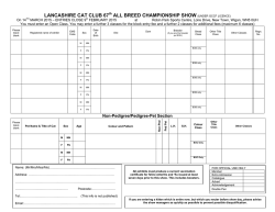
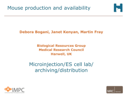
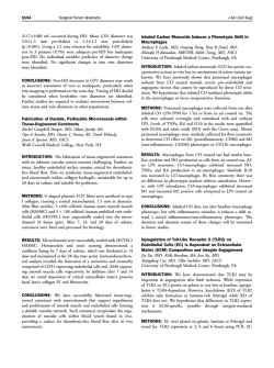
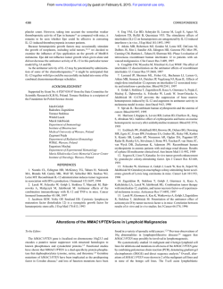
Edinburgh Research Explorer
Cloning and expression of feline colony stimulating factor
receptor (CSF-1R) and analysis of the species specificity of
stimulation by colony stimulating factor-1 (CSF-1) and
interleukin-34 (IL-34).
Citation for published version:
Gow, DJ, Garceau, V, Pridans, C, Gow, A, Simpson, KE, Gunn-Moore, D & Hume, D 2013, 'Cloning and
expression of feline colony stimulating factor receptor (CSF-1R) and analysis of the species specificity of
stimulation by colony stimulating factor-1 (CSF-1) and interleukin-34 (IL-34).' Cytokine, vol 61, no. 2, pp.
630-638., 10.1016/j.cyto.2012.11.014
Digital Object Identifier (DOI):
10.1016/j.cyto.2012.11.014
Link:
Link to publication record in Edinburgh Research Explorer
Document Version:
Publisher final version (usually the publisher pdf)
Published In:
Cytokine
Publisher Rights Statement:
Open Access funded by Biotechnology and Biological Sciences Research Council
General rights
Copyright for the publications made accessible via the Edinburgh Research Explorer is retained by the author(s)
and / or other copyright owners and it is a condition of accessing these publications that users recognise and
abide by the legal requirements associated with these rights.
Take down policy
The University of Edinburgh has made every reasonable effort to ensure that Edinburgh Research Explorer
content complies with UK legislation. If you believe that the public display of this file breaches copyright please
contact [email protected] providing details, and we will remove access to the work immediately and
investigate your claim.
Download date: 06. Feb. 2015
Cytokine 61 (2013) 630–638
Contents lists available at SciVerse ScienceDirect
Cytokine
journal homepage: www.journals.elsevier.com/cytokine
Cloning and expression of feline colony stimulating factor receptor (CSF-1R)
and analysis of the species specificity of stimulation by colony stimulating factor-1
(CSF-1) and interleukin-34 (IL-34)
Deborah J. Gow, Valerie Garceau, Clare Pridans, Adam G. Gow, Kerry E. Simpson, Danielle Gunn-Moore,
David A. Hume ⇑
The Roslin Institute and Royal (Dick) School of Veterinary Studies, University of Edinburgh, Easter Bush, Midlothian EH25 9RG Scotland, UK
a r t i c l e
i n f o
Article history:
Received 18 October 2012
Accepted 22 November 2012
Available online 19 December 2012
Keywords:
Macrophage
Ba/F3
Bone marrow
Species specificity
Renal
a b s t r a c t
Colony stimulating factor (CSF-1) and its receptor, CSF-1R, have been previously well studied in humans
and rodents to dissect the role they play in development of cells of the mononuclear phagocyte system. A
second ligand for the CSF-1R, IL-34 has been described in several species. In this study, we have cloned
and expressed the feline CSF-1R and examined the responsiveness to CSF-1 and IL-34 from a range of species. The results indicate that pig and human CSF-1 and human IL-34 are equally effective in cats, where
both mouse CSF-1 and IL-34 are significantly less active. Recombinant human CSF-1 can be used to generate populations of feline bone marrow and monocyte derived macrophages that can be used to further
dissect macrophage-specific gene expression in this species, and to compare it to data derived from
mouse, human and pig. These results set the scene for therapeutic use of CSF-1 and IL-34 in cats.
Ó 2012 Elsevier Ltd. All rights reserved.
1. Introduction
Macrophage colony-stimulating factor (CSF-1) controls the proliferation and differentiation of cells of the mononuclear phagocyte
lineage [1–3]. The receptor for CSF-1 (CSF-1R, MCSFR, CD115 or
FMS) is expressed in all cells of the mononuclear phagocyte lineage
including progenitor cells, osteoclasts and dendritic cells [4]. CSF1R is the proto-oncogene form of the transforming gene of Feline
McDonough Sarcoma (SM-FeSV), hence its name FMS [5]. A second
ligand for the CSF-1R was first described in humans [6] and subsequent studies confirmed that the two-ligand, one-receptor, system
is conserved in birds [7]. Binding of either ligand to the CSF-1R produces receptor dimerization, auto-phosphorylation, activation of
down-stream signalling (ERK1/2, Akt) and expression of genes involved in survival and proliferation of the mononuclear phagocyte
lineage cells [6,8–10].
Human CSF-1 cDNA was cloned and expressed in the 1980s, and
when injected into mice, it promoted an increase in blood monocyte and tissue macrophage numbers [11]. Human CSF-1 is almost
as effective as mouse CSF-1 in stimulating mouse macrophage proliferation in vitro [12,13] and indeed is equally active on all mammalian species tested (mouse, cat, sheep, dog, and pig). Conversely,
mouse CSF-1 bioactivity is restricted to non-primate species
[2,7,14–18]. IL-34 has even more restricted species cross-reactivity;
⇑ Corresponding author. Tel.: +44 131 6519181.
E-mail address: [email protected] (D.A. Hume).
1043-4666/$ - see front matter Ó 2012 Elsevier Ltd. All rights reserved.
http://dx.doi.org/10.1016/j.cyto.2012.11.014
with the human and mouse ligands much less active on the other
species, although both were active on the pig CSF-1R [2]. CSF-1 has
been widely-used to drive proliferation and differentiation of
mature macrophages from bone marrow or blood monocyte
progenitors in multiple species. In general, CSF-1 stimulated cells
are driven towards a more immunosuppressive function [1,19],
where another colony-stimulating factor, GM-CSF or CSF-2,
drives differentiation of phagocytic cells with antigen-presenting
function [20,21]. GM-CSF has been used to produce antigen presenting cells (APCs) from feline bone marrow [22–24], but there
are no reports of using CSF-1 to generate feline monocyte derived
macrophages.
Recombinant colony stimulating factors and/or antibodies directed against the ligand or the receptor have been tested in a
range of animal models and human patients [1,25–27]. Macrophages play an important role in tissue repair in a number of tissues including kidney, liver, heart, brain and lung [28–32] and
CSF-1 administration has been shown to promote regeneration in
a number of models. For example, in ischaemia reperfusion in
mice, a model of acute renal injury, recombinant human CSF-1
administration was able to stimulate macrophage infiltration to
promote epithelial repair and to prevent interstitial fibrosis [33].
Acute renal failure in cats can arise from numerous causes [34–
40] and has a very poor prognosis [36,41]. Many cats who survive
to discharge are azotaemic and, as a result, are likely to have high
morbidity and greatly-reduced lifespan, while a second ‘‘wave’’ of
apoptosis seems to occur in the recovery phase which may limit
D.J. Gow et al. / Cytokine 61 (2013) 630–638
further regeneration [42]. The studies in the mouse model suggest
that CSF-1 could have a therapeutic benefit in cats with acute renal
injury. For this purpose, we need to know whether it would be necessary to produce feline-specific agents. In the present study, we
have produced a cat CSF-1R-expressing factor-dependent cell line
and evaluated responsiveness of the cat receptor to CSF-1 and IL34 from multiple species.
2. Materials and methods
2.1. Cell culture and reagents
The Ba/F3 cell line, transfected Ba/F3 cells and primary bone
marrow cells were cultured in RPMI 1640 medium (Sigma) containing 10% HI-FCS, 2 mM L-glutamine, 100 lg/ml streptomycin,
and 100 Units/ml penicillin. Untransfected Ba/F3 cells were maintained in medium containing 10% IL-3 from X63 Ag8-653 myeloma
cells carrying an expression vector for IL-3 [43,44]. Unless otherwise stated, transfected Ba/F3 cells were maintained in medium
containing 104 Units/ml rhCSF-1 (a gift from Chiron Corp., Emeryville, CA, USA). Both Ba/F3 cells and primary bone marrow cells
(BMCs) were incubated at 37 °C with 5% CO2.
2.2. Total RNA extraction and cDNA synthesis
With owner’s consent, tissues were collected from a 5-year-old
male Siamese cat that was being euthanized for medical reasons.
Tissues were placed immediately in RNA Later (QIAGEN) and
stored at room temperature for 24 h until RNA extraction was performed. RNA was extracted from skin, testes, sub-mandibular
lymph node, uterus and spleen. Total RNA was prepared using an
RNeasy kit (QIAGEN) according to the manufacturer’s instructions,
including a DNase digestion step. Feline-specific cDNA was produced using 1 lg of total RNA and reversed transcribed using ImProm-II (Promega). Successful cDNA production without genomic
DNA contamination was demonstrated using feline HPRT primers
(Table 1) [45].
2.3. Expression cloning of feline CSF-1R
Feline PCR primer pairs (Table 1) were designed for amplification of full-length CSF-1R from the incomplete published feline
CSF-1R cDNA sequence (Ensembl ENSFCAP00000003348). Amplification was achieved using feline cDNA and expand high-fidelity
enzyme (Roche) with 3 mM MgCl2 using an initial cycle of 94 °C
for 3 min followed by 35 cycles of 94 °C for 30 s, 60 °C for 30 s
72 °C for 3 min and one cycle of 72 °C for 10 min. PCR products
were gel purified using a QIAquick gel extraction kit (QIAGEN)
and cloned in frame with V5-His C-terminal tag of pEF6/V5-His
expression construct using TOPO cloning kit (Invitrogen). DNA
sequencing was performed by DNA Sequencing and Services
(MRCPPU, College of Life Sciences, University of Dundee, Scotland,
www.dnaseq.co.uk) using Applied Biosystems Big-Dye Ver. 3.1
Table 1
Table of primers used for production of feline cDNA and cloning of feline CSF-1R. A
kozak sequence (highlighted in bold) was included in CSF-1R forward primer for
optimal translation initiation. Feline HRPT primers from Penning et al. [45].
Primer name
Primer sequence 50 –30
Tm
(°C)
Size
(bp)
Feline
Feline
Feline
Feline
ACTGTAATGACCAGTCAACAGGGG
TGTATCCAACACTTCGAGGAGTCC
GCC ATG GGC CCA AGG GCT
GCA GAA CTG GTA GTT GTT GGG CTG
60.0
60.0
61.5
61.9
210
–
2946
–
HPRT Forward
HPRT Reverse
CSF-1R Forward
CSF-1R Reverse
631
chemistry on an Applied Biosystems model 3730 automated capillary DNA sequence.
2.4. Generation of stable cell lines
For generation of stable Ba/F3 cells expressing feline CSF-1R,
5 106 Ba/F3 cells were electroporated (300 V, 975 lF) with
10 lg DNA (pEF6_fCSF-1R or empty pEF6 DNA), and selected with
30 lg/ml blasticidin (Invitrogen) and 10% IL-3 for 6 days prior to
further selection with 30 lg/ml blasticidin and 104 Units/ml of
rhCSF-1.
2.5. Immunoblotting
Whole-cell lysate was prepared by lysing 0.5 106 cells in
10 mM Tris containing 2% SDS and boiling for 10 min at 100 °C.
Protein concentration was determined using DC protein assay
(Bio-Rad) with 10 lg of protein mixed with Laemmli buffer (Invitrogen) and 5 mM DTT. Samples were run on a 4–12% gradient precast SDS–PAGE gel (Bio-Rad) and transferred onto polyvinylidene
difluoride membrane, as per manufacturer’s directions (Bio-Rad).
The membrane was blocked with 5% skimmed milk powder in
TBS-Tween 20 at 4 °C overnight prior to being washed and probed
with 1:5000 dilution of mouse anti-v5 tag antibody (AbD Serotec
MCA1360G) and 1:5000 dilution of anti-mouse IgG HRP conjugated antibody (Cell Signalling Technology, 7076) and detected
using enhanced chemiluminescence (ECL) reagents (Amersham,
GE Healthcare, UK).
2.6. Isolation of feline peripheral blood mononuclear (PBMC) and bone
marrow cells (BMC)
A 6-year-old male neutered domestic short-haired cat was
euthanized by pentobarbitone for medical reasons and owner’s
consent obtained to collect 20 ml of blood and one femur. The femur was dissected, placed in a zip-lock bag and placed on ice.
20 ml of blood was collected into a syringe containing Acid-citrate-dextroseat 1:10 dilution and placed on ice following collection. For BMC collection, both the proximal and distal ends of the
femur were removed and using an 18 g needle, cells were flushed
with 10 ml RPMI (Sigma) containing 5 mM EDTA to prevent clotting. Cells were washed and re-suspended in red cell lysis buffer
(Bio Legend, San Diego, CA) for 5 min, followed by a further 2
washes in PBS. Feline PBMC were isolated using Lymphoprep
(Axis-Shield, Oslo, Norway) following manufacturer’s instructions,
including the addition of red cell lysis buffer as above.
2.7. Feline peripheral PBMC and BMC stimulation with recombinant
human CSF-1
Ten million PBMC and BMC were cultured in 60 mm bacteriological plates with 4 ml RPMI supplemented with 104 Units/ml
rhCSF-1, and incubated for 8 days at 37 °C, 5% CO2.
2.8. Preparation of cells for cytospin
After 8 days in culture, adherent cells from both blood and BMC
cultures were recovered by repeated flushing of medium over the
bottom of the culture dish until all adherent cells were removed.
Cells were counted and 0.5 106 cell/ml were collected with
100 ll of cell suspension placed into the cytospin chamber
(Thermo) and centrifuged at 300 rpm for 3 min. Slides were
air-dried and fixed in 100% methanol for 5 min prior to staining
with Leishman’s stain (Sigma L6254) for the identification of macrophage nuclei. Cytospins were examined under 20 magnification for cellular morphology.
632
D.J. Gow et al. / Cytokine 61 (2013) 630–638
2.9. Phagocytosis assay
Following harvest of day 8 adherent cells from both blood and
BMC cultures (as above), 1 106 cells/ml were plated/well of a 6well plate in duplicate and cultured overnight at 37 °C, 5% CO2.
Phagocytosis was initiated by the addition of FITC conjugated
Zymosan bio-particles (Molecular Probes) at a particle:cell ratio
of 10:1, followed by further incubation for 1 h. Phagocytosis was
stopped by the addition of 500 ll/well of ice-cold PBS, followed
by 2 PBS washes. Cells were analysed for Zymosan particle uptake
using fluorescence microscopy (Zeiss LSM710).
the same complexes in other species. The contact residues for the
ligands are not conserved across species. The human and feline
CSF-1R binding sites for CSF-1 and IL-34 differ by 7 non-conserved
amino acids (Fig. 2A and B). Similarly, there are 10 contact amino
acid differences between mouse and feline CSF-1R for the CSF-1
and IL-34 binding sites (Fig. 2C and D). As noted previously in an
analysis of pig CSF-1R [2], it is difficult to predict from these
changes whether the cat receptor will bind CSF-1 or IL-34 from
other species, and the results may inform both therapeutic applications and structure–function predictions.
3.2. Production of feline CSF-1R expressing cells
2.10. Cell viability assays
Stable Ba/F3 cells expressing feline CSF-1R were maintained in
culture with RPMI supplemented with 104 Units/ml rhCSF-1 prior
to MTT assay. 2 104 cells/well were plated in quadruplicate and
appropriate treatment (serial dilutions of rhCSF-1, rpCSF-1,
rmCSF-1, (R&D Systems 416-ML), rhIL-34 (R&D Systems 5265), or
rmIL-34 (R&D Systems 5195) added to make a total volume of
100 ll per well. Cells were incubated for 48 h at 37 °C, 5% CO2, after
which 10 ll of MTT (Sigma–Aldrich M5655) was added directly to
each well (final concentration of 0.5 mg/ml) and incubated at 37 °C
for 3 h prior to solubilisation with 100 ll of solubilisation agent
(0.1 M HCl, 10% Triton x-100 and isopropanol) and overnight incubation. Plates were read at 570 nm with reference wavelength of
405 nm.
2.11. 3D modelling of contact amino acids
3D models in PDB format were generated with 3D-Jigsaw
(http://bmmcancerresearchuk.org/3djigsaw/) using structurebased alignments (performed by Domain Fishing). 3D models of
human CSF-1R (PDB 4DKD) and mouse CSF-1R (PDB 4EXP), were
obtained and viewed in FirstGlance in Jmol (http://firstglance.jmol.org). The 3D model of feline CSF-1R was generated using
3D-Jigsaw with the mouse CSF-1R structure as template (3EJJ).
Non-conserved contact amino acids for IL-34 and CSF-1 binding
of the CSF-1R were identified using recently-published data [46–
48] and highlighted.
3. Results
3.1. Cloning of feline CSF-1R
Woolford et al. [14] previously cloned feline CSF-1R cDNA from
splenic cDNA template but we identified a greater abundance of
CSF-1R cDNA using the lymph node template. Agarose gel electrophoresis of the PCR products revealed the expected single band of
approximately 3000 base pairs in this tissue. Following gel purification, feline CSF-1R was successfully cloned in frame with V5His C-terminal tag of pEF6 V5-His TOPO plasmid. The cDNA and
protein sequences were confirmed and multiple species alignments of CSF-1R performed (Fig. 1). The feline CSF-1R extracellular
domain shares 88% homology with the canine CSF-1R, 83% homology with human CSF-1R, and 75% with the mouse CSF-1R, while feline and pig share 80% homology. The cloned feline CSF-1R encodes
the full-length receptor of 982 amino acids, including a 19 amino
acid signal peptide (Met1 – Gly19) and a 963 amino acid mature
chain (Val20 to Cys982). The sequence is identical to the previously-published feline CSF-1R [14]. The earlier study emphasised
variation between feline c-fms and the transforming genes of the
feline sarcoma virus, v-fms.
The availability of the crystal structure for the human CSF-1/IL34 complexes with CSF-1R permits a structure-based modelling of
To enable studies of the cat CSF-1R binding specificity, we stably transfected the IL-3 dependent Ba/F3 cell line as previously described for the human, chicken and pig receptors with full-length
feline CSF-1R [2,7,49]. Stable clones were initially selected for their
survival in blasticidin, followed by further selection in rhCSF-1. The
presence of feline CSF-1R in Ba/F3 cells (Ba/F3fCSF-1R) permitted
proliferation in response to rhCSF-1 and removed the dependence
of these cells for IL-3. Western blot analysis of these cells demonstrated detectable expression of feline CSF-1R at a similar level to
porcine CSF-1R expressed in Ba/F3 cells [2] (Fig. 3).
3.3. Activation of feline CSF-1R with human, mouse and porcine CSF-1
The MTT bioassay for assessing the response of transfected Ba/
F3 cells to CSF-1 has been previously described and optimised [2].
Both the parent Ba/F3 and Ba/F3 cells expressing feline CSF-1R survived and proliferated in IL-3. Human, mouse and porcine recombinant CSF-1 allowed the Ba/F3 cells expressing feline CSF-1R to
survive, but with distinct efficacy. The actual EC50 differs between
experiments. Because the cells consume the factor, the assay is
sensitive to precise cell number and duration. In a side-by-side
comparison, mouse CSF-1 was substantially less active than human
CSF-1 (Fig. 4A) whereas human and pig CSF-1 demonstrated virtually identical activity on the feline CSF-1R (Fig. 4B), comparable to
their reported activity on the pig CSF-1R [2].
3.4. Cultivation of feline bone marrow and peripheral blood
mononuclear cells with human CSF-1
We have previously reported the production of bone marrowderived macrophages from pig marrow using human CSF-1 [50].
Similar to that study’s findings, feline BMC and PBMC cultivated
in recombinant human CSF-1 increased in number, granularity,
and size, and became adherent over an 8 day period, demonstrating clear macrophage-like morphology (Fig. 5A and B). Cells not
cultured with CSF-1 did not adhere, grow or survive. This was particularly evident for the bone marrow-derived-macrophages
(Fig. 5C and D). Both macrophage populations of primary cell populations were potently phagocytic and ingested particles after one
hour of incubation (Fig. 6). This result demonstrates that feline
CSF-1R in its native context is able to bind and respond to recombinant human CSF-1. By analogy to our previous study on the pig,
this method would allow the freezing of marrow progenitors from
euthanized cats for multiple future studies in vitro.
3.5. Activation of feline CSF-1R with IL-34
Human and mouse IL-34 have similar activity on the pig CSF-1R
[2]. Using the MTT bioassay with Ba/F3 cells expressing feline
CSF-1R, both human and mouse IL-34 were able to bind and activate the feline CSF-1R, in a dose-dependent manner (Fig. 7), but
the EC50 for mouse IL-34 on the feline CSF-1R was approximately
threefold higher than for human IL-34. Neither human, nor mouse
D.J. Gow et al. / Cytokine 61 (2013) 630–638
633
Fig. 1. Alignment of cloned feline CSF-1R extracellular domain with human, pig and mouse. Alignment was performed using Clustal W (http://www.ebi.ac.uk/Tools/msa/
clustalw2/). The alignment clearly demonstrates the high level of homology that exists in the CSF-1R extracellular domain containing the IL-34 and CSF-1 binding sites. Of
particular note is the high level of homology between both the feline and canine CSF-1R (88%) and feline and human CSF-1R (83%) while the homology between feline and the
mouse and pig CSF-1R share 75%and 80% homology respectively. The red colour represents small hydrophobic amino acids, blue colour represents acidic amino acids,
magenta denotes basic amino acids, and the green colour corresponds to hydrophilic or polar amino acids. Identical amino acids are represented by ‘‘⁄’’, conserved
substitutions are represented by ‘‘:’’ and semi-conserved substitutions are represented by ‘‘.’’.
634
D.J. Gow et al. / Cytokine 61 (2013) 630–638
Fig. 2. 3D models of non-conserved CSF-1 and IL-34 contact amino acids between human, mouse and cat CSF-1R. 3D models demonstrating the charged amino acid changes
between human (A) and cat (B) CSF-1R and between mouse (C) and cat (D) CSF-1R. Models were generated using the PDB files for mouse CSF-1R (3EJJ) and human CSF-1R
(4DKD chain X). The mouse PDB file (3EJJ) was used as a template to generate the cat CSF-1R. Published contact amino acids for both human and mouse CSF-1R binding sites
for CSF-1 and IL-34 were analysed and non-conserved contact amino acids highlighted using FirstGlance (http://firstglance.jmol.org). Non-conserved amino acids for CSF-1
binding are represented by black numbers; red numbers indicate CSF-1 and IL-34 binding, while blue numbers indicates IL-34 binding. Positively charged atoms are
represented by blue colour and negatively charged atoms by red colour. Medium blue coloured atoms denote partially charged atoms.
recombinant IL-34 was as active as human CSF-1 under the same
conditions (Fig. 8).
4. Discussion
Fig. 3. Western blot of transfected Ba/F3 cells expressing feline CSF-1R. A western
blot was performed on Ba/F3 cells expressing feline CSF-1R. Cells were cultured in
rhCSF-1 prior to collection of cell lysate. Cell lysate was collected from both Ba/F3
cells transfected with feline CSF-1R, but also Ba/F3 cell transfected with empty pEF6
vector as a negative control. A band at 150 kDa, corresponding to the predicted size
is clearly visible for the feline CSF-1R, with no band identified for the negative
control.
We confirmed the published cDNA and protein sequence of feline CSF-1R and stably cloned the cat receptor into the Ba/F3 cell
line. Activation of feline CSF-1R by recombinant human CSF-1
was previously tested by Woolford et al. [14]. Expression of oncogenic forms of feline CSF-1R in Rat-2 cells, which ordinarily are unable to grow in soft agar [51], permitted the formation of colonies
in soft agar upon the addition of human CSF-1 [14]. However, the
native receptor was insufficient to cause transformation even in
the presence of human CSF-1. This study also gave no insight into
relative efficacy. In the current study, the addition of either human
or mouse CSF-1 to Ba/F3 cells expressing full length feline CSF-1R
produced survival and proliferation of cells that are dependent
on CSF-1 for survival. Mouse CSF-1 was substantially less active
on the feline receptor than human CSF-1. In this respect, the cat
CSF-1R is idiosyncratic compared to the other species tested.
Mouse and pig CSF-1R respond equally to mouse, pig and human
CSF-1, human CSF-1R responds to human and pig, but not to mouse
CSF-1 [2].
Publication of the mouse CSF-1: CSF-1R contact amino acids
[48] permits a structure-based analysis of receptor specificity.
There are 6 non-conserved contact amino acids (His6, Asn13,
Phe55, Glu78, Arg79, and Asn85 in mouse) between mouse and
D.J. Gow et al. / Cytokine 61 (2013) 630–638
635
Fig. 4. Activity of human, mouse and porcine recombinant CSF-1 on feline CSF-1R expressed in Ba/F3 cells. An MTT cell viability assay was used to assess the biological
activity of recombinant human, mouse and porcine CSF-1 in the feline CSF-1R. (A) Both human and mouse CSF-1 are biologically active on the feline CSF-1R, with mouse CSF-1
demonstrating reduced activity compared to human CSF-1. (B) Comparing the effects of human and porcine CSF-1 on the feline CSF-1R demonstrates that these recombinant
proteins are identical in their activation.
Fig. 5. Feline monocyte derived and bone marrow derived macrophages. Feline peripheral blood monocytes and bone marrow cells progenitor cells were cultured with
104 Units/ml CSF-1 for 8 days. For both the peripheral blood mononuclear cell (A), and bone marrow cell (B), cultures with rh-CSF-1, cells were adherent, had increased in size
and granularity and divided, thus producing blood monocyte and bone marrow derived macrophages respectively. Cytospin analysis of the blood monocyte derived
macrophages (C) and bone marrow derived macrophages (D) demonstrate that both populations display macrophage-like morphology.
human CSF-1 [2]. His6 is conserved in cat, and is Asn in pigs, but is
the bulky aromatic amino acid, Tyr, in humans. This difference
could explain why mouse CSF-1 can bind the pig and cat CSF-1R,
but not the human receptor. Amino acids 78 and 79 of CSF-1 are
Glu and Arg (positive) in mice, Val and Thr in cat (neutral), and
Val and Gln (neutral) in both human and porcine CSF-1. These differences could explain the relatively lower efficacy of mouse CSF-1
on the feline CSF-1R.
Both human and mouse IL-34 were also able to activate the feline CSF-1R in vitro; again the mouse ligand was less active. IL-34 is
more species-specific than CSF-1 as identified by the reduced
activity of human IL-34 on the mouse CSF-1R [8]. Human IL-34
and CSF-1 were similarly active on the pig CSF-1R [2] but with
the cat receptor the EC50 for human IL-34 was substantially higher
than for CSF-1. Again, the differences between the species probably
reflect differences in CSF-1R contact amino acids for IL-34 that may
account for the reduced activity of mouse IL-34 on the cat CSF-1R.
The difference that is most likely to disrupt function is Gln258 (human and cat) which is substituted with a charged Lys in the mouse
CSF-1R.
636
D.J. Gow et al. / Cytokine 61 (2013) 630–638
Fig. 6. Phagocytic activity of feline monocyte and bone marrow derived macrophages. Cells were cultured for 10 days in rh-CSF-1, prior to the addition of FITC-labelled
Zymosan particles for 1 h. Both cell populations were highly phagocytic and ingested numerous particles of Zymosan after the 1 h incubation. (A) BMC and (B) PBMC.
Fig. 7. Activity of recombinant human and mouse IL-34 on feline CSF-1R expressed
in Ba/F3 cells. An MTT cell viability was used to assess the biological activity of
human and mouse IL-34 on expressed feline CSF-1R. Both human and mouse IL-34
are biologically active on the feline CSF-1R in a dose dependant manner. The EC50
for mouse IL-34 on the feline CSF-1R was approximately 3-fold higher than for
human IL-34.
The growth of macrophages from bone marrow cells using recombinant CSF-1 has been described previously for human, mouse,
chicken and pig bone marrow cells [7,17,50,52–54]. Culture of
feline macrophages is a valuable tool to allow the in vitro study
of the effects of drugs, e.g. chemotherapeutic agents, which may
cause immune-suppression or immune-modulation in cats.
Equally, the mechanisms of feline infectious diseases that can infect macrophages, e.g. FIV or mycobacteria, may be further investigated. It will be of interest to compare the response of feline
BMDMs to LPS in a similar fashion to the analysis performed for
mice, human and porcine BMDMs [50,55].
We have demonstrated previously that both recombinant human and porcine CSF-1 proteins can activate feline and canine
CSF-1Rs using bone marrow aspirates [2]. The primary marrow
cells used in the present study were harvested post-mortem, by repeated flushing from the femur. An earlier study Daniel et al. [56]
noted that feline primary bone marrow cultures could be successfully maintained without the addition of exogenous CSF-1 due to
the production of CSF-1 by the adherent bone-marrow cell population. This is a rather less-reproducible and inefficient approach.
Bone-marrow cells can be frozen and thawed, so that cells preserved from a small number of animals can provide a long term resource for studies of macrophage biology.
Recent studies investigating the effects of mesenchymal stem
cell (MSC) therapy in rodent models including chronic renal failure
and glomerulonephritis have demonstrated that MSC therapy can
result in beneficial effects [57,58]. In cats, autologous intra-renal
injections of either adipose tissue-derived or bone-marrow-derived mesenchymal stem cells demonstrated modest improvements in glomerular filtration rate (GFR) and serum biochemical
Fig. 8. Comparing the activity of human and mouse IL-34 and CSF-1 on expressed feline CSF-1R. An MTT cell viability assay was used to compare the biological activity of
human and mouse IL-34 with human and mouse CSF-1. For both human (A) and mouse (B), IL-34 has demonstrated approximately half the biological activity compared to
human and mouse CSF-1.
D.J. Gow et al. / Cytokine 61 (2013) 630–638
markers of renal disease [59]. MSCs are highly proliferative, undifferentiated cells that can self-renew [60,61]. Due to these properties, MSCs are being suggested as therapeutic options for a range
of diseases including chronic renal failure in cats [59,62]. CSF-1
mRNA and protein are constitutively expressed [63–65] and, given
the trophic functions, would be candidate mediators of some of the
effects of MSC. Previous studies using non-species-specific colony
stimulating factors for therapy have been hampered by the development of auto-antibodies. For example, the administration of
rhGM-CSF to healthy dogs has been reported to produce neutralising antibodies after 10–12 days [66]. Similarly, the administration
of rhGM-CSF to cats with FIV triggered neutralising antibodies in
75% of the cats, 35 days after a 2-week treatment protocol [67]. A
practical therapy for cats based upon CSF-1R agonist is likely to involve the production of the species-specific protein, but this is
costly. For therapeutic trials, our data shows that human and pig
CSF-1 or IL-34 would have similar efficacy and could be considered
for potential therapy. For acute therapy, the generation of neutralising antibodies may not be a significant issue.
In conclusion, we have developed an in vitro system for the
study of feline macrophages which will allow further investigation
of macrophage related diseases and the effects of therapy on these
cells. The feline CSF-1R has been cloned and expressed in Ba/F3
cells and used to assess the activity of non-species-specific CSF-1
and IL-34. This assay also offers the possibility of screening for
antagonists, including blocking antibodies, which might have
applications in inflammatory disease and malignancy [1].
Acknowledgements
This work forms part of a BBSRC Case Studentship (BBSRC Grant
Number: 338BCB R40954) undertaken at the Roslin Institute and
Royal (Dick) School of Veterinary Studies, in collaboration with
Pfizer Animal Health, Kalamazoo, USA.
References
[1] Hume DA, Macdonald KP. Therapeutic applications of macrophage colonystimulating factor-1 (CSF-1) and antagonists of CSF-1 receptor (CSF-1R)
signaling. Blood 2012;119:1810–20.
[2] Gow DJ, Garceau V, Kapetanovic R, Sester DP, Fici GJ, Shelly JA, et al. Cloning
and Expression of Porcine Colony Stimulating Factor-1 (CSF-1) and Colony
Stimulating Factor-1 Receptor (CSF-1R) and analysis of the species specificity
of stimulation by CSF-1 and Interleukin 34. Cytokine 2012. http://dx.doi.org/
10.1016/j.cyto.2012.08.008.
[3] Chitu V, Stanley ER. Colony-stimulating factor-1 in immunity and
inflammation. Curr Opin Immunol 2006;18:39–48.
[4] Bonifer C, Hume DA. The transcriptional regulation of the colony-stimulating
factor 1 receptor (csf1r) gene during hematopoiesis. Front. Biosci.
2008;13:549–60.
[5] Sherr CJ, Rettenmier CW, Sacca R, Roussel MF, Look AT, Stanley ER. The c-fms
proto-oncogene product is related to the receptor for the mononuclear
phagocyte growth factor, CSF-1. Cell 1985;41:665–76.
[6] Lin H, Lee E, Hestir K, Leo C, Huang M, Bosch E, et al. Discovery of a cytokine
and its receptor by functional screening of the extracellular proteome. Science
2008;320:807–11.
[7] Garceau V, Smith J, Paton IR, Davey M, Fares MA, Sester DP, et al. Pivotal
advance: avian colony-stimulating factor 1 (CSF-1), interleukin-34 (IL-34), and
CSF-1 receptor genes and gene products. J Leukocyte Biol 2010;45:45.
[8] Wei S, Nandi S, Chitu V, Yeung YG, Yu W, Huang M, et al. Functional overlap
but differential expression of CSF-1 and IL-34 in their CSF-1 receptor-mediated
regulation of myeloid cells. J Leukocyte Biol 2010;88:495–505.
[9] Droin N, Solary E. Editorial: CSF1R, CSF-1, and IL-34, a ‘‘menage a trois’’
conserved across vertebrates. J Leukocyte Biol 2010;87:745–7.
[10] Chihara T, Suzu S, Hassan R, Chutiwitoonchai N, Hiyoshi M, Motoyoshi K, et al.
IL-34 and M-CSF share the receptor Fms but are not identical in biological
activity and signal activation. Cell Death Differ 2010;17:1917–27.
[11] Hume DA, Pavli P, Donahue RE, Fidler IJ. The effect of human recombinant
macrophage colony-stimulating factor (CSF-1) on the murine mononuclear
phagocyte system in vivo. J Immunol 1988;141:3405–9.
[12] Das SK, Stanley ER, Guilbert LJ, Forman LW. Human colony-stimulating factor
(CSF-1) radioimmunoassay: resolution of three subclasses of human colonystimulating factors. Blood 1981;58:630–41.
637
[13] Das SK, Stanley ER, Guilbert LJ, Forman LW. Discrimination of a colony
stimulating factor subclass by a specific receptor on a macrophage cell line. J
Cell Physiol 1980;104:359–66.
[14] Woolford J, McAuliffe A, Rohrschneider LR. Activation of the feline c-fms protooncogene: multiple alterations are required to generate a fully transformed
phenotype. Cell 1988;55:965–77.
[15] Yoshihara K, Inumaru S, Hirota Y, Momotani E. Cloning and sequencing of
cDNA encoding bovine macrophage colony-stimulating factor (bM-CSF) and
expression of recombinant bM-CSF using baculovirus. Vet Immunol
Immunopathol 1998;63:381–91.
[16] Abrams K, Yunusov MY, Slichter S, Moore P, Nelp WB, Burstein SA, et al.
Recombinant human macrophage colony-stimulating factor-induced
thrombocytopenia in dogs. Br J Haematol 2003;121:614–22.
[17] Stanley ER, Guilbert LJ. Methods for the purification, assay, characterization
and target cell binding of a Colony Stimulating Factor (CSF-1). J Immunol
Methods 1981;42:253–84.
[18] Francey T, Jungi TW, Rey O, Peterhans E. Culture of ovine bone marrow-derived
macrophages and evidence for serum factors distinct from M-CSF contributing
to their propagation in vitro. J Leukocyte Biol 1992;51:525–34.
[19] Hume DA. Macrophages as APC and the dendritic cell myth. J Immunol
2008;181:5829–35.
[20] Mabbott NA, Kenneth Baillie J, Hume DA, Freeman TC. Meta-analysis of
lineage-specific gene expression signatures in mouse leukocyte populations.
Immunobiology 2010;215:724–36.
[21] Lacey DC, Achuthan A, Fleetwood AJ, Dinh H, Roiniotis J, Scholz GM, et al.
Defining GM-CSF- and macrophage-CSF-dependent macrophage responses by
in vitro models. J Immunol 2012;188:5752–65.
[22] Bienzle D, Reggeti F, Clark ME, Chow C. Immunophenotype and functional
properties of feline dendritic cells derived from blood and bone marrow. Vet
Immunol Immunopathol 2003;96:19–30.
[23] Sprague WS, Pope M, Hoover EA. Culture and comparison of feline myeloid
dendritic cells vs macrophages. J Comp Pathol 2005;133:136–45.
[24] Vermeulen BL, Gleich SE, Dedeurwaerder A, Olyslaegers DA, Desmarets LM,
Dewerchin HL, et al. In vitro assessment of the feline cell-mediated immune
response against feline panleukopeniavirus, calicivirus and felid herpesvirus 1
using 5-bromo-2’-deoxyuridine labeling. Vet Immunol Immunopathol
2012;146:177–84.
[25] Nemunaitis J, Shannon-Dorcy K, Appelbaum FR, Meyers J, Owens A, Day R,
et al. Long-term follow-up of patients with invasive fungal disease who
received adjunctive therapy with recombinant human macrophage colonystimulating factor. Blood 1993;82:1422–7.
[26] Wei S, Lightwood D, Ladyman H, Cross S, Neale H, Griffiths M, et al. Modulation
of CSF-1-regulated post-natal development with anti-CSF-1 antibody.
Immunobiology 2005;210:109–19.
[27] Martinez M, Ono N, Planutiene M, Planutis K, Nelson EL, Holcombe RF.
Granulocyte–macrophage stimulating factor (GM-CSF) increases circulating
dendritic cells but does not abrogate suppression of adaptive cellular
immunity in patients with metastatic colorectal cancer receiving
chemotherapy. Cancer Cell Int 2012;12:2.
[28] Vinuesa E, Hotter G, Jung M, Herrero-Fresneda I, Torras J, Sola A. Macrophage
involvement in the kidney repair phase after ischaemia/reperfusion injury. J
Pathol 2008;214:104–13.
[29] Nakamura T, Abu-Dahab R, Menger MD, Schafer U, Vollmar B, Wada H, et al.
Depletion of alveolar macrophages by clodronate–liposomes aggravates
ischemia–reperfusion injury of the lung. J Heart Lung Transplant
2005;24:38–45.
[30] Leor J, Rozen L, Zuloff-Shani A, Feinberg MS, Amsalem Y, Barbash IM, et al. Ex
vivo activated human macrophages improve healing, remodeling, and function
of the infarcted heart. Circulation 2006;114:100–94.
[31] Duffield JS, Forbes SJ, Constandinou CM, Clay S, Partolina M, Vuthoori S, et al.
Selective depletion of macrophages reveals distinct, opposing roles during
liver injury and repair. J Clin Invest 2005;115:56–65.
[32] Ricardo SD, van Goor H, Eddy AA. Macrophage diversity in renal injury and
repair. J Clin Invest 2008;118:3522–30.
[33] Alikhan MA, Jones CV, Williams TM, Beckhouse AG, Fletcher AL, Kett MM, et al.
Colony-stimulating factor-1 promotes kidney growth and repair via alteration
of macrophage responses. Am J Pathol 2011;179:1243–56.
[34] Fitzgerald KT. Lily toxicity in the cat. Top Companion Anim Med
2010;25:213–7.
[35] Gowan RA, Lingard AE, Johnston L, Stansen W, Brown SA, Malik R.
Retrospective case-control study of the effects of long-term dosing with
meloxicam on renal function in aged cats with degenerative joint disease. J
Feline Med Surg 2011;13:752–61.
[36] Ross L. Acute kidney injury in dogs and cats. Vet Clin N Am Small Anim Pract
2011;41:1–14.
[37] Bischoff K, Rumbeiha WK. Pet food recalls and pet food contaminants in small
animals. Vet Clin N Am Small Anim Pract 2012;42:237–50.
[38] O’Keefe DA, Schaeffer DJ. Hematologic toxicosis associated with doxorubicin
administration in cats. J Vet Int Med 1992;6:276–82.
[39] O’Keefe DA, Sisson DD, Gelberg HB, Schaeffer DJ, Krawiec DR. Systemic toxicity
associated with doxorubicin administration in cats. J Vet Int Med
1993;7:309–17.
[40] Reiman RA, Mauldin GE, Neal Mauldin G. A comparison of toxicity of two
dosing schemes for doxorubicin in the cat. J Feline Med Surg 2008;10:324–31.
[41] Worwag S, Langston CE. Acute intrinsic renal failure in cats: 32 cases (1997–
2004). J Am Vet Med Assoc 2008;232:728–32.
638
D.J. Gow et al. / Cytokine 61 (2013) 630–638
[42] Awad AS, Rouse M, Huang L, Vergis AL, Reutershan J, Cathro HP, et al.
Compartmentalization of neutrophils in the kidney and lung following acute
ischemic kidney injury. Kidney Int 2009;75:689–98.
[43] Karasuyama H, Melchers F. Establishment of mouse cell lines which
constitutively secrete large quantities of interleukin 2, 3, 4 or 5, using
modified cDNA expression vectors. Eur J Immunol 1988;18:97–104.
[44] Tagoh H, Himes R, Clarke D, Leenen PJ, Riggs AD, Hume D, et al. Transcription
factor complex formation and chromatin fine structure alterations at the
murine c-fms (CSF-1 receptor) locus during maturation of myeloid precursor
cells. Genes Dev 2002;16:1721–37.
[45] Penning LC, Vrieling HE, Brinkhof B, Riemers FM, Rothuizen J, Rutteman GR,
et al. A validation of 10 feline reference genes for gene expression
measurements in snap-frozen tissues. Vet Immunol Immunopathol
2007;120:212–22.
[46] Ma X, Lin WY, Chen Y, Stawicki S, Mukhyala K, Wu Y, et al. Structural basis for
the dual recognition of helical cytokines IL-34 and CSF-1 by CSF-1R. Structure
2012;20:676–87.
[47] Liu H, Leo C, Chen X, Wong BR, Williams LT, Lin H, et al. The mechanism of
shared but distinct CSF-1R signaling by the non-homologous cytokines IL-34
and CSF-1. Biochim Biophys Acta 2012;1824:938–45.
[48] Chen X, Liu H, Focia PJ, Shim AH, He X. Structure of macrophage colony
stimulating factor bound to FMS: diverse signaling assemblies of class III
receptor tyrosine kinases. Proc Natl Acad Sci USA 2008.
[49] Irvine KM, Andrews MR, Fernandez-Rojo MA, Schroder K, Burns CJ, Su S, et al.
Colony-stimulating factor-1 (CSF-1) delivers a proatherogenic signal to human
macrophages. J Leukocyte Biol 2008;34:45.
[50] Kapetanovic R, Fairbairn L, Beraldi D, Sester DP, Archibald AL, Tuggle CK, et al.
Pig bone marrow-derived macrophages resemble human macrophages in their
response to bacterial lipopolysaccharide. J Immunol 2012;89:90.
[51] Lania L, Griffiths M, Cooke B, Ito Y, Fried M. Untransformed rat cells containing
free and integrated DNA of a polyoma nontransforming (Hr-t) mutant. Cell
1979;18:793–802.
[52] Hume DA, Gordon S. Optimal conditions for proliferation of bone marrowderived mouse macrophages in culture: the roles of CSF-1, serum, Ca2+, and
adherence. J Cell Physiol 1983;117:189–94.
[53] Hume DA, Allan W, Golder J, Stephens RW, Doe WF, Warren HS. Preparation
and characterization of human bone marrow-derived macrophages. J
Leukocyte Biol 1985;38:541–52.
[54] Hume DA, Gordon S. The correlation between plasminogen activator activity
and thymidine incorporation in mouse bone marrow-derived macrophages.
Opposing actions of colony-stimulating factor, phorbol myristate acetate,
dexamethasone and prostaglandin E. Exp Cell Res 1984;150:347–55.
[55] Schroder K, Irvine KM, Taylor MS, Bokil NJ, Le Cao KA, Masterman KA, et al.
Conservation and divergence in Toll-like receptor 4-regulated gene expression
in primary human versus mouse macrophages. Proc Natl Acad Sci USA
2012;109:E944–53.
[56] Daniel SL, Legendre AM, Moore RN, Rouse BT. Isolation and functional studies
on feline bone marrow derived macrophages. Vet Immunol Immunopathol
1993;36:107–22.
[57] Choi S, Park M, Kim J, Hwang S, Park S, Lee Y. The role of mesenchymal stem
cells in the functional improvement of chronic renal failure. Stem Cells Dev
2009;18:521–9.
[58] Guillot PV, Cook HT, Pusey CD, Fisk NM, Harten S, Moss J, et al. Transplantation
of human fetal mesenchymal stem cells improves glomerulopathy in a
collagen type I alpha 2-deficient mouse. J Pathol 2008;214:627–36.
[59] Quimby JM, Webb TL, Gibbons DS, Dow SW. Evaluation of intrarenal
mesenchymal stem cell injection for treatment of chronic kidney disease in
cats: a pilot study. J Feline Med Surg 2011;13:418–26.
[60] Kern S, Eichler H, Stoeve J, Kluter H, Bieback K. Comparative analysis of
mesenchymal stem cells from bone marrow, umbilical cord blood, or adipose
tissue. Stem Cells 2006;24:1294–301.
[61] Pittenger MF, Mackay AM, Beck SC, Jaiswal RK, Douglas R, Mosca JD, et al.
Multilineage potential of adult human mesenchymal stem cells. Science
1999;284:143–7.
[62] Webb TL, Quimby JM, Dow SW. In vitro comparison of feline bone marrowderived and adipose tissue-derived mesenchymal stem cells. J Feline Med Surg
2012;14:165–8.
[63] Kilroy GE, Foster SJ, Wu X, Ruiz J, Sherwood S, Heifetz A, et al. Cytokine profile
of human adipose-derived stem cells: expression of angiogenic,
hematopoietic, and pro-inflammatory factors. J Cell Physiol 2007;212:702–9.
[64] Majumdar MK, Thiede MA, Haynesworth SE, Bruder SP, Gerson SL. Human
marrow-derived mesenchymal stem cells (MSCs) express hematopoietic
cytokines and support long-term hematopoiesis when differentiated toward
stromal and osteogenic lineages. J Hematother Stem Cell Res 2000;9:841–8.
[65] Majumdar MK, Thiede MA, Mosca JD, Moorman M, Gerson SL. Phenotypic and
functional comparison of cultures of marrow-derived mesenchymal stem cells
(MSCs) and stromal cells. J Cell Physiol 1998;176:57–66.
[66] Mayer P, Werner FJ, Lam C, Besemer J. In vitro and in vivo activity of human
recombinant granulocyte-macrophage colony-stimulating factor in dogs. Exp
Hematol 1990;18:1026–33.
[67] Arai M, Darman J, Lewis A, Yamamoto JK. The use of human hematopoietic
growth factors (rhGM-CSF and rhEPO) as a supportive therapy for FIV-infected
cats. Vet Immunol Immunopathol 2000;77:71–92.
© Copyright 2026