
p16 Gene Homozygous Deletions in Acute Lymphoblastic
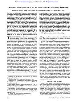
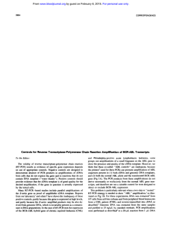
From www.bloodjournal.org by guest on February 6, 2015. For personal use only.
RAPID COMMUNICATION
p16 Gene Homozygous Deletions in Acute Lymphoblastic Leukemia
By Bruno Quesnel, Claude Preudhomme, Nathalie Philippe, Mickael Vanrumbeke, Isabelle Dervite, Jean Luc Lai,
Francis Bauters. Eric Wattel. and Pierre Fenaux
The p16 protein is a cyclin inhibitor encoded by a gene located in 9p21, which may have antioncogenic properties,
and isinactivated by homozygous p16gene deletion or, less
often, point mutation in several types of solid tumors often
associated t o cytogenetic evidence of 9p21 deletion. We
looked for homozygous deletion and point mutationof the
p16 gene in acute lymphoblastic leukemia (ALL), where 9p21
deletion or rearrangement are also nonrandom cytogenetic
findings. Other hematologic malignancies including acute
myeloid leukemia (AML), myelodysplastic syndromes
(MDS), chronic lymphocytic leukemia (CLL), and myeloma
were also studied. Homozygous deletion of the p16 gene
was seen in 9 of the 63 (14%) ALL analyzed, including 6/39
precursor B-ALL, 3/12 T-ALL, and 0/12 Burkitt's ALL. Three
of the 7 ALL with 9p rearrangement (including 3 of the 5
patients where this rearrangement was clearly associated
t o 9p21 monosomy) had homozygous deletion compared to
5 of the 55 patients with normal 9p (the last patient with
homozygous deletion was not successfully karyotyped). Single stranded conformation polymorphismanalysis of exons
1 and 2 of the p16 gene was performed in 88 cases of ALL,
including the 63 patients analyzed by Southern blot. Twentysix of the cases had 9p rearrangement, associated t o 9p21
monosomy in at least 12 cases. A missense point mutation,
at codon 49 (nucleotide 1641, was seen in only 1 of the 88
patients. No homozygous deletion and no point mutation
of the p16 gene was seen in AML, MDS, CLL, and myeloma.
Homozygous deletion of interferon a genes (situated close
t o p16 gene in 9 ~ 2 1was
) seen in only 3 of the9 ALL patients
with p16 gene homozygous deletion, and none of the ALL
without p16 gene homozygous deletion. Our findings suggest that homozygous deletion of the p16 gene is seen in
about 15% of ALL cases, is not restricted
t o cases with cytogenetically detectable 9p deletion, and could have a pathogenetic role in this malignancy. On theother hand, p16 point
p16 homomutations are very rare in ALL, and we found no
zygous deletions or mutations in the other
hematologic malignancies studied.
0 1995 by The American Societyof Hematology.
P
arrangements of the p1 6 gene by Southern blot analysis, and
for point mutations of the p16 gene by polymerase chain
reaction-single stranded conformation polymorphism (PCRSSCP) analysis of exons 1 and 2 of the gene (which correspond to 97% of the total coding region) in ALL and other
hematologic malignancies, including acute myeloid leukemia (AML), myelodysplastic syndromes (MDS), chronic
lymphocytic leukemia (CLL), and multiple myeloma (MM).
Because interferon a (IFNa) genes, which are located about
400 kb 3' of the p16 gene on 9p21, have been reported to
be deleted in some ALL,"."we also performed Southern
analysis of those genes in ALL.
ROGRESS THROUGH the cell cycle appears to be regulated by a number of cyclin-dependent protein kinases, the CdKs. The p16 protein is an inhibitor of CdK 4,
encoded by a gene (multiple tumor suppressor 1, MTS 1
gene or p16 gene) localized on chromosome 9~21." By
binding to and inhibiting CdK 4, p16 could suppress cell
division in a similar fashion as p21, whose synthesis is stimulated by p53 and which inhibits other CdKs! Therefore, the
p16 gene is considered as a potential tumor suppressor
gene.'-3
The human 9p21 region contains chromosomal inversions,
translocations, deletions, and loss of heterozygosity in a large
variety of solid tumors, including malignant melanoma, glioma, lung cancer, bladder, pancreatic, and renal cancer, and
homozygous deletions of the p16 gene have recently been
found in 30% to 85% of cell lines established from those
turn or^.^.^.^ Furthermore, melanoma and pancreatic cell lines
frequently carry nonsense, missense, or frameshift mutations
of the p16 gene, predominantly in exon However, in fresh
solid tumors, the incidence of homozygous deletion of the
p16 gene seems to be lower (10% to 20%), and point mutations very rare except in melanoma and pancreatic carciGermline p16 mutations may also be associated to
familial melanoma.'
Deletions or unbalanced translocations of the short arm
of chromosome 9 leading to 9p21 monosomy are also found
in 7% to 13% of acute lymphoblastic leukemias (ALL) but
are uncommon in other hematologic malignancies.*.' Homozygous deletion of the p16 gene was found in 9 of 14 and
1 of 4 leukemic cell lines, respectively, in two
However, the lineage of those leukemia cell lines was not
mentioned and no study of the p16 gene in uncultured ALL
or other hematologic malignancies has been published, to
our knowledge.
Therefore, we looked for homozygous deletions and reBlood, Vol 85,No 3 (February l ) , 1995:pp 657-663
MATERIALS AND METHODS
Materials
DNA from cases of newly diagnosed ALL and other hematologic
malignancies, including AML, MDS, CLL, and myeloma, was studied by Southern analysis and PCR-SSCP analysis of exons 1 and 2
of the p16 gene. Diagnosis of ALL, AML,andMDSwasmade
From U,,, Inserm Institut de Recherches sur le Cancer; Service
des Maladies du Sang; Laboratoire d'Hkmtologie; and Service de
Cytogknktique, C.H. U. Lille, France.
Submitted October I , 1994; accepted November 6, 1994.
Supported by the Comitk du Nord of the Ligue contre le Cancer,
the GEFLUC and the Centre HospitalierUniversitaire of Lille,
France.
Address reprint requests to PierreFenaux, MD, Service des Maladies du Sang, C.H.U. I , Place de Verdun, 59037 Lille. France.
The publication costsof this article were defrayedin part by page
chargepayment. This article must therefore be hereby marked
"advertisement" in accordance with 18 U.S.C. section 1734 solely to
indicate this fact.
0 1995 by The American Society of Hematology.
0006-4971/95/8503-0037$3.00/0
657
From www.bloodjournal.org by guest on February 6, 2015. For personal use only.
658
QUESNEL ET AL
Table 1. Primers Used for the PCR of Exons 1 and 2
of the p16 Gene
DNAFragmentFragment
Amplified
Size
Region 1
(Containing
exon 1)
Region 2
(Containing
exon 2)
Sequence
343 bp
2F
394 bp
1108R 5'GCGCTACCTGATTCCAAiTC 3'
P36
S'TTCCTTTCCGTCATGCCGG 3'
M1
S'GAAGAAAGAGGAGGGGCTG 3'
S'GTACAAATTCTCAGATCATCAGTCCTC 3'
according to French-American-British
and an immunophenotype was performed in all ALL cases. All patients, except one
case of ALL, were successfully karyotyped by conventional banding
technique^.'^
In the 63 ALL patients who underwent Southern blot analysis,
DNA was extracted from blood or bone marrow (BM) cell samples
containing greater than 80% blasts. SSCP analysis was made in
those 63 patients, and in 25 additional cases of ALL that generally
had cytogenetic rearrangements involving 9p and where DNA could
only be obtained by scraping cells from marrow slides, as previously
described." This method, in our experience, generally provides sufficient DNA for PCR-SSCP analysis, but not for Southern blot analysis
DNA for Southern blot analysis was obtained from blood samples
in CLL (containing 63% to 90% lymphocytes), marrow samples in
MDS (containing 74% to 92% myeloid cells), and myeloma (containing 50% to 63% of plasma cells), and blood or marrow samples
inAML (containing at least 70% blasts). In additional cases of
AML, MDS, CLL, and myeloma, DNA was obtained for PCR-SSCP
analysis by scraping marrow cells, as for ALL.
Southern blot analysis of IFNa genes was also performed in the
63 cases of ALL studied by Southern analysis of the p16 gene.
Finally, Southern blot analysis of the p16 gene was made in HSB2
and CEM cell lines, two T-ALL cell lines obtained from American
Type Culture Collection (ATCC; references CLL 120.1 and CCL
119, respectively).
Methods
Southern blot analysis. DNA was digested with EcoRI and Hindl11 restriction enzymes, separated by electrophoresis in 0.8% agarose gel and transferred to nylon membranes, according to conventional methods.I6
The p16 gene probe used was a 0.96 cDNA probe (kindly provided
by D. Beach, Cold Spring Harbor,NY). Southern blots of ALL cases
were subsequently rehybridized to an IFNa-l EcoRI-Xba I 642-bp
genomic probe (kindly provided by M. Tovey, CNRS UPR 274,
Villejuif, France) which cross-hybridizes with all IFNa genes. Finally, blots were hybridized to a probe for the actin gene, situated
on chromosome 7, which served as control. In a few cases of MDS
and AML with monosomy 7, a probe for the neurofibromatosis (NF,)
gene, which is not deleted in our experience in adult MDS and AML,
and which is situated on chromosome 17, was used instead of the
actin probe."
Homozygous deletion of the p16 and IFNa genes was determined
by visual inspection of the autoradiographs sequentially hybridized
with the p16, IFNa, and control probe. Suspected homozygous deletions were more objectively confirmed by measuring the intensity
of hybridization signals by a densitometer (Densylab; BIOPROBE
Systems, Montreuil, France).
PCR-SSCP analysis. Intronic oligonucleotide primers were purchased from BIOPROBE Systems. The names and nucleotide sequences of the primers used in this work are listed in Table 1. Two
genomic regions were amplified: region I , encompassing exon 1 and
measuring 343 bp; and region 2. encompassing exon 2 and measuring
394 bp. For region 1. weused the primers published by Kambet
al.' Because SSCP analysis seems to require fragments of no more
than 350 to 400 bp in length, region 2 was digested, after amplification and before SSCP analysis, by Sma I enzyme, because an Sma
I restriction site is present in exon 2. This led to two fragments,
region 2aand region 2b, each measuring 162 and 232 bp, respectively.
Genomic DNAs (0.1 pg) were subjected to PCR in a 50-pL solution containing 200 pmol/L each of dATP, dGTP, dTTP, dCTP, 0.1
pL of "P-dCTP (Amersham, Amersham, UK, I O mCi/mL), 25 pmol
of S' and 3' primer 5% dimethyl sulfoxide (DMSO), 10 mmol/L
TRIS-HCI (pH 9),SO mmol/L KCI, 1.5 mmoVL MgCI2,Triton X 100
(0.1%) and gelatin 0.2 mg/L, 0.4 U of Taq polymerase (Appligene,
Illkirch, France) in a thermocycler (Minicycler; M.J. Research, Watertown, MA). For exon I , PCRwas performed as follows: 10
minutes at 9 4 T , then 20 cycles with 94°C for 1 minute, 64°C for I
minute, 72°C for I minute with decrement of 0.2"C per cycle followed by IS cycles with 94°C for 1 minute, 60°C for 1 minute, 72°C
for 1 minute, then by final elongation at72°C for 5 minutes. For
exon 2, PCR was performed as follows: IO minutes at 9 4 T , then
20 cycles with 94°C for 1 minute, 56°C for I minute, 72°C for 1
minute, with a decrement of 0.2"C per cycle followed by 1 S cycles
with 94°C for 1 minute, 52°C for 1 minute, 72°C for 1 minute,
followed by final elongation at 72°C for 5 minutes. After amplification, 1 pL of the reaction mixture for region 1 was mixedwith19
pL of 0.1 % sodium dodecyl sulfate (SDS), 20 mmol/L EDTA solution. For region 2, 1 pL of the reaction mixture was first digested
by Sma I in I O pL, and diluted in I O pL of SDS-EDTA solution.
Then 3 pL of the diluted region 1 and of regions 2a and 2b, respectively, were mixedwith 3 pL of a solution of 95% formamide,
20 mmol/L EDTA, 0.05% bromophenol blue, 0.05% xyene cyanol,
heated 4 minutes at 80°C and applied (2 pWlane) to an MDE polyacrylamide gel (BIOPROBE Systems) containing 90 mmol/L TRISborate pH 8.3, 4 mmol/L EDTA. Electrophoresis was performed at
35 W for 6 hours at room temperature. with cooling using a fan.
Sequencing analysis. PCR amplification was performed as described above, using a biotinylated primer. Single-stranded DNA
template was obtained by binding the biotinylated PCR products
to streptavidin-coupled magnetic beads (Dynabeads; Dynal, Oslo,
Norway) and NaOH denaturation according to the manufacturer.
Sequencing reactions were performed following the Sequenase 2.0
protocol (US Biochemical Corp, Cleveland, OH). The sequencing
primer was the nonbiotinylated primer usedin the PCR reaction.
When ambiguities were present, the opposite strand was sequenced
using the reciprocal combination of biotinylated and nonbiotinylated
primers. The sequencing products were analyzed on a 6% polyacrylamide gel containing 7 mol/L urea.
RESULTS
Southern Blot Analysis
of the p16 Gene in ALL
The 63 ALL studied by Southern blot included 2 children
ages 5 1 5 years and 61 people ages > l 5 years, 24 females
and 39 males, 39 B-precursor ALL (37 early B-ALL, which
were CALLA' in 31 cases, and 2 pre B-ALL with intracytoplasmic Igs), 12 T-ALL, and 12 Burkitt's ALL (Table 2).
Cytogenetic analysis (successfully performed in 62 cases)
was normal in 16 cases. Four patients had a hyperdiploid
karyotype, 6 had t(9;22), 4 had t(4; l l), 12 had t(8; 14), 2
had t( 1 ;19), and the remaining patients had various other
rearrangements. Seven patients had 9p rearrangements, including unbalanced translocations between chromosome 9
From www.bloodjournal.org by guest on February 6, 2015. For personal use only.
659
p16 DELETION IN ACUTELEUKEMIAS
435
Table 2. Hematologic Characteristics of the Patients Studied by
Southern Analysis of the p16 Gene
Diagnosis
No. of
Patients
Studied
ALL (39 B precursor ALL,
12 T-ALL, 12 Burkitt’s
ALL)
AML (1 Ml, 3 M2, 1 M3,
2 M4, 3 M5, 1 M6)
MDS (8 RA, 9 RAEB,
11 CMML)
CLL
Myeloma
No. of Cases
With 9p
Rearrangement.
Gene
No. of Cases With
11 kb
Homozygous p16
Deletion
4.3 kb
715
900
920 1002 1269
1280
P16
3.2kb
63
7 (5)
9
11
2 (2)
0
28
20
18
1 (1)
0
2 (2)
0
0
0
1.6 kb
1.2W
1 1 kb
No. of cases with 9p21 monosomy is shown in parentheses.
4.3 kb
and another chromosome in 4 cases, i(9q) in 1 case, and del
(9p) in 2 cases (del (9)(p21) in 1 case, del (9)(p13p21) in
the other case). In 5 of those 7 cases, the rearrangement was
associated to 9p21 monosomy, and therefore to probable
hemizygosity for genes located on this band (including the
p16 gene). In the other cases [the 2 cases of del (9p)l, the
breakpoint was in 9 ~ 2 1and
, this did not allow us to determine if one allele of genes located in 9p21 band was lost.
Homozygous deletion of the p16 gene was seen in 9 cases
(14%). including 6/39 (15%) B-precursor ALL, 3/12 (25%)
T-ALL, and no Burkitt’s ALL (Table 3). In those 9 cases,
the ratio between the hybridization signals from the p 16 gene
probe and the actin probe, measured by densitometry, was
always less than 20% of that observed in controls (Fig I).
No abnormal band on Southern blots was seen in any case.
Homozygous deletion of the p16 gene was also present in
HSB2 and CEM cell lines.
In one of the patients with p 16 gene homozygous deletion,
cytogenetic analysis was a failure. Three of the 7 patients
with detectable 9p rearrangement, including 3 of the S patients with 9p21 monosomy, had homozygous deletion of
the p16 gene, compared to S of the 55 patients without
detectable 9p abnormality.
Eight of the 9 patients (89%) with p16 gene homozygous
deletion had one or several poor prognostic factors, including
IFN
3.2W
1.8 kb
1.2kb
ACTIN
Fig l. Southern blot analysis of thep16 and IFNa genes in 7 cases
of ALL after EcoRl digestion. Blots weresuccessively hybridized with
the p16 probe, an IFNa probe, and an actin probe. The absence of
signal for p16 gene in cases no. 435 and 715 demonstrates homozygous deletion of this gene in both cases. In case no. 435, the Southern
profile with the IFNa probe showed loss of one band, suggesting
homozygous loss of one or several of the IFN genes. In case no. 715
and in the 5 remaining cases, Southern analysis of the IFNa genes
appeared normal.
“bulky” disease in 8 cases, white blood cell (WBC) count
>SO X IO”/L in 7 cases, and “poor-risk” karyotype in 3
cases: t(9;22) ( I case), t(4; I 1 ) ( I case). and complex cytogenetic findings ( I case) (Table 3). All 9 patients obtained
complete remission (CR) with intensive chemotherapy but
Table 3. Hematologic Characteristics and Outcome of Patients With p16 Gene Homozygous Deletion or Point Mutation
Patient
No.
Bulky
1109/Ll
Diagnosis
DiseaseAge/Sex
WBC
Karyotype
Gene
30 Early B-ALL 11/M
Yes
84 Failure
Yes 45/F
124
198 Early46,XX
B-ALL
52 45,XY,der(5)t(5;9)(p14;q21),-9
305 Early B-ALL Yes
36/M
365 Early B-ALL
9/F
No
25 56,XX,+4,+6,+?8,+?10,+13,+18,+21x2,+2mar,inc
435 T-ALL
46,XY,del(13)(q14q22)
96
22/M
Yes
46,XY,de1(6)(q22)
380
520
T-ALL
27/M
Yes
569 Early B-ALL 54/M 191Yes
46,XY,t(4;11)(q21;q23)
150 Early B-ALL 53/M
14 Yes
46,XY,der(9)t(9;22)(q34;qll)t(9;16)(p13;q12)
60 Yes 30/M
T-ALL715
47,XY.-6,i(9)(q10),del(ll)(q21),+2mar
157 Early B-ALL 54/F 150Yes
46,XX,t(4;11)(q21;q23)
p16
Anomaly
Homozygous deletion
Homozygous deletion
Homozygous deletion
Homozygous deletion 25
Homozygous deletion 34
Homozygous deletion 18
Homozygous deletion
Homozygous deletion
Homozygous deletion
Missense mutation in exon
2, codon 49 nucleotide 164
(GCC GIC, Ala -Val)
-
CR
Duration Survival
(mod
(mas)
108+
3
59+
24
31
17
3
3
72+
3
109+
8
60+
4
4
73+
3
From www.bloodjournal.org by guest on February 6, 2015. For personal use only.
660
QUESNEL
Table 4. Hematologic Characteristics of thePatients Studiedby PCR-SSCP of Exon 1 and Exon2 of the p16 Gene
Diagnosis
No. of Patients
Studied
ALL (60 precursor B-ALL, 16 T-ALL,
12 Burkitt'ALL)
AML (7 M1.6 M2.3 M3.8 M4,
8 M5, 3 M6)
MDS (7 RA, 12 RAEB, 11 CMML)
CLL
Myeloma
No. of Cases With
9p Rearrangement.
No. of Cases With Point Mutation of t h e
p16 Gene
88
26 (12)
35
2 (2)
1 point mutation (5 polymorphisms
at nucleotide 436)
(3 polymorphisms at nucleotide 436)
30
30
30
1 (1)
0
2 (2)
0
(2 polymorphisms at nucleotide 436)
(2 polymorphismsat nucleotide 436)
No. of cases with 9p21 monosomy is shown in parentheses.
6 relapsed after 3 to 31 months. Median CR duration was
24 months, and median survival was 25 months (Table 3).
By comparison, 44 of the S4 ALL (81%) without p16 gene
homozygous deletion had one or several poor prognostic
factors. Fifty-one of the S4 cases were treated with intensive
chemotherapy and 38 (75%) achieved CR. Median CR duration was 18.5 months and median survival of the 5 l patients
treated intensively was 22 months. None of the differences
between patients with and withoutp16 homozygous deletion
were significant.
tients for exon l and in 82 cases for exon 2, by comparison
with controls. Six patients had an abnormal SSCP profile for
exon 2, which was identical in S of them,and differed in
the remaining abnormal case (Fig 2). Direct sequencing of
exon 2 showedthatthe S patients with similar abnormal
SSCP findings had a G + A transitionat nucleotide 436
Southern Blot Analvsis of IFNa Genes in ALL
Southern blots of the 63 ALL were rehybridized with the
IFNa gene probe. Complete disappearance of one or several
bands, suggesting homozygous deletion of at least part of
the IFNa genes, was found in 3 cases (patients no. 30, 43.5,
and 751). who also had homozygous deletion of the p16
gene (Fig I). No homozygous deletion of IFNa genes was
found in the S4 patients who had no homozygous deletion
of the p 16 gene.
PCR-SSCP Ana1.vsi.v of Exons I and 2 of the p16 Gene in
ALL
PCR-SSCP analysis of exons 1 and 2 of the p16 gene
(which cover 97% of the coding sequence) was performed
in the 63 cases analyzed by Southern blot, and in 25 additional cases of ALL where DNA was obtained by scraping
marrow smears from diagnosis samples. The 88 patients
studied by PCR-SSCP included 6 children and 82 adults, 39
females and 49 males, 60 B-precursor ALL (S8 early BALL and 2 pre B-ALL), 16 T-ALL, and 12 Burkitt's ALL
(Table 4). Cytogenetic analysis (successfully performed in
87 cases) was normal in 23 cases. Four cases had a hyperdiploid karyotype, 6 had t(9;22), 4 had t(4; 1 l), 2 had t( 1 ;19).
and the remaining patients had other variable anomalies.
Twenty-six patients had a detectable 9p rearrangement, including: 12 cases of unbalanced translocation between chromosome 9 and another chromosome, 9p deletion or monosomy 9, all leading to 9p21 monosomy and therefore to loss
of a p16 allele; 9 cases of de1(9)(p21) or de1(9)(p13p21)
where it could therefore not be determined if deletion of one
p16 allele was present; and S balanced translocations between9p21and another chromosome, with apparently no
material loss (Table 4).
Normal PCR-SSCP findings were observed in all 88 pa-
Fig 2. SSCP analysis of exon 2 of the p16 gene in 7 ALL cases.
The amplified PCR product for exon 2 was migrated after digestion
with Sma I enzyme (which separated it in two fragments). The 5'
fragment, in case no. 157,had an abnormal migration, corresponding
t o a point mutation at nucleotide 164. The 3' fragment, in patient
no. 1028, had a abnormal migration, corresponding t o a probable
polymorphism at nucleotide 436.
From www.bloodjournal.org by guest on February 6, 2015. For personal use only.
p16 DELETION IN ACUTE LEUKEMIAS
661
However, in those 7 cases which included 3 AML, 2 CLL.
and 2 myelomas, the SSCP profile was identical to that observed in the ALL cases with nucleotide 436 G + A mutation. Directsequencing, performed in two of them, confirmed
the presence of this mutation. which probably corresponded
to a polymorphism, as seen above.
/G
Codon 49
Ala
\ 'c'
DISCUSSION
I
Control
Patient 157
Fig 3. Sequence analysis of exon 2 of the p16 gene in patient no.
157, showing heterozygous mutation at codon 49, nucleotide 164
(GSC GICI.
-
(codon 140. Ala
Thr). Persistance of normal wild-type
bands by SSCP and sequence analysis, of approximately
similar intensity as abnormal bands, showed that the mutation was heterozygous. The remaining patient had a C T
missense mutation at nucleotide 1 6 4 , codon 49 (GCC
GTC) leading to Ala + Val substitution (Table 3). SSCP
and sequence findings also showed that the mutationwas
heterozygous and that the remaining p16 allele was not mutated or deleted (Figs 2 and 3). This patient had B-precursor
ALL, bulky disease. high WBC count, t(4; 1 1) translocation,
and a poor outcome. None of the patients with 9p rearrangement had detectable p16 gene mutation. Samples
from the 9 patients with p 16 gene homozygous deletion gave
faint but normally migrating SSCP bands for exons I and
2, certainly corresponding to the amplification of DNA from
residual marrow cells.
DNA from the S patients with the nucleotide 436 mutation
was reanalyzed by PCR-SSCP after the patients had reached
CR with combination chemotherapy. All S patients still had
the SSCP profile observed at diagnosis, strongly suggesting
that the G A mutation at nucleotide 436 was a polymorphic
variant.
+
+
+
Southem Blot and PCR-SSCP Analysis qf the p16 Gene in
Other Hematologic Malignancies
Eleven AML, 28 MDS, 20 CLL, and 18 myelomas were
studied by Southern blot (Table 2). Those patients, and additional patients, leading to a total number of 35 AML, 30
MDS, 30 CLL, and 30 myelomas, also underwentPCRSSCP analysis of exons 1 and 2 of the p16 gene (Table
4). All patients were successfully karyotyped and detectable
rearrangements leading to 9p21 monosomy were only seen
in 2 cases of AML, 1 case of MDS, and 2 cases of myeloma.
Those patients had monosomy 9, and were studied by both
Southern blot and SSCP.
No homozygous deletion of the p I6 gene was found by
Southern analysis in those patients. SSCP findings were normal for exon 1 in all cases, and for exon 2 in all but 7 cases.
This study represents, to our knowledge. the first analysis
of deletions and point mutations of the p16 gene in uncultured samples from hematologic malignancies. Homozygous
deletion was found in 14% of the ALL but in none of the 4
other malignancies studied, including AML, MDS, CLL. and
myeloma. In ALL, homozygous deletion was not found in
Burkitt's ALL, but was found in IS% of B-precursor ALL
and 25% of T-ALL. Also of note was the homozygous deletion observed in HSBz and CEM cell lines, which are both
derived from T-ALL.
Three of the S ALL cases with cytogenetic rearrangements
leading to9p21monosomyand
S of the SS patients with
cytogenetically normal 9p2 1 band had homozygous p 16 gene
deletion. This demonstrated submicroscopic deletion in band
9 ~ 2 1involving
,
one chromosome 9 in the former group, and
both chromosomes 9 in the latter group. In the 9 deleted
cases, no bands were seen by Southern analysis and, because
the probe we used covered the entire coding region. it can
be concluded that the entire p16 gene was deleted. In solid
tumor cell lines with homozygous p16 gene deletion, the
entire coding region was also generally deleted although, in
a few cell lines, the deletion involved only exon I (or only
exon 2).' No abnormal band suggesting a chromosome 9
breakpoint within the p16 gene was seen, especially in the
2 ALL cases with a 9p21 breakpoint that were studied
by
Southern blot.
In primary solid tumors. the incidence of p16 gene homozygous deletions seems to be one third to one half of that
observed in corresponding cell lines.'.'.' These figures could
also hold true for leukemias, because SS% of the 18 leukemia
cell lines studied in two previous reports showed p16 gene
homozygous deletion.*.' However, no details on these cell
lines were available to confirm if, as in uncultured leukemic
samples, homozygous deletions predominated or were exclusively seen in ALL cell lines, by comparison with AML cell
lines.
Using PCR-SSCP, a sensitive method for the detection of
point mutations in DNA fragments no longer than 300 to
400 bp,'Xwe could find only one point mutation in exon 1
and 2 of the p16 gene (which cover 97% of the coding
region of the gene) in 88 cases of ALL. This mutation was
a missense mutation at nucleotide 1 6 4 , codon 49. which had
not been previously reported
in tumors, to our knowledge.
SSCP and sequence results showed that this mutation was
heterozygous, the remaining p16 allele beingstillpresent
and nonmutated. Five other cases of ALL had a G + A
transition at nucleotide 436. This mutation had already been
reported in 2 of 31 bladder cancers,' I of 34 melanoma cell
lines,'and 4 of 75 various tumor types." In one of those
reports, the mutation was also present in the patients' leukocytes." This nucleotide 436 G + A transition was also often
From www.bloodjournal.org by guest on February 6, 2015. For personal use only.
662
QUESNEL ET AL
observed as a germline mutation in familial melanoma kindreds but, in those families, did not cosegregate withthe
disease, suggesting that it corresponded to a polymorphi~m.~
This conclusion was also supported by our findings, as the
nucleotide 436 G A transition was still present in the CR
marrow of the 5 ALL cases that carried it on diagnosis
samples. We also observed this probable polymorphism in
2 of the 30 myelomas, 3 of the 35 AML, and 2 of the 30
CLL analyzed by PCR-SSCP. In those 3 disorders, and in
CLL, no other point mutations were seen.
Thus, only 1 of 88 ALLstudiedhad a point mutation,
although 12 of them had 9p21 monosomy, leading to probable loss of one p 16 allele (and 9 had del 9p with a 9p21
breakpoint, possibly also associated to p16 allele loss in
several cases). Loss of one gene copy is a situation where for
tumor suppressor genes, a high incidence of point mutations
inactivating the other allele is ~ b s e r v e d . ~Point
* . ~ ~mutations
of the p16 gene, predominantly in exon 2, are seen in about
one third of melanomas and pancreatic carcinomas’,’but
appear tobe rare in the other solid tumors
In melanoma and pancreatic carcinoma, they are generally associated to loss of the normal residual allele.3*5However, in the
only case of ALL with a p16 point mutation reported here,
cytogenetic, SSCP, and sequence findings showed that the
normal residual p16 allele was still present. These findings
suggest that, in ALL and in many solid tumor types (with
the exception of melanoma and pancreatic adenocarcinoma),
p1 6 gene inactivation mainly occurs through deletion of both
copies of the gene, rather than by deletion of one allele and
point mutation of the other allele, as often seen with the P53
gene.
In a previous report, Diaz et al” showed that 7% and
22% of ALL had a homozygous and hemizygous deletion
of interferon cy genes, respectively, and that these deletions
often occurred in the absence of detectable of deletion 9p2 1
band, where IFNcy genes, like the p16 gene, are clustered.
Because IFNa genes are situated very close to the p16 gene
in 9p21, we also looked for homozygous deletions of these
genes in our ALL cases. Only 3 of the 9 patients with p16
gene homozygous deletion and none of the 54 patients without p 16 gene homozygous deletion had a homozygous deletion of one or several IFNa genes. This confirmed previous
results in 14 leukemia cell lines, where 9 had a homozygous
deletion of the p 16 gene, and only 4 a homozygous deletion
of IFNa 8 gene.3 Seven of those 14 cell lines had homozygous loss of the methylthioadenosine phosphorylase
(MTAP) gene, also situated close but in 5‘ of the p16 gene.
These findings suggest that, in ALL, homozygous loss of
the p 16 gene could have more pathogenetic importance than
loss of neighboring genes (especially IFN andMTAP genes).
In this study we only focused on p16 gene homozygous
deletions and did not determine the incidence of p16 gene
hemizygous deletions. One of the reasons was that in our
experience with Southern analysis, demonstrating of loss of
only one copy of a gene in tumor cells contaminated by up
to 20%oreven 30% of normal residual cells is difficult.
Furthermore, as for other tumor suppressor genes, inactivation of the p16 gene presumably requires the inactivation of
both alleles, and the relevance of hemizygous deletions to
the oncogenic process may be more hypothetical.
-+
gene homozygous
Finally, most of theALLwithp16
deletion or point mutations had one or several poor prognostic factors, including bulky disease andhighWBC count,
and most of them relapsed. However, these characteristics
were not significantly different from those observed in patients without p16 homozygous deletion. Although larger
numbers of cases of ALL with p16 gene homozygous deletion will be required before drawing conclusions, it does not
appear that deletion of this gene is associated to specific
hematologic and prognostic features in ALL.
In conclusion, our findings suggest that homozygous deletions of the p16 gene are seen in a small proportion of ALL,
but is not observed or mustbe very rare in AML, MDS,
CLL, and myeloma, whereas point mutations potentially inactivating p16 are very rare in those five disorders. In ALL,
homozygous deletion does not appear tobe associated to
a specific immunophenotype and predominates butis not
exclusively seen in ALL with 9p2 1 deletion. A pathogenetic
role for p16 gene homozygous deletion in the development
or progression of those ALL, suspected because of the potentially antiproliferative properties of p16,will have to be
clearly shown.
REFERENCES
I . Serrano M, Hannon GJ, Beach D: A new regulation motif in
cell-cycle control causing specific inhibition of cyclin D/CDK4. Nature 366:704, 1993
2. Nobori T, Miura K, Wu DJ, Lois A, Takabayashi K, Carson
DA: Deletions of the cyclin-dependent kinase-4 inhibitor gene in
multiple human cancers. Nature 368:7S3, 1994
3. Kamb A, Gruis NA, Weaver-Feldhaus J, Liu Q, Harshman K,
Tavtigian SV, Stockert E, Day RS 111, Johnson BE, Skolnick MH:
A cell cycle regulator potentially involved in genesis of many tumor
types. Science 264:436, 1994
4. Pines J: P21 inhibits cyclin shock. Nature 369520, 1994
S. Caldas C, Hahn SA, Da Costa LT, Redston MS, Schutte M,
Seymour AB, Weinstein CL, Hruban RH, Yeo CJ, Kern SE: Frequent somatic mutations and homozygous deletions of the p16
(MTSI) gene in pancreatic adenocarcinoma. Nature Genet 8:27.
1994
6. Spruck CH, Gonzalez-Zulueta M, Shibata A, Simoneau AR.
Lin MF, Gonzales F, Tsal YC, Jones PA: p16 gene in uncultured
tumours. Nature 370: 183, 1994
7. Hussussian C, Struewing JP, Goldstein AM, Higgins PAT,
Ally DS, Sheahan MD, Clark WH, Tucker MA, Dracopoli NC:
Germline p16 mutations in familial melanoma. Nature Genet 8:15,
I994
8. Chilcote RR, Brown E, Rowley JD: Lymphoblastic leukemia
with lymphomatous features associated with abnormalities ofthe
short arm of chromosome 9. N Engl J Med 313:286, 1985
9. Carroll AJ, Castleberry RP, Crist WM: Lack of association
between abnormalities of the chromosome 9 short arm and either
“lymphomatous” features or T cell phenotype in childhood acute
lymphocytic leukemia. Blood 69:735, 1987
10. Diaz MO, Ziemin S, Le Beau MM, Pitha P, Smith SD, Chilcote RR, Rowley JD: Homozygous deletion in the alpha- and beta
I interferon genes in human leukemia and derived cell lines. Proc
Natl Acad Sci USA 855259, 1988
I I . Diaz MO, Rubin CM, Harden A, Ziemin S, Larson RA,
Le Beau MM, Rowley JD: Deletions of interferon genes in acute
lymphoblastic leukemia. N Engl J Med 322:77, 1990
From www.bloodjournal.org by guest on February 6, 2015. For personal use only.
p16 DELETION IN ACUTE LEUKEMIAS
12. Bennett JM, Catovsky D, Daniel MT. Flandrin G, Galton
D, Gralnick H, Sultan C: Proposals for the classification of acute
leukemias. Br J Haematol 33:451, 1976
13. Bennett JM, Catovsky D, Daniel MT, Flandrin G, Galton
D, Gralnick H, Sultan C: Proposals for the classification of the
myelodysplastic syndromes. Br J Haematol 51:189, 1982
14. ISCN: Guidelines for cancer cytogenetics, supplement to an
international system for human cytogenetic nomenclature, in Mitelman F (ed). Basel, Switzerland, Karger, 1991
15. Van Kamp H, De Pijper C, Verlaan de Vries M, Bos J,
Leeksma C, Kerkhofs H, Willemze R, Fibbe W, Landegent J: Longitudinal analysis of point mutations of the N-ras proto-oncogene in
patients with myelodysplasia using archived blood smears. Blood
79:1266, 1992
663
16. Maniatis T, Fritsh E, Sambrook J: Molecular Cloning: A
Laboratory Manual. Cold Spring Harbor, NY, Cold Spring Harbor
Laboratory, 1982
17. Quesnel B, Preudhomme C, Vanrumbeke M, Vachee A, Lai
JL, Fenaux P: Absence of rearrangement of the neurofibromatosis
1 (NF1) gene in myelodysplastic syndromes and acute myeloid leukemia. Leukemia 8:878, 1994
18. Fenaux P, Jonveaux P, Quiquandon I, Lai JL, Pignon JM,
Loucheux-Lefebvre MH, Bauters F, Berger R, Kerckaert JP: p53
gene mutations in acute myeloid leukemia with 17p monosomy.
Blood 78:1652, 1991
19. Harris CC, Hollstein M: Clinical implications of the p53 tumor-suppressor gene. N Engl J Med 329:1318, 1993
From www.bloodjournal.org by guest on February 6, 2015. For personal use only.
1995 85: 657-663
p16 gene homozygous deletions in acute lymphoblastic leukemia
B Quesnel, C Preudhomme, N Philippe, M Vanrumbeke, I Dervite, JL Lai, F Bauters, E Wattel
and P Fenaux
Updated information and services can be found at:
http://www.bloodjournal.org/content/85/3/657.full.html
Articles on similar topics can be found in the following Blood collections
Information about reproducing this article in parts or in its entirety may be found online at:
http://www.bloodjournal.org/site/misc/rights.xhtml#repub_requests
Information about ordering reprints may be found online at:
http://www.bloodjournal.org/site/misc/rights.xhtml#reprints
Information about subscriptions and ASH membership may be found online at:
http://www.bloodjournal.org/site/subscriptions/index.xhtml
Blood (print ISSN 0006-4971, online ISSN 1528-0020), is published weekly by the American
Society of Hematology, 2021 L St, NW, Suite 900, Washington DC 20036.
Copyright 2011 by The American Society of Hematology; all rights reserved.
© Copyright 2026