
Shedding and Uptake - revised with figs
BBS General Subjects, Special Issue on Glycoproteomics, 2006
Shedding and Uptake of Gangliosides and GlycosylphosphatidylinositolAnchored Proteins
Gordan Lauca, b,* and Marija Heffer-Laucc
a
Department of Chemistry and Biochemistry, University of Osijek School of Medicine, J.
Huttlera 4, 31000 Osijek, Croatia.
b
Department of Biochemistry and Molecular Biology, Faculty of Pharmacy and
Biochemistry, University of Zagreb, A. Kovačića 1, 10000 Zagreb, Croatia.
c
Department of Biology, University of Osijek School of Medicine, J. Huttlera 4, 31000
Osijek, Croatia
* Corresponding author
Dr. Gordan Lauc, Department of Chemistry and Biochemistry, University of Osijek School of
Medicine, J. Huttlera 4, 31000 Osijek, Croatia. fax: +385 31 505 615; email:
[email protected]
Keywords:
Shedding and Uptake, Gangliosides, Glycosylphosphatidylinositol-Anchored Proteins, Lipid
Rafts, Saposins, Immunohistochemistry
1
Summary:
Gangliosides and glycosylphosphatidylinositol (GPI)-anchored proteins have very different
biosynthetic origin, but they have one thing in common: they are both comprised of a
relatively large hydrophilic moiety tethered to a membrane by a relatively small lipid tail.
Both gangliosides and GPI-anchored proteins can be actively shed from the membrane of one
cell and taken up by other cells by insertion of their lipid anchors into the cell membrane. The
process of shedding and uptake of gangliosides and GPI-anchored proteins has been
independently discovered in several disciplines during the last few decades, but these
discoveries were largely ignored by people working in other areas of science. By bringing
together results from these, sometimes very distant disciplines, in this review we give an
overview of current knowledge about shedding and uptake of gangliosides and GPI-anchored
proteins. Tumor cells and some pathogens apparently misuse this process for their own
advantage, but its real physiological functions remain to be discovered.
2
Gangliosides and glycosylphosphatidylinositol (GPI)-anchored proteins come from different
biochemical pathways, but they have the same general assembly plan: they both have lipid
anchors that tether them to the cell membrane and relatively large hydrophilic domains that
protrude in the extracellular space. At the cell membrane gangliosides and GPI-anchored
proteins form membrane microdomains called lipid rafts that are involved in the regulation
and modulation of numerous cellular processes. Many reviews have been recently published
on GPI-anchored proteins [1-4], gangliosides [5-9] and lipid rafts [10-14] and they should be
consulted for more detailed consideration of these molecules and their functions. In this
review we would like to look at them through a different perspective, not addressing the role
of gangliosides and GPI-linked proteins in specific cellular processes, but focusing on their
one peculiar characteristic: the ability to be released from the membrane of one cell and
incorporated into the membrane of another cell. This process has been independently
discovered and rediscovered in several research areas during the past decades, and named
with different names, including shedding, release, incorporation, uptake, jumping and even
cell-painting. In this manuscript we shall refer to as “shedding and uptake”, as originally
suggested by Ladisch and colleagues in 1983 [15].
Gangliosides
Gangliosides are a large family of complex glycosphingolipids that contain one or more sialic
acid residues (Fig. 1). Based on variations in carbohydrate and ceramide structures, the
ganglioside family comprises hundreds of molecular species [16]. Their nomenclature is very
complex (IUPAC–IUB Commission on Biochemical Nomenclature 1978), but a simplified
system developed by Lars Svennerholm is widely accepted [17]. In this system, the ‘ganglio’
core is designated by the capital letter G, which is followed by a capital letter designating the
total number of sialic acids (M – mono; D – di; T – tri; Q – tetra; P – penta; A – asialo). This
is followed by a number designating the length of the neutral ‘ganglio’ core, with 1
representing the full four-saccharide core and shorter structures having higher numbers. The
number of sialic acids linked to the internal Gal residue is designated by a lower case letter (a
= 1, b = 2, etc.) and the number of sialic acids linked to the GalNAc residue is designated by a
Greek letter (α = 1, β = 2, etc.).
(Fig. 1)
3
Gangliosides are biosynthesized stepwise as shown in Fig. 2, starting with the addition of
galactose in β-linkage to ceramide [18]. A key branch point in brain ganglioside biosynthesis
is the addition of sialic acid(s) and/or GalNAc to the growing saccharide chain. Once GalNAc
is added, no further sialic acids can be added to the internal galactose residue of the ‘ganglio’
core. This leads to the generation of ganglioside ‘series’ bearing no, one, two, or three sialic
acids on the internal galactose residue. These have been designated the 0-series, the a-series,
the b-series, and the c-series, respectively. Gangliosides exist in all cells, but they are most
prominent in the brain whey they represent over 25% of conjugated carbohydrates [16, 19]. A
large number of different gangliosides has been isolated from brains of various organisms, but
only four structures, GM1a (from traditional reasons, name GM1 will subsequently be used
instead of GM1a), GD1a, GD1b, and GT1b (Fig. 1B) constitute the great majority (>90%) of
gangliosides in brains from various mammalian species [20]. However, although gangliosides
dominate the complex carbohydrate coat (glycocalyx) of nerve cells, their physiological
functions are largely undefined.
(Fig. 2)
In principle, gangliosides can function on the cell membrane in three ways: (i) as specific
ligands for binding proteins such as MAG [21, 22] and cholera toxin [23]; as glycan arrays
that interact with other glycan arrays on adjacent cells [24, 25]; and (iii) as organizers of lipid
rafts that modulate activity of various proteins through lateral interactions in the same
membrane [26]. In general, cell–cell recognition may occur when glycans on one cell surface
bind specifically to complementary binding proteins (lectins) or carbohydrates on an apposing
cell surface, whereas cellular regulation may occur through lateral interactions between
glycans and signaling molecules on the same membrane [25].
Patterns of ganglioside expression change with cell growth, differentiation, viral
transformation, ontogenesis and oncogenesis [24]. In the brain, gangliosides are expressed in
cell-type and developmentally specific patterns [27-33]. The same is true in the peripheral
nervous system [34], indicating that there is a tight regulation of ganglioside biosynthesis,
degradation and intracellular transport [35]. Gangliosides were also reported to be involved in
decisions regarding neural growth and myelination (reviewed in [7]), as well as in the
development of new axons [36]. Consequently, the expression of specific brain gangliosides
was considered to be essential for neuronal differentiation and brain development, but the
4
unexpectedly mild phenotype of mice deficient for complex gangliosides was a large surprise
that significantly amended that hypothesis (reviewed in [9]).
Apparently when ganglioside biosynthetic pathways are blocked by deletion of a specific
enzyme, the quantity of total gangliosides is often retained even though the structures are
different [37]. Despite major changes in the expression of a particular ganglioside species
associated, phenotypic alterations were found to be only subtle [38-40], indicating that none
of the specific ganglioside structures is essential, and that different gangliosides (at least those
more complex than GM3) can compensate for each other. In that context it is surprising that
major brain ganglioside patterns are highly conserved among mammalian species [20, 41] and
that major ganglioside polymorphisms have not been reported in the brain, although they have
been described in other tissues [42]. Furthermore, recent observation that glycosphingolipid
are essential for the development of Drosophila melanogaster [43], indicates that the lack of
severe phenotype in mice deficient for complex gangliosides might be the consequence of
some kind of a backup mechanisms for ganglioside functions that developed later in
evolution.
Gangliosides modulate transmembrane signaling
An attractive new line of research of ganglioside function was opened in the nineties when it
was found that gangliosides play an important role in the formation and maintenance of lipid
rafts, which are supposed to mediate many signaling processes in the cell membrane. Lipid
rafts have been extensively reviewed in the last few years [10-13, 44-48], and only some of
their aspects will be presented here.
The first indication that gangliosides and other glycosphingolipids can associate and form
patches in the cell membrane came from two independent lines of experiments in 1984.
Spiegel and colleagues were studying fluorescently labeled gangliosides inserted into cell
membranes and observed concerted moving of different gangliosides, that was actually a
manifestation of association of gangliosides into lipid rafts [49]. The second line of evidence
came from studies of Okada and colleagues who were investigating effects of detergents on
membranes and concluded that some gangliosides are located within detergent-insoluble
fraction of the membrane [50]. The hypothesis of glycosphingolipid-enriched membrane
microdomains (lipid rafts) was formulated by Simons and van Meer in 1988 [51], but more
convincing evidence that supported the hypothesis was provided nearly a decade later [26,
5
52]. Even though the existence and importance of lipid rafts in living cells is still being
actively debated [53, 54], several lines of evidence strongly support this hypothesis:
fluorescence resonance energy transfer measurements using fluorescent folate showed
interactions of folate receptors when they are in proximity in rafts in living cells [55];
biochemical crosslinking demonstrated that GPI-anchored proteins are in proximity in rafts
[56]; antibody crosslinking segregated raft proteins from non-raft proteins [57]; photonic
force microscopy was used to determine the size of rafts in living cells [58], and electron
microscopy was used to visualize clustering of rafts in IgE signaling [58]. However, it was
recently shown that crosslinking or proteins inserted into the outer leaflet of the cell
membrane through artificially attached lipid anchors can also induce activation of Jurkat Tcell-signaling responses, indicating that at least in some cases, the formation of artificial raftlike patches on the cell membrane might be sufficient to trigger signaling events [59]. In
some signaling processes the formation of protein clusters in the membrane was reported to
depend on protein-protein, and not protein-lipid interactions [60], thus although lipid rafts
apparently play an important role in mediating many signal transduction processes (Table 1),
they might be only one of several similar mechanisms.
(Table 1)
Glycosphingolipids in the plasma membrane are able to interact laterally with other
membrane molecules modulating their properties (cis-interactions), and the dynamic
clustering of sphingolipids and cholesterol in membrane microdomains represent the basis of
lipid raft formation [26]. These structures move within the fluid bilayer and function as
platforms for the attachment of proteins when membranes are moved around the cell and
during signal transduction [61, 62]. The first convincing evidence for the involvement of
gangliosides in the modulation of transmembrane signaling through the formation of lipid
rafts came from studies of FcεRI, the receptor for IgE on basophils and mast cells. IgE binds
constitutively to cell-surface FcεRI. Aggregation of FcεRI after binding of antigen to FcεRIbound IgE activates the associated Src-family kinase, Lyn, and initiates a signaling cascade
that culminates in degranulation. Colocalization experiments showed that the microdomains
where tyrosine phosphorylation occurred were enriched in GM1. Fluorescently labeled FcεRI
was found to be uniformly distributed in the plasma membrane of unstimulated cells and only
transiently translocated to GM1-rich microdomains after antigen addition [63]
6
The role of gangliosides in the function of receptors for epidermal growth factor (EGF) and
platlet-derived growth factor (PDGF) has been extensively studied (reviewed in [5]). GM3
was reported to inhibit dimerization of EGF receptor (EGFR), while GD1a was reported to
induce dimerization of the same receptor [64]. Interestingly, the addition of GD1a caused
significant EGFR dimerization even in the absence of the growth factor. GD1a apparently
creates some kind of a “primed” state of the fibroblast cell membrane and sets the stage for
enhanced responsiveness to EGF (Fig. 3). For PDGFR (PDGF receptor), the situation was
exactly the opposite; GD1a was found to inhibit dimerization of PDGFR, while GM3 did not
have any effect [65]. Because different rafts exist with unique ganglioside composition,
specific gangliosides might target the respective receptors through direct interaction to unique
rafts. It is also possible that different gangliosides compete to segregate receptors into
different rafts resulting in different effects on their activity. Modifying membrane
gangliosides through action of a membrane sialidase was reported to be essential for the
development of new axons [36], so in addition to shedding, ganglioside composition on the
membrane can also be rapidly altered by action of the membrane sialidase.
(Fig. 3)
Uptake of exogenous gangliosides into the cell membrane
Uptake of exogenous gangliosides into cells was first reported by Keenan and colleagues
more than 30 years ago [66]. During the subsequent years exogenous gangliosides were
administered to fibroblasts [67], astrocytes [68], HeLa cels [69], neuroblastoma cells [70],
glioma cells [71], red and white human blood cells [72], as well as normal and leukemic
lymphocytes [73]. Exogenously administered gangliosides showed a variety of biological
effects depending upon the type of ganglioside and the target cell (for a review see [74]).
Initially it was assumed that all exogenous gangliosides that became associated with cells
were inserted into outer leaflet of the cell membrane, but subsequent studies demonstrated
that exogenously administered gangliosides can be taken up by cells in three different ways:
(i) as loosely associated micelles removable by serum proteins; (ii) as a protein-bound serumresistant, but protease-sensitive ganglioside fraction; and (iii) as gangliosides associated in a
protease-resistant manner [75-77].
7
Gangliosides form aggregates in aqueous media which, depending on their carbohydrate part
and ceramide composition, form micelles of different sizes and shapes [78], or in the case of
ganglioside GM3 even bilayer structures [79]. Ganglioside micelles appear to be quite stable
structures and different micelles do not readily exchange their molecules [80] due to a low
off-rate from micelles or membranes at room temperature [81]. When exogenous gangliosides
are added to the cells, approximately 20% of micelles that had adhered to the cell surface can
be removed by extensive washes with serum-containing media while another 30% of micelles
are tightly bound to proteins and can only be released by treatment with proteases like pronase
or trypsin [82]. In a relatively slow process most of bound ganglioside molecules eventually
escape micelles and, after diffusion through the aqueous phase, insert into the cell membrane,
where they behave as endogenous gangliosides [49]. From the analysis of the electron spin
resonance spectra it could be shown that over 70% of the incorporated spin-labeled
gangliosides are intermixed with other lipids of the host membranes, thus the proteaseresistant fraction represent gangliosides incorporated in the cell membrane [77, 83]. The
remaining (approx. 20%) could represent either ganglioside molecules clustered in
microdomains or ganglioside micelles endocytosed by the cells. The rate of transfer depends
on various parameters like ganglioside concentration, temperature, cell type and duration of
incubation and can roughly be predicted using a formula developed by Saqr and colleagues
[84]. When applied for 24–72 h at 37 ºC ganglioside GM1 incorporates into cultured
fibroblasts in a protease-stable fashion in the range of a few nanomoles per mg cellular
protein. From this it can be estimated that about 109 GM1 molecules can be inserted into the
cell membrane of a single cell, corresponding to roughly 3% of total membrane lipid content.
Several lines of evidence suggest that the uptake of gangliosides into cell membrane involves
action of some specific proteins. Gangliosides added exogenously to epithelial cell cultures
are taken up by the apical membranes, but do not pass the tight junction to the basolateral
membranes of the cell [85]. Pretreatment of cells with trypsin reduces ganglioside uptake [86,
87] and prevents adhesion of cells to GM1-coated wells [88]. The recovery of gangliosideuptake ability requires de novo synthesis of proteins [70]. Several proteins were reported to be
implicated in binding of gangliosides at the cell surface [89-91], but their identity or exact
functions were not determined.
8
Shedding and uptake of gangliosides at the cell surface
The first indications that gangliosides can be shed from the cell surface and exit into
extracellular space came from studies of Kloppel and colleagues who found increased
concentrations of gangliosides in sera of humans and mice bearing mammary carcinomas
[92]. This was soon followed by the first demonstration of efficient shedding and uptake of
gangliosides by Portoukalian and colleagues who reported increased amounts of gangliosides
taken up by erythrocytes of melanoma patients [93]. Interestingly, even though plasma
concentration of GM3 was increased only by 30%, the concentration of GM3 in erythrocytes
increased nearly three times. In the same time, GD3 whose level in serum increased four-fold
was undetectable in erythrocytes, clearly indicating that shedding and uptake differently affect
different gangliosides.
Many tumor cell lines overexpress gangliosides. For example, malignant melanomas and
neuroblastomas overexpress GD3, GD2, and GM2 [94-96], while increased expression of
GD1a, GM1, and GM2 was observed in renal cell carcinomas [97]. The process of
ganglioside shedding has been intensively studied by S. Ladisch and colleagues in the past 20
years. They found that tumor cells can shed up to 0.5% of their membrane gangliosides per
hour [98]. Interestingly, mouse ascites hepatoma cells cultivated at lower cell density were
shedding 3 times more gangliosides then cells grown at higher density [99].
Olshevski and Ladisch demonstrated that gangliosides can be effectively transferred from one
cell to another in combined cultures separated by a membrane that prevented direct contact
between donor and acceptor cells [100]. Inhibition of ganglioside synthesis in donor cells
effectively blocked this transfer [101]. Up to 107 individual ganglioside molecules were found
to insert into a single cell in a co-culture medium with total gangliosides concentration of
7×10⁹. The fact that transfer of gangliosides from the lymphoma cells to the fibroblasts
occurred at a relatively low concentration of shed gangliosides [100] indicates the potential
biological importance of this process. In tumor cells shedding of gangliosides apparently help
to suppress the immune response [15, 102], and the inhibition of NF-kappa B in T-cells by
shed gangliosides has been proposed as one of the possible mechanisms [103]
Gangliosides are able to spontaneously transfer between membranes at elevated temperatures
[104] and the rate of transfer is dependant on both temperature and the physical state of donor
and acceptor membranes [105]. Different gangliosides have significantly different
9
physicochemical properties and it should not be expected that all gangliosides behave in the
same way. However, it is likely that under physiological conditions the effective exchange of
most gangliosides, or at least monosialogangliosides, requires the intervention of specific
exchange proteins [104].
Cells can shed gangliosides both as large membrane vesicles and by preferential release of
particular glycolipids [106]. A certain degree of specificity was reported to exist in both
shedding
and
uptake.
Young
and
colleagues
reported
preferential
release
of
glycosphingolipids with shorter fatty acyl chains, over the corresponding glycosphingolipids
with longer fatty acil chains [106, 107]. Shorter forms of ceramide apparently also enables
more efficient uptake of gangliosides from the medium as reported by Ladisch and Olson
[108]. However, the composition of shed gangliosides was generally found to mirror the
composition of gangliosides in donor cells [101, 109], indicating lack of preference for
specific carbohydrate structures of gangliosides in the process of shedding.
Kong et al. reported that shed gangliosides mostly exist as monomers in the medium [110].
This is very unusual because when exogenous gangliosides were added to the culture medium
at same concentrations (10-8-10-7 M) they mainly existed in micelles, suggesting that the
naturally shed gangliosides are somehow different in their aggregation properties from
exogenously added purified gangliosides. This observation is supported by a fact that uptake
of shed gangliosides is much more efficient than the uptake of the purified exogenously added
gangliosides [100]. Glomerular mesangial cells, neuroblastoma and melanoma cells
undergoing apoptosis shed gangliosides in a process that appears to be regulated and occurs in
the early stages of the apoptotic process [109]. On the other hand, nearly no shedding was
found in cultured Cos7 cells [111]. Taken together, all these results strongly suggest that
shedding and uptake of gangliosides is a regulated physiological process that proceeds
through action of some specific membrane and/or transfer proteins. Although the identity of
these proteins is not known, there are some likely candidates.
Prosaposin is a potential catalyst of ganglioside shedding and uptake
Saposins (also called SAPs – Sphingolipid Activator Proteins) are a group of four highly
homologous small heat-stable glycoproteins (called saposins A, B, C, and D) that are required
for lysosomal degradation of sphingolipids (for a review see [112]). The first saposin (now
called saposin B) was described by Jatzkewitz and his colleagues in 1964 as a heat-stable
10
factor required for hydrolysis of sulfatides by arylsulfatase A [113]. Cloning of the
corresponding cDNA [114] indicated that all four saposins are being synthesized as a single
precursor, a 53-kDa protein prosaposin that can be differentially glycosylated into 65-kDa or
70 kDa forms [115]. Prosaposin of 65 kDa is associated with Golgi membranes and targeted
to lysosomes where four saposins (A, B, C and D) are generated by its partial proteolysis.
Interestingly, the targeting of the 65-kDa protein to lyzosomes is not mediated by the
mannose 6-phosphate receptor, but the Golgi apparatus appears to accomplish molecular
sorting of the 65-kDa prosaposin by decoding a signal from its amino acid backbone [116].
Each mature saposin contains about 80 amino acid residues and has six equally placed
cysteines, two prolines, and a glycosylation site (two in saposin A, one each in saposins B, C,
and D). These residues are also completely conserved in saposins from different animal
species [117].
In addition to being targeted to lysosomes and cleaved to saposins, prosaposin can be secreted
in an uncleaved form and retained at the outer side of the cell membrane [116]. It has been
suggested that its association with the cell membrane proceeds through the interaction with
membrane gangliosides [118, 119]. Prosaposin can also be found in many biological fluids
such as seminal plasma, human milk, and cerebrospinal fluid (reviewed in [120]). Prosaposin
is abundant in the brain where it is localized exclusively in certain neurons [121]. Its presence
on the neuronal surface was first reported by Fu and colleagues in 1994 [122] and since then
many functions have been attributed to the secreted form of prosaposin. Among other effects,
it was reported to be neurotrophic [123], to promote myelination after nerve injury [124], to
prevent apoptosis of neuronal cells in tissue culture [125], and to act as a neuroprotective and
neuroregenerative agent in vivo [126].
Prosaposin is the predominant form of saposins in neurons [127] and the majority of effects of
prosaposin were observed in neuronal cells. However, recently Misasi and her colleagues
reported that prosaposin also prevents TNFα -induced cell death in human histiocytes and
demonstrated that this occurs through stimulation of signal cascades in which signal-regulated
protein kinases are involved [128]. In a similar way, saposin C itself was shown to prevent
apoptosis in prostate cancer cells [128]. These effects are consistent with the observations that
prosaposin addition rescues neuroblastoma cells, primary hippocampal neurons [129],
Schwann cells [125], and PC12 pheochromocytoma cells [119] from apoptosis induced by
various agens. Neurotrophic, neuroregenerative and anti-apoptotic effects of prosaposin are
11
apparently mediated by modification of signaling pathways and prosaposin was shown to be
involved in ERK phosphorylation [130]. Apparently it activates the MAPK pathway by a Gprotein-dependent mechanism [131], and through a same or similar mechanism it also
stimulates growth, migration, and invasion of prostate cancer cells [132].
A mouse knockout model for prosaposin has been created, but since prosaposin is a precursor
of saposins, in addition to affecting membrane functions of prosaposin, the disruption of its
gene also abrogates functions of saposins in the endosomal pathway and results in complex
phenotype including severe progressive central nervous system disease and early death [133].
In addition to the nervous system, the mostly affected system in mice deficient for prosaposin
was the reproductive tract [134, 135]. The prosaposin gene contains 15 exons that can be
transcribed into several mRNAs, resulting from alternative splicing of the 9-bp exon 8 [136,
137]. A splicing variant of prosaposin without exon 8 is preferentially expressed in the brain
following injury [138], and alternative splicing of the prosaposin gene was assumed to be the
mechanism responsible for differential sorting of the different prosaposin forms [139].
However, targeted disruption of this specific splicing variant did not show any specific
phenotype, and the levels of secreted prosaposin in serum were similar to those of wild-type
mice, indicating that both splicing variants of prosaposin are being secreted to the membrane
[140].
Prosaposin and saposins bind different gangliosides differently, with each protein showing
preference for specific structures [141]. Different splicing variants of prosaposin were also
shown to differentially bind different gangliosides [142]. In vitro, prosaposin, as well as
saposins, promoted the transfer of gangliosides from donor liposomes to acceptor erythrocyte
ghosts [141]. Transfer rates were found to be concentration dependent, and up to 50% of
gangliosides were found to be transferred in 60 minutes. On the membrane of neural cells
prosaposin was reported to be in complex with gangliosides [119], and neuroblastoma cells
incubated in the presence of prosaposin were found to have increased levels of gangliosides
on the cell membrane [143]. Saposin is able to extract monomeric lipids from the membrane
and functional significance of prosaposin-ganglioside interactions was recently demonstrated
in the process of lipid presentation by CD1 proteins during immune recognition [144].
Hiraiwa and colleagues reported that prosaposin purified from milk or medium forms
oligomers of varied masses [145] and this was recently confirmed by analysis of recombinant
prosaposin expressed in the bakulovirus system [146]. Direct observation by atomic force
12
microscopy of saposin C effects on membrane bilayers demonstrated ability of saposins to
induce membrane reorganization and form raft-like structures [147].
Membrane rafts are places where receptor signaling and processing occurs (for review see
[45, 148]). Because different rafts exist with unique ganglioside composition, specific
gangliosides might target the respective receptors through direct interaction to unique rafts
and it was suggested that the duration and localization of the signal is controlled by the
proportion of rafts with unique ganglioside compositions to the number of target receptors [5].
Both gangliosides and prosaposin function through the formation and modulation of lipid
rafts, and it is appealing to hypothesize that a possible function of prosaposin on the cell
membrane is the regulation of formation and modulation of lipid rafts by insertion or removal
of specific gangliosides. Even though there is no direct evidence for functional significance of
interactions between saposins and gangliosides, circumstantial evidence seems quite
convincing. Prosaposin and gangliosides both exist in rafts at the cell surface [26, 147].
Prosaposin can bind gangliosides, and is able to catalyze their transfer between different
vesicles in vitro [141]. Both prosaposin and shed gangliosides were reported to be present in
milk and cerebrospinal fluid [120, 149, 150]. Gangliosides are being actively shed from the
membranes [15], and this process appears to be regulated, indicating that it includes specific
protein activity. Another line of evidence comes from the fact that both gangliosides and
prosaposin are involved in the same cellular processes. They were both shown to modify
signal cascades in which signal-regulated protein kinases are involved [130, 131, 151], they
both mediate apoptosis [6, 125, 128, 129], and are involved in decisions regarding neural
growth and myelination [7, 123, 124]. Their distribution and expression changes with
development [152-154] and in response to brain injury [124, 155]. Both gangliosides and
prosaposin are being secreted by tumor cells [15, 156] and were shown to promote tumor
development [132, 157]. Mice deficient for prosaposin and mice deficient for complex
gangliosides are both infertile [39, 134, 135]. Taken together, all these data suggest that
prosaposin has an active role in the regulation of ganglioside shedding and uptake, and
consequently functions as modifier of lipid rafts. Although three-dimensional structure of
prosaposin is not known, since it has multiple glycolipid binding sites, it is quite possible that
at the cell membrane it functions analogously to GM2 activator protein [158, 159] and
shuttles gangliosides between neighboring cells, or cells and the extracellular medium.
13
Glycolipid transfer protein may also be involved in ganglioside shedding and uptake
Glycolipid transfer protein (GLTP) is a soluble protein that selectively accelerates
intermembrane transfer of glycolipids in vitro. After the initial discovery in the membranefree cytosolic extract of bovine spleen [160], proteins with similar activities were found in a
wide variety of tissues, including bovine and porcine brain, liver and kidney, as well as in
plants [161]. Purified GLTPs from animal spleen and brain consist of single polypeptides of
23-24 kDa and have basic isoelectric points and absolute specificity for glycolipids [162-164].
Even though GLTP transfers glycolipids with shorter sugars more efficiently, it also
significantly facilitates exchange of gangliosides between membrane vesicles [165].
Molecular cloning indicated that GLTP is highly conserved among mammals and that bovine
and porcine brain cDNAs encode identical 209 amino acid sequences [166]. The structure of
GLTP distinctly differ from structures of saposin B [167], saposin C [168], and GM2activator protein [158]. As recently revealed by x-ray diffraction [169], GLTP is characterized
by a novel folding motif among proteins that transfer or bind lipids. The structural data show
that complexation of lactosylceramide by GLTP involves a single glycolipid liganding site.
The glycolipid liganding site of GLTP is composed of a surface recognition center for the
sugar headgroup and a molded-to-fit, hydrophobic tunnel that accommodates the hydrocarbon
chains of the ceramide moiety via a cleft-like conformational gating mechanism [169].
Extensive analysis of its transfer properties by Rao and colleagues concluded that GLTP
might act as a freely transporting shuttle that carries glycolipids back and forth between the
donor and acceptor vesicles [170]. Mutational analysis confirmed that GLTP forms a soluble,
stable complex with glycolipids that can be released from the GTLP/complex in the presence
of acceptor membranes. Interestingly, the release of glycolipids into artificial membranes was
not very efficient, indicating that some acceptor specificity might be involved in the release
process [171]. Recent in vitro study also concluded that GLTP’s ability to both capture
glycolipids from the membrane and insert them into the other membrane significantly
depends on structure and composition of both membranes, and the authors concluded that this
suggests that GLTP might be involved in the assembly of lipid rafts [172].
Even though it is assumed that GLTP is a cytosolic protein, its distribution was never studied
in detail, and its physiological functions are mostly unknown. Lin and colleagues suggested
that GLTP might function as cytosolic transporter of glycosphingolipids to the membrane
14
[166], but since glycosphingolipids are generally found on inner leaflet of intracellular
vesicles and outer leaflet of the cell membrane, this function does not seem very probable.
GLTP orthologs in plants and fungi have been implicated in apoptosis and regulation of vital
cellular processes [173, 174], indicating that it might have a similar function in mammalian
cells. Even though there is no direct evidence that GLTP is involved in ganglioside shedding
and uptake in vivo, its ability to perform these functions in vitro [164, 165] puts it high on the
list of potential candidates.
On the basis of currently published results prosaposin and GLTP appear to be the best
candidates for proteins involved in the regulation of ganglioside shedding and uptake, but it is
of course possible that some other known or unknown proteins are actually performing this
task in vivo. Possible alternative candidates might be some of the nonspecific lipid transfer
proteins that were reported to be able to transfer different glycosphingolipids [175].
Glycosylphosphatidylinositol-anchored proteins
First indications that proteins might be attached to the cell membrane by lipid anchors
appeared in 1963 with the finding that bacterial phospholipase can release alkaline
phosphatase from cells [176]. The presence of inositol-containing phospholipid protein
anchors was postulated by Ikezawa and colleagues in 1976 [177], but their hypothesis was not
widely accepted until 1985, when a body of compositional data about Torpedo electric-organ
acetylcholinesterase (AChE) [178], human erythrocyte AChE [179], rat brain and thymocyte
Thy-1 [180], and Trypanosoma brucei variant surface glycoprotein (VSG) [181, 182] became
available. All glycosylphosphatidylinositols (GPIs) share a common core structure [183].
Phosphatidylinositol is glycosidically linked through carbon 6 of the inositol ring to the
reducing end of a nonacetylated glucosamine moiety. Interestingly, GPIs are one of the rare
instances in nature where glucosamine is found without either an acetyl group (present in
most glycoconjugates) or a sulfate moiety (present in heparin) attached to the amino-group at
the 2-position. Three mannosyl residues, linked α1 4, α1 6, and α1 2, respectively, are
attached to the glucosamine. The terminal α1 2 linked mannose is linked to
phosphoethanolamine by a phosphodiester linkage. The GPI is attached to the carboxyterminal carboxyl group of the protein by an amide linkage to the amino group of
phosphoethanolamine (Fig. 4). This common core structure can be further modified in a way
that depends on both the organism and cell type in which it is synthesized [1].
15
(Fig. 4)
The whole process of GPI biosynthesis is carried out in the endoplasmic reticulum [184] and
nearly 20 enzymes participate in this pathway. Corresponding genes have been cloned from
mammals, yeast and protozoa [185]. Once it is completed, the pre-formed anchor is
transferred to a specific site upstream of the C-terminal end of the protein in the ER lumen by
the action of a transamidase complex, which simultaneously cleaves off the remaining Cterminal peptide [2]. The C-terminal sequence of the protein thus acts as a signal for GPI
attachment. It is encoded in the sequences of genomic and cDNA, but does not appear in the
final processed protein.
The initial step of GPI synthesis, attachment of N-acetylglucosamine to phosphatidylinositol,
depends on the product of a X chromosome gene termed phosphatidylinositol glycan class A
(PIG-A in humans, Pig-a in mice) [186]. A deficiency in PIG-A results in rare human disease
named paroxysmal nocturnal hemoglobinuria (PNH) [186-188]. Patients with PNH have
abnormal cells of various hematopoietic lineages that are defective in the biosynthesis of GPIanchored proteins. These include the complement-regulatory proteins, CD55 and CD59,
whose absence results in enhanced complement-mediated lysis [189, 190]. Since deficiency of
GPI is embryonically lethal [191-193], all PNH patients reported to date acquired a somatic
mutation in PIG-A [194]. The exact mechanism how one or a few of the large number of
pluripotent hematopoietic stem cells that bear mutation in PIG-A achieve dominance in the
bone marrow and the peripheral blood is not known [195], but it has been recently shown that
PIG-A deficient cells have lower susceptibility to TNF-α and IFN-γ, what might contribute to
their clonal dominance [196].
(Table 2)
Today, hundreds of GPI-anchored proteins are known (see examples in Table 2) and it is
estimated that approximately 0.5% of all proteins in lower and higher eukaryotes are being
modified in this manner [197]. Although GPI-anchored proteins do not apparently share
common features, the presence of the anchor itself appears to confer some important
functional and behavioral attributes on proteins to which it is attached. In particular,
localization to lipid raft microdomains and cleavage by endogenous and exogenous
phospholipases appears to play a major role in transduction of signals across the plasma
16
membrane (for a recent review see [4]). Recent observation that prion protein and Thy-1 exist
in separate lipid rafts, and that the composition of membrane lipids in rafts containing prion
protein is different from the composition of lipids in rafts containing Thy-1 [198] suggests
that interplay of lipids and GPI-linked proteins in lipid rafts is very specific and carefully
regulated.
Release of GPI-anchored proteins by GPI cleavage
The hypothesis that one of the functions of the GPI anchor may be to offer a site for
degradation by specific endogenous phospholipases resulting in a release of the protein from
the cell surface has been postulated very soon after the existence of GPI-anchors was widely
accepted [199]. The removal of the GPI lipid moiety in vitro was reported to cause significant
alterations in enzymatic activities [200-203] and ligand binding properties [204-206], thus it is
quite likely that some GPI-anchored proteins in the membrane are actually reservoirs of
inactive proteins that can be activated and rapidly released by GPI cleavage.
Two types of GPI-specific phospholipases, GPI-phospholipase C (GPI-PLC) and GPIphospholipase D (GPI-PLD) cleave GPI on different sides of the phosphodiester bond
between inositol and the lipid part of the anchor (Fig. 4). Very recently, it was demonstrated
that angiotensin-converting enzyme (ACE) can also specifically cleave GPI [207]. Several
bacterial species secrete PI-specific type C phospholipases, including Bacillus cereus,
Bacillus thuringiensis, Staphylococcus aureus, Listeria monocytogenes, and Clostridium
novii. These enzymes are able to hydrolyze mammalian GPI anchors, and have been
extensively used in the study of structure and function of GPI-linked proteins. Several
parasitic protozoans, for example, Tripanosoma brucei and Leishmania, contain endogenous
GPI-PLC that converts membrane-bound proteins to hydrophilic soluble forms (reviewed in
[4]).
Since the first discovery of bacterial PI-PLC, endogenous mammalian GPI-PLC have been
postulated to serve as important regulatory factors, reducing surface expression of GPIanchored proteins, while simultaneously increasing the levels of soluble protein. Chan and
colleagues reported that lipoprotein lipase was released from insulin treated 3T3-L1
adipocytes by cleavage of its GPI anchor and they proposed that activation of an insulindependent PI-PLC was responsible [208]. Alkaline phosphatase was also reported to be
released in soluble form from myocytes and adipocytes upon insulin stimulation (Romero et
17
al., 1988), again suggesting the action of a phospholipase C [209, 210]. Park and colleagues
reported that endogenous GPI-PLC releases renal dipeptidase from kidney proximal tubules in
vitro [211] and in vivo [212], but mammalian GPI-PLC has yet to be identified.
Mammalian GPI-PLD was discovered in human serum by Davitz and colleagues in 1987
[213]. Despite its high concentration in mammalian serum [214] and relatively wellcharacterized molecular biology [215] and biochemistry [216] the physiological role of GPIPLD is not clear. In serum, GPI-PLD is associated with HDL and is apparently not active
[217]. Initial reports indicated that GPI-PLD was active against GPI-anchored proteins only in
the presence of detergent, and was not able to cleave the anchors of proteins in a native
membrane context [218]. Overexpression experiments indicated that it is active in
endoplasmic reticulum during GPI synthesis, but also in lipid rafts [219]. Lipid fluidity and
packing are the most important modulators of bacterial phospholipase ability to cleave GPI
anchors [220] and modulation of membrane lipids were reported to affect GPI-PLD activity in
vitro [221], so it is quite possible that mammalian GPI-PLD also requires particular
membrane composition for activity. The fact that endogenous GPI-PLD was reported to
specifically release NCAM from differentiating myoblast cells [222], receptor for urokinasetype plasminogen activator from ovarian cancer cells [223] and carcinoembryonic antigen
from human colon cancer cells [224] strongly support this hypothesis.
The angiotensin-converting enzyme (ACE) is a well-characterized zinc peptidase that
regulates blood pressure by hydrolyzing bioactive peptides such as angiotensin I and
bradykinin [225]. There are two ACE isoforms: a somatic form of around 150-180 kDa,
which bears two catalytically active sites, and a smaller isoform (90-110 kDa) found in the
testes, which contains a single active site [226]. Kondoh and colleagues recently reported that
testicular ACE can specifically release GPI-anchored proteins from the cell membrane [207].
Even when the peptidase activity is abolished by either mutation or inactivation, the enzyme
could still cleave GPI-anchored proteins and restore fertility to ACE-deficient sperm. This
activity is not protein-specific because it cleaves a variety of GPI-anchored proteins, and its
cleavage site is located between the second and the third residue of the conserved mannose
core (Fig. 4). GPI-anchor-releasing activity of ACE requires removal of cholesterol from cell
membranes, and similarly to GPI-PLD that is also widely present, but mostly inactive, ACE
apparently also requires a particular form of substrate presentation on the membrane for
activity.
18
Release of intact GPI-anchored proteins from the cell membrane
In addition to release by enzymatic cleavage, GPI-linked proteins can be released from the
cell membrane with their GPI-anchors intact. This release can be in the form of membrane
vesicles (exosomes), but also as small aggregates that contain some membrane lipids in
addition to GPI-linked proteins [227]. Exosomes are small (50–200 nm) membrane vesicles
first described in studies of reticulocyte maturation about 20 years ago [228, 229], that were
subsequently demonstrated to be released from various cell types [230-235]. Exosomes were
initially thought to correspond to internal vesicles of multivesicular bodies being released in
the extracellular space upon their fusion with the cell membrane, but this is apparently only
one way how exosomes can be formed since glycolipids and GPI-anchored proteins already
embedded in the outer leaflet of the cell membrane can also be efficiently secreted in the form
of exosomes [236]. Various GPI-linked proteins, including the prion protein [237] are being
actively secreted in exosomes. This process can be quite extensive as demonstrated by
reticulocytes that release approximately 50% of acetylcholinesterase in exosomes during
differentiation into erythrocytes [238]. Similar vesicles named prostasomes exist in seminal
plasma where they assist sperm function [239]. GPI-anchored CD59, CD55 and CD52 were
found on prostasomes [240], but also in a form of small aggregates in seminal plasma [227].
While prostasomes bind to target cells and are later internalized, the kinetics of transfer of
GPI-anchored molecules from aggregates into cells is consistent with direct incorporation into
cell membranes [227].
Shedding and uptake of GPI-anchored proteins
The phenomenon of shedding and uptake of a GPI-linked protein was reported even before
GPI-anchors were discovered. While investigating phospholipid exchange between cells and
artificial vesicles, Bouma and colleagues showed that acetylcholinesterase and some other
erythrocyte proteins were transferred from erythrocytes to the vesicles and that this process
was reversible [241]. The rate, direction, and extent of such intermembrane transfers was
found to depend on the relative lipid composition and fluidity of the donor and acceptor
membranes [242]
Contrary to the release of GPI-anchored proteins by phospholipases C and D that removes
GPI and yields soluble protein, shedding releases proteins with intact GPI that are still able to
insert into membranes of other cells. Cell-to-cell transfer of GPI-anchored protein has been
19
reported in a variety of in vitro and in vivo systems. CD59 was transferred from seminal
plasma to erythrocytes and other cells [240], as well as from erythrocytes to endothelial cells
in mice made transgenic for this GPI-anchored protein [243]. Thy-1 was transferred between
cells in chimeric murine embryoid bodies composed of normal and PIG-A “knock-out” cells
[244] and trypanosomal variant surface glycoprotein (VSG) was found to be incorporated into
erythrocytes of infected patients [245]. High-density lipoproteins (HDL) may act as carriers of
CD59 and are capable of transferring this protein to erythrocytes [246]. Transfer between
membranes can occur without actual membrane fusion [227] and GPI-anchored proteins are
apparently transferred through vesicles or liposomes released from the donor cell [247].
GPI-anchored molecules are clustered in lipid raft membrane microdomains and they actively
take part in membrane vesicle formation, resulting in vesicles enriched in GPI-anchored
proteins [247]. Storage of erythrocytes results in loss of both CD55 and CD59 from the
erythrocyte membrane [248] and creation of erythrocyte microvesicles that are enriched in
GPI-linked proteins including CD55 and CD59 [249]. When erythrocytes from PNH patients
that were deficient in GPI-anchored proteins were incubated with HDL preparations or
erythrocyte microvesicles, there was significant transfer of CD55 and CD59 to the cell
surface. Pretreatment of microvesicles and HDL with phosphatidylinositol-specific
phospholipase C abrogated protein transfer to deficient cells, indicating that increased cellassociated CD55 and CD59 levels were related to the insertion of an intact GPI moiety, rather
than to simple adhesion [250].
In a recent elegant experiment Sloand and colleagues confirmed the ability of GPI-linked
proteins to transfer between cells in vivo [251]. PNH patients of group A1 blood type were
given transfusions of compatible, washed group O blood. Patient’s group A1 cells were
distinguished from the transfused group O cells by staining with a Dolichos biflorus lectin
that specifically binds to group A1 erythrocytes. Significant transfer of GPI-linked proteins
from donor cells to patient’s erythrocytes could be demonstrated as early as 1 day following
transfusion and persisted for several days.
GPI-linked proteins transferred from cell to cell appear to be stable and biologically
functional [227, 243, 252-254]. For example, transfer of CD55 and CD59 to erythrocytes
confers resistance to complement-mediated lysis [250]. For effective transfer to occur, both
the GPI anchor and the protein moiety must be intact [255]. Transferred molecules are
20
inserted into the outer leaflet of the plasma membrane by lipid chains on the GPI moiety and
soluble CD59 (that lacks GPI anchor) was found to have only 1/200th the ability of GPIlinked CD59 to inactivate complement [256].
Incubation of rat Thy-1 antigen with murine lymphocytes showed that the rat protein could
incorporate into murine cells, and that after the membrane uptake the exogenous protein
migrated with the same lateral mobility as endogenous murine Thy-1 protein [257]. Similarly,
incorporation of Trypanosoma brucei variant surface glycoproteins (VSG) into baby hamster
kidney cells showed that the inserted VSG exhibited lateral mobility equivalent to that of
endogenous VSG in T. brucei [258]. Interestingly neither Thy-1 inserted into lymphocytes
[257] nor CD59 incorporated into neutrophils [259] supported transmembrane signaling
immediately following transfer. However, CD59 incorporated into U937 monocytic cells and
allowed to equilibrate for 2 h at 37°C showed a redistribution into lipid rafts and signaled
intracellular Ca2+ fluxes [260]. Therefore, exogenously introduced GPI-anchored molecules
appear to become functional within the target cell membrane once they have acquired a
distribution similar to that of endogenous GPI-anchored proteins, but this process is slow and
can take even more than 24 h [255, 261].
GPI-linked proteins were reported not to transfer spontaneously from erythrocytes to
liposomes, and it was suggested that in vivo GPI-linked membrane proteins do not
spontaneously transfer between cell membranes, but that some catalyst is needed [247]. This
hypothesis is also supported by the observation that CD4 engineered to have GPI anchor can
be efficiently transferred between cell membranes in one type of cells [262], while another
cell line expressing CD4-GPI fusion protein failed to release it in any form [263]. However,
the identity of a potential protein catalyst of GPI shedding and uptake is not known.
What is a physiological function for shedding and uptake of gangliosides and GPIanchored proteins?
Tumor cells use shedding and uptake to evade destruction by immune cells [15, 102, 157],
and retroviruses exploit shedding for spreading to other cells [264], but these extensively
studied mechanisms are actually only examples of a misuse of shedding and uptake, and the
real reason why this process developed in the course of evolution still has to be discovered.
21
One reported function of shedding and uptake is the transfer of GPI-linked proteins and
gangliosides from prostasomes and GPI-lipid aggregates released by prostate epithelium to
spermatozoa [227]. Since spermatozoa do not synthesize proteins, shedding and uptake here
represent an important mechanism by which spermatozoa can acquire new proteins and alter
their antigenicity, resistance to immune attack, or other surface properties. Another rather
probable function of shedding and uptake is the modulation of lipid rafts and signal
transduction. Exogenously added GM1 was reported to inhibit fibroblast growth factor 2
(FGF2)-mediated proliferation in endothelial cells by binding to FGF2 and preventing its
interaction with the receptor [265]. In the same time, endogenous GM1 in the cell membrane
was found to promote FGF2-mediated fibroblast proliferation [266]. Apparently GM1 in the
medium binds to FGF2 in an inhibitory manner, while GM1 in the cell membrane binds FGF2
in a way that promotes its interaction with the receptor [267]. Shedding of GM1 from the cell
membrane in the same time decreases promoting activity and increases inhibitory activity of
GM1, thus providing a very efficient way of modifying effects of FGF2 on the cell. Shedding
of gangliosides from one cell and their uptake by a neighboring cell might also be a way how
different cells in a tissue could coordinate reaction to hormonal signals.
Exogenous administration of gangliosides affects membrane distribution of GPI-anchored
proteins in lipid rafts [268, 269]. Both GPI-PLD and ACE were reported to require some kind
of specific membrane environment to become active, and it is tempting to speculate that
modification of lipid rafts by removal or addition of specific gangliosides might create
favorable conditions for activity of these enzymes and consequential release of GPI-anchored
proteins. In addition to its role in the modulation of lipid rafts, shedding and uptake of
gangliosides and GPI-linked proteins might be involved in some other processes. For example
it was hypothesized that shed gangliosides might be involved in cell synchronization [270,
271]. The fact that shed ganglioside suppress immune response to cancer cells suggest that
this mechanism could actually be used to suppress autoimmune response in some situations.
For example, gangliosides are especially enriched in the brain and shed gangliosides in the
cerebrospinal fluid might be responsible for suppressing autoimune activity of T-cells that
pass blood-brain barrier.
Impact of shedding and uptake of gangliosides and GPI-anchored proteins on the
analysis of their distribution by immunohistochemical analysis
22
The ability of gangliosides and GPI-anchored proteins to move between cells in physiological
conditions has profound effects on their behavior in various assay systems in vitro.
Immunohistochemistry is a very important tool that enables precise localization of various
types of biological molecules and structures. However, this method is prone to serious
artifacts, and significant care is needed to avoid false interpretation of experimental data
[272]. Gangliosides appear to be particularly problematic for immunohistochemical
evaluation. This field was for years hampered by the inadequate specificity of antibodies and
fixation artifacts [273]. Most of these problems were resolved when adequate fixation
techniques were developed and when high-affinity IgG antibodies were raised in mice
deficient for complex gangliosides, but recently we reported another serious pitfall of
ganglioside immunohistochemistry [274]. Many immunostaining procedures include addition
of detergents, either to aid detection of some proteins, or to reduce background staining.
However, even when all steps in the procedure are being performed at +4º, the inclusion of
even small amount of detergents in the immunostaining buffers causes significant
redistribution of gangliosides and GPI-linked proteins from one brain region into another (Fig.
5).
Fig. 5
In addition, tissue sections can not be stored for a long time before immunostaining, nor can
they be incubated at 37ºC. Even in detergent-free solutions kept at +4ºC gangliosides are
being lost from the tissues during storage. In addition to shedding, interconversion of
gangliosides might also be a significant factor in this process. GT1b and GD1b can be easily
converted to GD1a by simple removal of one sialic acid (Fig. 2), a process that can occur
either through the remaining activity of endogenous membrane sialidases [111] or by
spontaneous hydrolysis. Recently we observed that gangliosides can redistribute even in
mounted immunostained slides (unpublished results). This phenomenon was observed both
for fluorescently labeled antibodies and enzyme-conjugated antibodies. It is somewhat
difficult to comprehend that precipitated substrate could move from one place to another, but
this apparently happens. One possible explanation for this phenomenon might be the fact that
the large proportion of colored product actually precipitates on the complex of primary
antibody, secondary antibody, and conjugated enzyme. This complex can be up to a million
daltons large and is being anchored to the membrane with a single ganglioside ceramide part.
Thus it is easily conceivable that this bulky hydrophilic mass can pull the ceramide out of the
23
membrane and allow it to move into more hydrophobic environment like myelin rich neuronal
fibers in the white matter. The fact that immunostaining of non-fixed cells results in very little
or no staining [273], is in the accordance with the hypothesis that attachment of antibodies to
a ganglioside can simply pull the ganglioside out of the membrane, in a kind of an in vitro
shedding process enhanced by addition of antibodies. Fixation apparently creates some kind
of mesh on the membrane what makes this more complicated.
A grim consequence of these observations is the fact that tissue sections have to be
immunostained for gangliosides and GPI-anchored proteins in detergent-free buffers and that
all steps have to be performed at +4ºC. Immunostained sections have to be examined and
photographed immediately after mounting onto slides. Unfortunately this was frequently not
the case, and a significant amount of previous work on the distribution of these two classes of
molecules may need to be re-evaluated.
Fig. 6
Conclusions
The phenomenon of shedding and uptake of gangliosides and GPI-linked proteins have been
discovered, forgotten and again discovered several times during the past few decades. In this
review we have presented evidence from several nearly completely separated scientific areas
that clearly demonstrated the ability of gangliosides and GPI-linked proteins to be actively
released from membrane of one cell and inserted in a functional form into membranes of other
cells (Fig. 6). This process appears to be regulated, and most probably involves catalytic
activity of some proteins that still have to be identified. Functional significance of this
phenomenon is not known and it will be very interesting to learn how this complicated
process aids in the integration of individual cells into complex organisms.
24
Figure legends
Fig 1. Gangliosides.
A) GM1 ganglioside consists of neutral core structure Gal β3 GalNAc β4 Gal β4 Glc β1 Cer
and one N-acetylneuraminic acid attached to the inner galactose. B) Schematic representation
of major gangliosides in vertebrate brain: GM1, GD1a, GD1b and GT1b.
Fig. 2. Schematic representation of the biosynthetic pathway of major gangliosides.
Gangliosides are being synthesized by sequential addition of monosaccharides to ceramide.
Key enzymes depicted in the pathway are as follows: A: UDP-glucose:ceramide
glucosyltransferase;
(galactosyltransferase
B:
I);
UDP-galactose:glucosylceramide
C:
β1,4-galactosyltransferase
CMP-NeuAc:lactosylceramide
α2,3-sialyltransferase
(sialyltransferase I); D: CMP-NeuAc:GM3 α2,8-sialyltransferase (sialyltransferase II); E:
UDP-GalNAc:GM3 β1,4-N-acetylgalactosaminyltransferase (GalNAc transferase); F: UDPGal:GM2 β1,3-galactosyltransferase (galactosyltransferase II); G: CMP-NeuAc:GM1 α2,3sialyltransferase (sialyltransferase IV).
Fig. 3. Gangliosides function as modulators of lipid rafts. Gangliosides are specifically
enriched in lipid raft domains where they function as modulators of signal transduction
through the cell membrane. Effects of gangliosides on the receptor for epidermal growth
factor (EGFR) are presented as an example of ganglioside function. Signal transduction
through EGFR requires receptor dimerization. The presence of GM3 inhibit dimerization and
diminish EGF signaling, while the presence GD1a induces dimerization facilitates EGF
signaling [64]. The reaction of cell to EFG can be diminished or enhanced by selective
incorporation of EGFR into rafts enriched in GM3, or GD1a, respectively.
Figure 4. Structure of a GPI anchor. All characterized GPI anchors share a common core
consisting of ethanolamine-PO4-6Manα1-2Manα1-6Manα1-4GlcNα1-6myo-Ino-1-PO4-lipid.
Heterogeneity in GPI anchors is derived from various substitutions of this core structure that
are represented as R groups. Various glycans can be attached to R1, phosphoetanolamine is
frequently found at R2, and additional fatty acids can be attached at R3. Cleavage sites of GPIphospholipase C (GPI-PLC), GPI-phospholipase D (GPI-PLD) and angiotensin-converting
enzyme (ACE) are marked by arrows.
25
Fig 5. Effects of Triton X-100 on the distribution of GD1a ganglioside.
Tissue slices of cerebellum from a wild-type mouse (A) and telencephalon from mice
deficient for complex gangliosides (B) were immunostained with antibodies against GD1a.
During immunostaining both tissue slices were incubated together in single microtiter wells in
the presence of increasing concentrations of Triton X-100 at +4ºC. In the absence of
detergents white matter of wild type mouse cerebellum (wm) and telencephalon of mouse
deficient for complex gangliosides were completely devoid of GD1a. However, when
increasing concentration of Triton X-100 in immunostaining solutions were used, more and
more GD1a was transferred from other brain regions of wild type mouse and inserted into
corpus callosum (cc) of mouse deficient for complex gangliosides and cerebellar white matter
of wild type mouse (for experimental details and more examples of this phenomenon see
[274]).
Fig. 6. Shedding and uptake
Gangliosides and GPI-linked proteins can be transferred from cell to cell either directly (A),
with help of specific carrier proteins (B), or through small vesicles or micelles (C). The
identity of specific proteins that catalyze shedding and uptake on the cell membrane is not
known, but experimental data strongly support their existence.
Table 1. Examples of signal transduction processes that involve lipid rafts
Table 2. Examples of GPI-anchored proteins (for a more complete list see a recent review
by H. Ikezawa [1])
26
References
[1]
[2]
[3]
[4]
[5]
[6]
[7]
[8]
[9]
[10]
[11]
[12]
[13]
[14]
[15]
[16]
[17]
[18]
[19]
[20]
[21]
H. Ikezawa, Glycosylphosphatidylinositol (GPI)-anchored proteins, Biol Pharm Bull
25 (2002) 409-17.
B. Eisenhaber, S. Maurer-Stroh, M. Novatchkova, G. Schneider, F. Eisenhaber,
Enzymes and auxiliary factors for GPI lipid anchor biosynthesis and post-translational
transfer to proteins, Bioessays 25 (2003) 367-85.
S. Mayor, H. Riezman, Sorting GPI-anchored proteins, Nat. Rev. Mol. Cell Biol. 5
(2004) 110-20.
F.J. Sharom, G. Radeva, GPI-anchored protein cleavage in the regulation of
transmembrane signals, Subcell. Biochem. 37 (2004) 285-315.
E.A. Miljan, E.G. Bremer, Regulation of growth factor receptors by gangliosides,
Science's STKE 2002 (2002) RE15.
M. Bektas, S. Spiegel, Glycosphingolipids and cell death, Glycoconjugate J. 20 (2004)
39-47.
L. Colombaioni, M. Garcia-Gil, Sphingolipid metabolites in neural signalling and
function, Brain Res. Rev. 46 (2004) 328-55.
S. Degroote, J. Wolthoorn, G. van Meer, The cell biology of glycosphingolipids,
Semin. Cell Dev. Biol. 15 (2004) 375-87.
R.L. Schnaar, Brain glycolipids: insights from genetic modifications of biosynthetic
enzymes, in (Fukuda, M., Rutishauser, U. and Schnaar, R.L., eds.) Neuroglycobiology
(Molecular and Cellular Neurobiology), Oxford University Press, New York 2005, pp.
95-113.
M. Dykstra, A. Cherukuri, H.W. Sohn, S.J. Tzeng, S.K. Pierce, Location is
everything: lipid rafts and immune cell signaling, Annu. Rev. Immunol. 21 (2003)
457-81.
D. Allende, A. Vidal, T.J. McIntosh, Jumping to rafts: gatekeeper role of bilayer
elasticity, Trends Biochem. Sci. 29 (2004) 325-30.
L.J. Pike, Lipid rafts: heterogeneity on the high seas, Biochem. J. 378 (2004) 281-92.
R. Rao, B. Logan, K. Forrest, T.L. Roszman, J. Goebel, Lipid rafts in cytokine
signaling, Cytokine Growth Factor Rev. 15 (2004) 103-10.
K. Simons, W.L. Vaz, Model systems, lipid rafts, and cell membranes, Annu. Rev.
Biophys. Biomol. Struc. 33 (2004) 269-95.
S. Ladisch, B. Gillard, C. Wong, L. Ulsh, Shedding and immunoregulatory activity of
YAC-1 lymphoma cell gangliosides, Cancer. Res. 43 (1983) 3808-13.
C.L. Stults, C.C. Sweeley, B.A. Macher, Glycosphingolipids: structure, biological
source, and properties, Methods Enzymol. 179 (1989) 167-214.
L. Svennerholm, Ganglioside designation, Adv. Exp. Med. Biol. 125 (1980) 11.
K. Sandhoff, T. Kolter, Biosynthesis and degradation of mammalian
glycosphingolipids, Philos. Trans. R. Soc. Lond. B Biol. Sci. 358 (2003) 847-61.
R.L. Schnaar, Glycobiology of the nervous system, in (Ernst, B., Hart, G.W. and Sina,
P., eds.) Carbohydrates in Chemistry and Biology, Part II: Biology of Saccharides,
Wiley-VCH, Weinheim 2000, pp. 1013-1027.
G. Tettamanti, F. Bonali, S. Marchesini, V. Zambotti, A new procedure for the
extraction, purification and fractionation of brain gangliosides, Biochem. Biophys.
Acta 296 (1973) 160-70.
R.L. Schnaar, B.E. Collins, L.P. Wright, M. Kiso, M.B. Tropak, J.C. Roder, P.R.
Crocker, Myelin-associated glycoprotein binding to gangliosides. Structural specificity
and functional implications, Ann. N. Y. Acad. Sci. 845 (1998) 92-105.
27
[22]
[23]
[24]
[25]
[26]
[27]
[28]
[29]
[30]
[31]
[32]
[33]
[34]
[35]
[36]
[37]
[38]
A.A. Vyas, H.V. Patel, S.E. Fromholt, M. Heffer-Lauc, K.A. Vyas, J. Dang, M.
Schachner, R.L. Schnaar, Gangliosides are functional nerve cell ligands for myelinassociated glycoprotein (MAG), an inhibitor of nerve regeneration, Proc. Natl. Acad.
Sci. U.S.A. 99 (2002) 8412-7.
J. Holmgren, I. Lonnroth, L. Svennerholm, Tissue receptor for cholera exotoxin:
postulated structure from studies with GM1 ganglioside and related glycolipids, Infect.
Immun. 8 (1973) 208-14.
S. Hakomori, Glycosphingolipids in cellular interaction, differentiation, and
oncogenesis, Annu. Rev. Biochem. 50 (1981) 733-64.
S. Hakomori, K. Handa, Glycosphingolipid-dependent cross-talk between
glycosynapses interfacing tumor cells with their host cells: essential basis to define
tumor malignancy, FEBS Lett. 531 (2002) 88-92.
K. Simons, E. Ikonen, Functional rafts in cell membranes, Nature 387 (1997) 569-72.
H. Rösner, Developmental expression and possible roles of gangliosides in brain
development, Prog. Mol. Subcell. Biol. 32 (2003) 49-73.
H. Rösner, Developmental expression of gangliosides in vivo and in vitro, in (Roth, J.,
Rutishauser, U. and Troy, F.A., eds.) In Polysialic Acid, Birkhäuser Verlag, Basel
1993, pp. 279-297.
M. Heffer-Lauc, M. Cacic, M. Judas, J. Muthing, Anti-GM3 (II3Neu5Aclactosylceramide) ganglioside antibody labels human fetal Purkinje neurons during the
critical stage of cerebellar development, Neurosci. Lett. 213 (1996) 91-4.
A. Schwarz, A.H. Futerman, The localization of gangliosides in neurons of the central
nervous system: the use of anti-ganglioside antibodies, Biochem. Biophys. Acta 1286
(1996) 247-67.
M. Heffer-Lauc, M. Cacic, D. Serman, C-series polysialogangliosides are expressed
on stellate neurons of adult human cerebellum, Glycoconjugate J. 15 (1998) 423-6.
K. Letinic, M. Heffer-Lauc, H. Rosner, I. Kostovic, C-pathway polysialogangliosides
are transiently expressed in the human cerebrum during fetal development,
Neuroscience 86 (1998) 1-5.
M. Judas, N.J. Milosevic, M.R. Rasin, M. Heffer-Lauc, I. Kostovic, Complex patterns
and simple architects: molecular guidance cues for developing axonal pathways in the
telencephalon, Prog. Mol. Subcell. Biol. 32 (2003) 1-32.
Y. Gong, Y. Tagawa, M.P. Lunn, W. Laroy, M. Heffer-Lauc, C.Y. Li, J.W. Griffin,
R.L. Schnaar, K.A. Sheikh, Localization of major gangliosides in the PNS:
implications for immune neuropathies, Brain 125 (2002) 2491-506.
G. van Meer, Q. Lisman, Sphingolipid transport: rafts and translocators, J. Biol.
Chem. 277 (2002) 25855-8.
J.S. Da Silva, T. Hasegawa, T. Miyagi, C.G. Dotti, J. Abad-Rodriguez, Asymmetric
membrane ganglioside sialidase activity specifies axonal fate, Nat. Neurosci. 8 (2005)
606-15.
H. Kawai, M.L. Allende, R. Wada, M. Kono, K. Sango, C. Deng, T. Miyakawa, J.N.
Crawley, N. Werth, U. Bierfreund, K. Sandhoff, R.L. Proia, Mice expressing only
monosialoganglioside GM3 exhibit lethal audiogenic seizures, J. Biol. Chem. 276
(2001) 6885-8.
K. Takamiya, A. Yamamoto, K. Furukawa, S. Yamashiro, M. Shin, M. Okada, S.
Fukumoto, M. Haraguchi, N. Takeda, K. Fujimura, M. Sakae, M. Kishikawa, H.
Shiku, S. Aizawa, Mice with disrupted GM2/GD2 synthase gene lack complex
gangliosides but exhibit only subtle defects in their nervous system, Proc. Natl. Acad.
Sci. U.S.A. 93 (1996) 10662-7.
28
[39]
[40]
[41]
[42]
[43]
[44]
[45]
[46]
[47]
[48]
[49]
[50]
[51]
[52]
[53]
[54]
[55]
[56]
[57]
[58]
K. Takamiya, A. Yamamoto, K. Furukawa, J. Zhao, S. Fukumoto, S. Yamashiro, M.
Okada, M. Haraguchi, M. Shin, M. Kishikawa, H. Shiku, S. Aizawa, Complex
gangliosides are essential in spermatogenesis of mice: possible roles in the transport of
testosterone, Proc. Natl. Acad. Sci. U.S.A. 95 (1998) 12147-52.
K.A. Sheikh, J. Sun, Y. Liu, H. Kawai, T.O. Crawford, R.L. Proia, J.W. Griffin, R.L.
Schnaar, Mice lacking complex gangliosides develop Wallerian degeneration and
myelination defects, Proc. Natl. Acad. Sci. U.S.A. 96 (1999) 7532-7.
S. Ando, N.C. Chang, R.K. Yu, High-performance thin-layer chromatography and
densitometric determination of brain ganglioside compositions of several species,
Anal. Biochem. 89 (1978) 437-50.
K. Nakamura, Y. Hashimoto, T. Yamakawa, A. Suzuki, Genetic polymorphism of
ganglioside expression in mouse organs, J. Biochem. (Tokyo) 103 (1988) 201-8.
H.H. Wandall, S. Pizette, J.W. Pedersen, H. Eichert, S.B. Levery, U. Mandel, S.M.
Cohen, H. Clausen, Egghead and brainiac are essential for glycosphingolipid
biosynthesis in vivo, J. Biol. Chem. 280 (2005) 4858-63.
O.O. Glebov, B.J. Nichols, Lipid raft proteins have a random distribution during
localized activation of the T-cell receptor, Nat. Cell Biol. 6 (2004) 238-43.
T. Golub, S. Wacha, P. Caroni, Spatial and temporal control of signaling through lipid
rafts, Curr. Opin. Neurobiol. 14 (2004) 542-50.
J.R. Muppidi, J. Tschopp, R.M. Siegel, Life and death decisions: secondary complexes
and lipid rafts in TNF receptor family signal transduction, Immunity 21 (2004) 461-5.
R.G. Parton, J.F. Hancock, Lipid rafts and plasma membrane microorganization:
insights from Ras, Trends Cell Biol. 14 (2004) 141-7.
V. Horejsi, Lipid rafts and their roles in T-cell activation, Microbes Infect. 7 (2005)
310-6.
S. Spiegel, S. Kassis, M. Wilchek, P.H. Fishman, Direct visualization of redistribution
and capping of fluorescent gangliosides on lymphocytes, J. Cell Biol. 99 (1984) 157581.
Y. Okada, G. Mugnai, E.G. Bremer, S. Hakomori, Glycosphingolipids in detergentinsoluble substrate attachment matrix (DISAM) prepared from substrate attachment
material (SAM). Their possible role in regulating cell adhesion, Exp. Cell Res. 155
(1984) 448-56.
K. Simons, G. van Meer, Lipid sorting in epithelial cells, Biochemistry 27 (1988)
6197-202.
M. Sorice, I. Parolini, T. Sansolini, T. Garofalo, V. Dolo, M. Sargiacomo, T. Tai, C.
Peschle, M.R. Torrisi, A. Pavan, Evidence for the existence of ganglioside-enriched
plasma membrane domains in human peripheral lymphocytes, J. Lipid Res. 38 (1997)
969-80.
S. Munro, Lipid rafts: elusive or illusive?, Cell 115 (2003) 377-88.
E.C. Lai, Lipid rafts make for slippery platforms, J. Cell Biol. 162 (2003) 365-70.
R. Varma, S. Mayor, GPI-anchored proteins are organized in submicron domains at
the cell surface, Nature 394 (1998) 798-801.
T. Friedrichson, T.V. Kurzchalia, Microdomains of GPI-anchored proteins in living
cells revealed by crosslinking, Nature 394 (1998) 802-5.
T. Harder, P. Scheiffele, P. Verkade, K. Simons, Lipid domain structure of the plasma
membrane revealed by patching of membrane components, J. Cell Biol. 141 (1998)
929-42.
A. Pralle, P. Keller, E.L. Florin, K. Simons, J.K. Horber, Sphingolipid-cholesterol
rafts diffuse as small entities in the plasma membrane of mammalian cells, Journal of
Cell Biology 148 (2000) 997-1008.
29
[59]
[60]
[61]
[62]
[63]
[64]
[65]
[66]
[67]
[68]
[69]
[70]
[71]
[72]
[73]
[74]
[75]
[76]
[77]
T.Y. Wang, R. Leventis, J.R. Silvius, Artificially lipid-anchored proteins can elicit
clustering-induced intracellular signaling events in Jurkat T-lymphocytes independent
of lipid raft association, J. Biol. Chem. 280 (2005) 22839-46.
A.D. Douglass, R.D. Vale, Single-molecule microscopy reveals plasma membrane
microdomains created by protein-protein networks that exclude or trap signaling
molecules in T cells, Cell 121 (2005) 937-50.
R. Li, Y. Liu, S. Ladisch, Enhancement of epidermal growth factor signaling and
activation of SRC kinase by gangliosides, J. Biol. Chem. 276 (2001) 42782-92.
K. Simons, D. Toomre, Lipid rafts and signal transduction, Nat. Rev. Mol. Cell Biol. 1
(2000) 31-9.
T.P. Stauffer, T. Meyer, Compartmentalized IgE receptor-mediated signal transduction
in living cells, J. Cell Biol. 139 (1997) 1447-54.
Y. Liu, R. Li, S. Ladisch, Exogenous ganglioside GD1a enhances epidermal growth
factor receptor binding and dimerization, J. Biol. Chem. 279 (2004) 36481-9.
J. Van Brocklyn, E.G. Bremer, A.J. Yates, Gangliosides inhibit platelet-derived
growth factor-stimulated receptor dimerization in human glioma U-1242MG and
Swiss 3T3 cells, J. Neurochem. 61 (1993) 371-4.
T.W. Keenan, W.W. Franke, H. Wiegandt, Ganglioside accumulation by transformed
murine fibroblasts (3T3) cells and canine erythrocytes, Hoppe Seylers Z. Physiol.
Chem. 355 (1974) 1543-8.
P.H. Fishman, J. Moss, V.C. Manganiello, Synthesis and uptake of gangliosides by
choleragen-responsive human fibroblasts, Biochemistry 16 (1977) 1871-5.
D. Masco, B. Flott, W. Seifert, Astrocytes in cell culture incorporate GM1
ganglioside, Glia 2 (1989) 231-40.
M. Masserini, P. Palestini, M. Pitto, V. Chigorno, M. Tomasi, G. Tettamanti, Cyclic
AMP accumulation in HeLa cells induced by cholera toxin. Involvement of the
ceramide moiety of GM1 ganglioside, Biochem. J. 271 (1990) 107-11.
K.C. Leskawa, R.E. Erwin, A. Leon, G. Toffano, E.L. Hogan, Incorporation of
exogenous ganglioside GM1 into neuroblastoma membranes: inhibition by calcium
ion and dependence upon membrane protein, Neurochem. Res. 14 (1989) 547-54.
P.H. Fishman, T. Pacuszka, B. Hom, J. Moss, Modification of ganglioside GM1.
Effect of lipid moiety on choleragen action, J. Biol. Chem. 255 (1980) 7657-64.
G.A. Ackerman, K.W. Wolken, F.B. Gelder, Surface distribution of
monosialoganglioside GM1 on human blood cells and the effect of exogenous GM1
and neuraminidase on cholera toxin surface labeling. A quantitative
immunocytochemical study, J. Histochem. Cytochem. 28 (1980) 1100-12.
R. Krishnaraj, Y.A. Saat, R.G. Kemp, Binding of monosialoganglioside by murine
thymus cells in vitro, Cancer. Res. 40 (1980) 2808-13.
G. Schwarzmann, Uptake and metabolism of exogenous glycosphingolipids by
cultured cells, Semin. Cell Dev. Biol. 12 (2001) 163-71.
R. Callies, G. Schwarzmann, K. Radsak, R. Siegert, H. Wiegandt, Characterization of
the cellular binding of exogenous gangliosides, Eur. J. Biochem. 80 (1977) 425-32.
K. Radsak, G. Schwarzmann, H. Wiegandt, Studies on the cell association of
exogenously added sialo-glycolipids, Hoppe Seylers Z. Physiol. Chem. 363 (1982)
263-72.
G. Schwarzmann, P. Hoffmann-Bleihauer, J. Schubert, K. Sandhoff, D. Marsh,
Incorporation of ganglioside analogues into fibroblast cell membranes. A spin-label
study, Biochemistry 22 (1983) 5041-8.
30
[78]
[79]
[80]
[81]
[82]
[83]
[84]
[85]
[86]
[87]
[88]
[89]
[90]
[91]
[92]
[93]
[94]
[95]
L. Cantu, M. Corti, S. Sonnino, G. Tettamanti, Light scattering measurements on
gangliosides: dependence of micellar properties on molecular structure and
temperature, Chem. Phys. Lipids 41 (1986) 315-28.
S. Sonnino, L. Cantu, D. Acquotti, M. Corti, G. Tettamanti, Aggregation properties of
GM3 ganglioside (II3Neu5AcLacCer) in aqueous solutions, Chem. Phys. Lipids 52
(1990) 231-41.
E. Heuser, K. Lipp, H. Wiegandt, Detection of sialic acid containing compounds and
the behaviour of gangliosides in polyacrylamide disc electrophoresis, Anal. Biochem.
60 (1974) 382-8.
R.E. Brown, F.A. Stephenson, T. Markello, Y. Barenholz, T.E. Thompson, Properties
of a specific glycolipid transfer protein from bovine brain, Chem. Phys. Lipids 38
(1985) 79-93.
F.J. Sharom, T.E. Ross, Association of gangliosides with the lymphocyte plasma
membrane studied using radiolabels and spin labels, Biochem. Biophys. Acta 854
(1986) 287-97.
S. Kanda, K. Inoue, S. Nojima, H. Utsumi, H. Wiegandt, Incorporation of spin-labeled
ganglioside analogues into cell and liposomal membranes, J. Biochem. (Tokyo) 91
(1982) 1707-18.
H.E. Saqr, D.K. Pearl, A.J. Yates, A review and predictive models of ganglioside
uptake by biological membranes, J. Neurochem. 61 (1993) 395-411.
S. Spiegel, R. Blumenthal, P.H. Fishman, J.S. Handler, Gangliosides do not move
from apical to basolateral plasma membrane in cultured epithelial cells, Biochem.
Biophys. Acta 821 (1985) 310-8.
G. Boltz-Nitulescu, B. Ortel, M. Riedl, O. Forster, Ganglioside receptor of rat
macrophages. Modulation by enzyme treatment and evidence for its protein nature,
Immunology 51 (1984) 177-84.
O. Forster, G. Boltz-Nitulescu, C. Holzinger, C. Wiltschke, M. Riedl, B. Ortel, A.
Fellinger, H. Bernheimer, Specificity of ganglioside binding to rat macrophages, Mol.
Immunol. 23 (1986) 1267-73.
S.M. Fueshko, C.L. Schengrund, Murine neuroblastoma cells express ganglioside
binding sites on their cell surface, J. Neurochem. 54 (1990) 1791-7.
C.A. Lingwood, S. Hakomori, T.H. Ji, A glycolipid and its associated proteins:
evidence by crosslinking of human erythrocyte surface components, FEBS Lett. 112
(1980) 265-8.
S. Sonnino, V. Chigorno, D. Acquotti, M. Pitto, G. Kirschner, G. Tettamanti, A
photoreactive derivative of radiolabeled GM1 ganglioside: preparation and use to
establish the involvement of specific proteins in GM1 uptake by human fibroblasts in
culture, Biochemistry 28 (1989) 77-84.
S.M. Fueshko, C.L. Schengrund, Identification of a GM1-binding protein on the
surface of murine neuroblastoma cells, J. Neurochem. 59 (1992) 527-35.
T.M. Kloppel, T.W. Keenan, M.J. Freeman, D.J. Morre, Glycolipid-bound sialic acid
in serum: increased levels in mice and humans bearing mammary carcinomas, Proc.
Natl. Acad. Sci. U.S.A. 74 (1977) 3011-3.
J. Portoukalian, G. Zwingelstein, N. Abdul-Malak, J.F. Dore, Alteration of
gangliosides in plasma and red cells of humans bearing melanoma tumors, Biochem.
Biophys. Res. Comm. 85 (1978) 916-20.
S. Ladisch, Z.L. Wu, Detection of a tumour-associated ganglioside in plasma of
patients with neuroblastoma, Lancet 1 (1985) 136-8.
Z.L. Wu, E. Schwartz, R. Seeger, S. Ladisch, Expression of GD2 ganglioside by
untreated primary human neuroblastomas, Cancer. Res. 46 (1986) 440-3.
31
[96]
[97]
[98]
[99]
[100]
[101]
[102]
[103]
[104]
[105]
[106]
[107]
[108]
[109]
[110]
[111]
[112]
[113]
[114]
G. Ritter, P.O. Livingston, Ganglioside antigens expressed by human cancer cells,
Semin. Cancer Biol. 2 (1991) 401-9.
D.S. Hoon, E. Okun, H. Neuwirth, D.L. Morton, R.F. Irie, Aberrant expression of
gangliosides in human renal cell carcinomas, J. Urol. 150 (1993) 2013-8.
R.X. Li, S. Ladisch, Shedding of human neuroblastoma gangliosides, Biochem.
Biophys. Acta 1083 (1991) 57-64.
G.I. Shaposhnikova, N.V. Prokazova, G.A. Buznikov, N.D. Zvezdina, N.A. Teplitz,
L.D. Bergelson, Shedding of gangliosides from tumor cells depends on cell density,
Eur. J. Biochem. 140 (1984) 567-70.
R. Olshefski, S. Ladisch, Intercellular transfer of shed tumor cell gangliosides, FEBS
Lett. 386 (1996) 11-4.
R. Olshefski, S. Ladisch, Synthesis, shedding, and intercellular transfer of human
medulloblastoma gangliosides: abrogation by a new inhibitor of glucosylceramide
synthase, J. Neurochem. 70 (1998) 467-72.
P. Lu, F.J. Sharom, Immunosuppression by YAC-1 lymphoma: role of shed
gangliosides, Cell Immunol. 173 (1996) 22-32.
M.V. Thornton, D. Kudo, P. Rayman, C. Horton, L. Molto, M.K. Cathcart, C. Ng, E.
Paszkiewicz-Kozik, R. Bukowski, I. Derweesh, C.S. Tannenbaum, J.H. Finke,
Degradation of NF-kappa B in T cells by gangliosides expressed on renal cell
carcinomas, J. Immunol. 172 (2004) 3480-90.
M. Masserini, E. Freire, Kinetics of ganglioside transfer between liposomal and
synaptosomal membranes, Biochemistry 26 (1987) 237-42.
P. Palestini, M. Pitto, S. Sonnino, M.F. Omodeo-Sale, M. Masserini, Spontaneous
transfer of GM3 ganglioside between vesicles, Chem. Phys. Lipids 77 (1995) 253-60.
W.W. Young, Jr., C.A. Borgman, D.M. Wolock, Modes of shedding of
glycosphingolipids from mouse lymphoma cells, J. Biol. Chem. 261 (1986) 2279-83.
F. Chang, R. Li, S. Ladisch, Shedding of gangliosides by human medulloblastoma
cells, Exp. Cell Res. 234 (1997) 341-6.
S. Ladisch, R. Li, E. Olson, Ceramide structure predicts tumor ganglioside
immunosuppressive activity, Proc. Natl. Acad. Sci. U.S.A. 91 (1994) 1974-8.
N. Tsuboi, Y. Utsunomiya, T. Kawamura, T. Kikuchi, T. Hosoya, T. Ohno, H.
Yamada, Shedding of growth-suppressive gangliosides from glomerular mesangial
cells undergoing apoptosis, Kidney Int. 63 (2003) 936-46.
Y. Kong, R. Li, S. Ladisch, Natural forms of shed tumor gangliosides, Biochem.
Biophys. Acta 1394 (1998) 43-56.
N. Papini, L. Anastasia, C. Tringali, G. Croci, R. Bresciani, K. Yamaguchi, T. Miyagi,
A. Preti, A. Prinetti, S. Prioni, S. Sonnino, G. Tettamanti, B. Venerando, E. Monti,
The plasma membrane-associated sialidase MmNEU3 modifies the ganglioside
pattern of adjacent cells supporting its involvement in cell-to-cell interactions, J. Biol.
Chem. 279 (2004) 16989-95.
T. Kolter, K. Sandhoff, Principles of Lysosomal Membrane Digestion Stimulation of
Sphingolipid Degradation by Sphingolipid Activator Proteins and Anionic Lysosomal
Lipids, Annu. Rev. Cell Dev. Biol. (2005) Published
online(doi:10.1146/annurev.cellbio.21.122303.120013).
E. Mehl, H. Jatzkewitz, Eine cerebrosidsulfatase aus schweineniere, Hoppe Seylers Z.
Physiol. Chem. 339 (1964) 260-76.
N.N. Dewji, D.A. Wenger, J.S. O'Brien, Nucleotide sequence of cloned cDNA for
human sphingolipid activator protein 1 precursor, Proc. Natl. Acad. Sci. U.S.A. 84
(1987) 8652-6.
32
[115] S. Fujibayashi, D.A. Wenger, Biosynthesis of the sulfatide/GM1 activator protein
(SAP-1) in control and mutant cultured skin fibroblasts, Biochem. Biophys. Acta 875
(1986) 554-62.
[116] S. Rijnboutt, H.M. Aerts, H.J. Geuze, J.M. Tager, G.J. Strous, Mannose 6-phosphateindependent membrane association of cathepsin D, glucocerebrosidase, and
sphingolipid-activating protein in HepG2 cells, J. Biol. Chem. 266 (1991) 4862-8.
[117] J.S. O'Brien, Y. Kishimoto, Saposin proteins: structure, function, and role in human
lysosomal storage disorders, FASEB J. 5 (1991) 301-8.
[118] Y. Kishimoto, M. Hiraiwa, J.S. O'Brien, Saposins: structure, function, distribution,
and molecular genetics, J. Lipid Res. 33 (1992) 1255-67.
[119] R. Misasi, M. Sorice, T. Garofalo, T. Griggi, W.M. Campana, M. Giammatteo, A.
Pavan, M. Hiraiwa, G.M. Pontieri, J.S. O'Brien, Colocalization and complex
formation between prosaposin and monosialoganglioside GM3 in neural cells, J.
Neurochem. 71 (1998) 2313-21.
[120] C.G. Schuette, B. Pierstorff, S. Huettler, K. Sandhoff, Sphingolipid activator proteins:
proteins with complex functions in lipid degradation and skin biogenesis,
Glycobiology 11 (2001) 81R-90R.
[121] K. Kondoh, A. Sano, Y. Kakimoto, S. Matsuda, M. Sakanaka, Distribution of
prosaposin-like immunoreactivity in rat brain, J Comp Neurol 334 (1993) 590-602.
[122] Q. Fu, G.S. Carson, M. Hiraiwa, M. Grafe, Y. Kishimoto, J.S. O'Brien, Occurrence of
prosaposin as a neuronal surface membrane component, J. Mol. Neurosci. 5 (1994)
59-67.
[123] J.S. O'Brien, G.S. Carson, H.C. Seo, M. Hiraiwa, Y. Kishimoto, Identification of
prosaposin as a neurotrophic factor, Proc. Natl. Acad. Sci. U.S.A. 91 (1994) 9593-6.
[124] M. Hiraiwa, W.M. Campana, A.P. Mizisin, L. Mohiuddin, J.S. O'Brien, Prosaposin: a
myelinotrophic protein that promotes expression of myelin constituents and is secreted
after nerve injury, Glia 26 (1999) 353-60.
[125] M. Hiraiwa, E.M. Taylor, W.M. Campana, S.J. Darin, J.S. O'Brien, Cell death
prevention, mitogen-activated protein kinase stimulation, and increased sulfatide
concentrations in Schwann cells and oligodendrocytes by prosaposin and prosaptides,
Proc. Natl. Acad. Sci. U.S.A. 94 (1997) 4778-81.
[126] Y. Kotani, S. Matsuda, M. Sakanaka, K. Kondoh, S. Ueno, A. Sano, Prosaposin
facilitates sciatic nerve regeneration in vivo, J. Neurochem. 66 (1996) 2019-25.
[127] A. Sano, T. Hineno, T. Mizuno, K. Kondoh, S. Ueno, Y. Kakimoto, K. Inui,
Sphingolipid hydrolase activator proteins and their precursors, Biochem. Biophys.
Res. Comm. 165 (1989) 1191-7.
[128] T.J. Lee, O. Sartor, R.B. Luftig, S. Koochekpour, Saposin C promotes survival and
prevents apoptosis via PI3K/Akt-dependent pathway in prostate cancer cells, Mol.
Cancer 3 (2004) 31.
[129] A. Sano, S. Matsuda, T.C. Wen, Y. Kotani, K. Kondoh, S. Ueno, Y. Kakimoto, H.
Yoshimura, M. Sakanaka, Protection by prosaposin against ischemia-induced learning
disability and neuronal loss, Biochem. Biophys. Res. Comm. 204 (1994) 994-1000.
[130] W.M. Campana, M. Hiraiwa, K.C. Addison, J.S. O'Brien, Induction of MAPK
phosphorylation by prosaposin and prosaptide in PC12 cells, Biochem. Biophys. Res.
Comm. 229 (1996) 706-12.
[131] W.M. Campana, M. Hiraiwa, J.S. O'Brien, Prosaptide activates the MAPK pathway by
a G-protein-dependent mechanism essential for enhanced sulfatide synthesis by
Schwann cells, FASEB J. 12 (1998) 307-14.
[132] S. Koochekpour, O. Sartor, T.J. Lee, A. Zieske, D.Y. Patten, M. Hiraiwa, K. Sandhoff,
N. Remmel, A. Minokadeh, Prosaptide TX14A stimulates growth, migration, and
33
[133]
[134]
[135]
[136]
[137]
[138]
[139]
[140]
[141]
[142]
[143]
[144]
[145]
[146]
invasion and activates the Raf-MEK-ERK-RSK-Elk-1 signaling pathway in prostate
cancer cells, Prostate 61 (2004) 114-23.
N. Fujita, K. Suzuki, M.T. Vanier, B. Popko, N. Maeda, A. Klein, M. Henseler, K.
Sandhoff, H. Nakayasu, Targeted disruption of the mouse sphingolipid activator
protein gene: a complex phenotype, including severe leukodystrophy and wide-spread
storage of multiple sphingolipids, Hum. Mol. Genet. 5 (1996) 711-25.
C.R. Morales, Q. Zhao, S. Lefrancois, D. Ham, Role of prosaposin in the male
reproductive system: effect of prosaposin inactivation on the testis, epididymis,
prostate, and seminal vesicles, Arch. Androl. 44 (2000) 173-86.
C.R. Morales, Q. Zhao, M. El-Alfy, K. Suzuki, Targeted disruption of the mouse
prosaposin gene affects the development of the prostate gland and other male
reproductive organs, J. Androl. 21 (2000) 765-75.
H. Holtschmidt, K. Sandhoff, H.Y. Kwon, K. Harzer, T. Nakano, K. Suzuki, Sulfatide
activator protein. Alternative splicing that generates three mRNAs and a newly found
mutation responsible for a clinical disease, J. Biol. Chem. 266 (1991) 7556-60.
T. Nakano, K. Sandhoff, J. Stumper, H. Christomanou, K. Suzuki, Structure of fulllength cDNA coding for sulfatide activator, a Co-beta-glucosidase and two other
homologous proteins: two alternate forms of the sulfatide activator, J. Biochem.
(Tokyo) 105 (1989) 152-4.
M. Hiraiwa, J. Liu, A.G. Lu, C.Y. Wang, R. Misasi, T. Yamauchi, I. Hozumi, T.
Inuzuka, J.S. O'Brien, Regulation of gene expression in response to brain injury:
enhanced expression and alternative splicing of rat prosaposin (SGP-1) mRNA in
injured brain, J. Neurotrauma 20 (2003) 755-65.
L. Madar-Shapiro, M. Pasmanik-Chor, A.M. Vaccaro, T. Dinur, A. Dagan, S. Gatt, M.
Horowitz, Importance of splicing for prosaposin sorting, Biochem. J. 337 ( Pt 3)
(1999) 433-43.
T. Cohen, W. Auerbach, L. Ravid, J. Bodennec, A. Fein, A.H. Futerman, A.L. Joyner,
M. Horowitz, The exon 8-containing prosaposin gene splice variant is dispensable for
mouse development, lysosomal function, and secretion, Mol. Cell Biol. 25 (2005)
2431-40.
M. Hiraiwa, S. Soeda, Y. Kishimoto, J.S. O'Brien, Binding and transport of
gangliosides by prosaposin, Proc. Natl. Acad. Sci. U.S.A. 89 (1992) 11254-8.
S. Lamontagne, M. Potier, Modulation of human saposin B sphingolipid-binding
specificity by alternative splicing. A study with saposin B-derived synthetic peptides,
J. Biol. Chem. 269 (1994) 20528-32.
R. Misasi, M. Sorice, G.S. Carson, T. Griggi, L. Lenti, G.M. Pontieri, J.S. O'Brien,
Prosaposin and prosaptide, a peptide from prosaposin, induce an increase in
ganglioside content on NS20Y neuroblastoma cells, Glycoconjugate J. 13 (1996) 195202.
D. Zhou, C. Cantu, 3rd, Y. Sagiv, N. Schrantz, A.B. Kulkarni, X. Qi, D.J. Mahuran,
C.R. Morales, G.A. Grabowski, K. Benlagha, P. Savage, A. Bendelac, L. Teyton,
Editing of CD1d-bound lipid antigens by endosomal lipid transfer proteins, Science
303 (2004) 523-7.
M. Hiraiwa, J.S. O'Brien, Y. Kishimoto, M. Galdzicka, A.L. Fluharty, E.I. Ginns,
B.M. Martin, Isolation, characterization, and proteolysis of human prosaposin, the
precursor of saposins (sphingolipid activator proteins), Arch. Biochem. Biophys. 304
(1993) 110-6.
M.M. Gopalakrishnan, H.W. Grosch, S. Locatelli-Hoops, N. Werth, E. Smolenova, M.
Nettersheim, K. Sandhoff, A. Hasilik, Purified recombinant human prosaposin forms
34
[147]
[148]
[149]
[150]
[151]
[152]
[153]
[154]
[155]
[156]
[157]
[158]
[159]
[160]
[161]
[162]
[163]
[164]
[165]
[166]
oligomers that bind procathepsin D and affect its autoactivation, Biochem. J. 383
(2004) 507-15.
H.X. You, X. Qi, L. Yu, Direct AFM observation of saposin C-induced membrane
domains in lipid bilayers: from simple to complex lipid mixtures, Chem. Phys. Lipids
132 (2004) 15-22.
R. Morris, H. Cox, E. Mombelli, P.J. Quinn, Rafts, little caves and large potholes: how
lipid structure interacts with membrane proteins to create functionally diverse
membrane environments, Subcell. Biochem. 37 (2004) 35-118.
M. Trbojevic-Cepe, I. Kracun, Determination of gangliosides in human cerebrospinal
fluid by high-performance thin-layer chromatography and direct densitometry, J. Clin.
Chem. Clin. Biochem. 28 (1990) 863-72.
X.L. Pan, T. Izumi, Variation of the ganglioside compositions of human milk, cow's
milk and infant formulas, Early Hum. Dev. 57 (2000) 25-31.
E.G. Bremer, J. Schlessinger, S. Hakomori, Ganglioside-mediated modulation of cell
growth. Specific effects of GM3 on tyrosine phosphorylation of the epidermal growth
factor receptor, J. Biol. Chem. 261 (1986) 2434-40.
I. Kracun, H. Rosner, V. Drnovsek, M. Heffer-Lauc, C. Cosovic, G. Lauc, Human
brain gangliosides in development, aging and disease, Int. J. Dev. Biol. 35 (1991) 28995.
Y. Sun, D.P. Witte, G.A. Grabowski, Developmental and tissue-specific expression of
prosaposin mRNA in murine tissues, Am. J. Pathol. 145 (1994) 1390-8.
S.M. Kreda, N. Fujita, K. Suzuki, Expression of sphingolipid activator protein gene in
brain and systemic organs of developing mice, Dev. Neurosci. 16 (1994) 90-9.
S.E. Karpiak, S.P. Mahadik, C.G. Wakade, Ganglioside reduction of ischemic injury,
Crit. Rev. Neurobiol. 5 (1990) 221-37.
W.M. Campana, J.S. O'Brien, M. Hiraiwa, S. Patton, Secretion of prosaposin, a
multifunctional protein, by breast cancer cells, Biochem. Biophys. Acta 1427 (1999)
392-400.
W. Deng, R. Li, S. Ladisch, Influence of cellular ganglioside depletion on tumor
formation, J. Natl. Cancer Inst. 92 (2000) 912-7.
C.S. Wright, Q. Zhao, F. Rastinejad, Structural analysis of lipid complexes of GM2activator protein, J. Mol. Biol. 331 (2003) 951-64.
M. Wendeler, J. Hoernschemeyer, D. Hoffmann, T. Kolter, G. Schwarzmann, K.
Sandhoff, Photoaffinity labelling of the human GM2-activator protein. Mechanistic
insight into ganglioside GM2 degradation, Eur. J. Biochem. 271 (2004) 614-27.
R.J. Metz, N.S. Radin, Glucosylceramide uptake protein from spleen cytosol, J. Biol.
Chem. 255 (1980) 4463-7.
T. Sasaki, Glycolipid transfer protein and intracellular traffic of glucosylceramide,
Experientia 46 (1990) 611-6.
R.J. Metz, N.S. Radin, Purification and properties of a cerebroside transfer protein, J.
Biol. Chem. 257 (1982) 12901-7.
A. Abe, T. Sasaki, Purification and some properties of the glycolipid transfer protein
from pig brain, J. Biol. Chem. 260 (1985) 11231-9.
R.E. Brown, K.L. Jarvis, K.J. Hyland, Purification and characterization of glycolipid
transfer protein from bovine brain, Biochem. Biophys. Acta 1044 (1990) 77-83.
K. Yamada, A. Abe, T. Sasaki, Specificity of the glycolipid transfer protein from pig
brain, J. Biol. Chem. 260 (1985) 4615-21.
X. Lin, P. Mattjus, H.M. Pike, A.J. Windebank, R.E. Brown, Cloning and expression
of glycolipid transfer protein from bovine and porcine brain, J. Biol. Chem. 275
(2000) 5104-10.
35
[167] V.E. Ahn, K.F. Faull, J.P. Whitelegge, A.L. Fluharty, G.G. Prive, Crystal structure of
saposin B reveals a dimeric shell for lipid binding, Proc. Natl. Acad. Sci. U.S.A. 100
(2003) 38-43.
[168] E. de Alba, S. Weiler, N. Tjandra, Solution structure of human saposin C: pHdependent interaction with phospholipid vesicles, Biochemistry 42 (2003) 14729-40.
[169] L. Malinina, M.L. Malakhova, A. Teplov, R.E. Brown, D.J. Patel, Structural basis for
glycosphingolipid transfer specificity, Nature 430 (2004) 1048-53.
[170] C.S. Rao, X. Lin, H.M. Pike, J.G. Molotkovsky, R.E. Brown, Glycolipid transfer
protein mediated transfer of glycosphingolipids between membranes: a model for
action based on kinetic and thermodynamic analyses, Biochemistry 43 (2004) 1380515.
[171] M.L. Malakhova, L. Malinina, H.M. Pike, A.T. Kanack, D.J. Patel, R.E. Brown, Point
mutational analysis of the liganding site in human glycolipid transfer protein.
Functionality of the complex, J. Biol. Chem. 280 (2005) 26312-20.
[172] M. Nylund, P. Mattjus, Protein mediated glycolipid transfer is inhibited FROM
sphingomyelin membranes but enhanced TO sphingomyelin containing raft like
membranes, Biochem. Biophys. Acta 1669 (2005) 87-94.
[173] P. Brodersen, M. Petersen, H.M. Pike, B. Olszak, S. Skov, N. Odum, L.B. Jorgensen,
R.E. Brown, J. Mundy, Knockout of Arabidopsis accelerated-cell-death11 encoding a
sphingosine transfer protein causes activation of programmed cell death and defense,
Genes Dev. 16 (2002) 490-502.
[174] P. Mattjus, B. Turcq, H.M. Pike, J.G. Molotkovsky, R.E. Brown, Glycolipid
intermembrane transfer is accelerated by HET-C2, a filamentous fungus gene product
involved in the cell-cell incompatibility response, Biochemistry 42 (2003) 535-42.
[175] B. Bloj, D.B. Zilversmit, Accelerated transfer of neutral glycosphingolipids and
ganglioside GM1 by a purified lipid transfer protein, J. Biol. Chem. 256 (1981) 598891.
[176] M.W. Slein, G.F. Logan, Jr., Partial purification and properties of two phospholipases
of Bacillus cereus, J. Bacteriol. 85 (1963) 369-81.
[177] H. Ikezawa, M. Yamanegi, R. Taguchi, T. Miyashita, T. Ohyabu, Studies on
phosphatidylinositol phosphodiesterase (phospholipase C type) of Bacillus cereus. I.
purification, properties and phosphatase-releasing activity, Biochem. Biophys. Acta
450 (1976) 154-64.
[178] A.H. Futerman, M.G. Low, K.E. Ackermann, W.R. Sherman, I. Silman, Identification
of covalently bound inositol in the hydrophobic membrane-anchoring domain of
Torpedo acetylcholinesterase, Biochem. Biophys. Res. Comm. 129 (1985) 312-7.
[179] W.L. Roberts, T.L. Rosenberry, Identification of covalently attached fatty acids in the
hydrophobic membrane-binding domain of human erythrocyte acetylcholinesterase,
Biochem. Biophys. Res. Comm. 133 (1985) 621-7.
[180] A.G. Tse, A.N. Barclay, A. Watts, A.F. Williams, A glycophospholipid tail at the
carboxyl terminus of the Thy-1 glycoprotein of neurons and thymocytes, Science 230
(1985) 1003-8.
[181] M.A. Ferguson, K. Haldar, G.A. Cross, Trypanosoma brucei variant surface
glycoprotein has a sn-1,2-dimyristyl glycerol membrane anchor at its COOH terminus,
J. Biol. Chem. 260 (1985) 4963-8.
[182] M.A. Ferguson, M.G. Low, G.A. Cross, Glycosyl-sn-1,2dimyristylphosphatidylinositol is covalently linked to Trypanosoma brucei variant
surface glycoprotein, J. Biol. Chem. 260 (1985) 14547-55.
[183] M.A. Ferguson, A.F. Williams, Cell-surface anchoring of proteins via glycosylphosphatidylinositol structures, Annu. Rev. Biochem. 57 (1988) 285-320.
36
[184] G.A. Cross, Glycolipid anchoring of plasma membrane proteins, Annu. Rev. Cell
Biol. 6 (1990) 1-39.
[185] T. Kinoshita, N. Inoue, Dissecting and manipulating the pathway for glycosylphosphatidylinositol-anchor biosynthesis, Curr. Opin. Struc. Biol. 4 (2000) 632-8.
[186] T. Miyata, J. Takeda, Y. Iida, N. Yamada, N. Inoue, M. Takahashi, K. Maeda, T.
Kitani, T. Kinoshita, The cloning of PIG-A, a component in the early step of GPIanchor biosynthesis, Science 259 (1993) 1318-20.
[187] J. Takeda, T. Miyata, K. Kawagoe, Y. Iida, Y. Endo, T. Fujita, M. Takahashi, T.
Kitani, T. Kinoshita, Deficiency of the GPI anchor caused by a somatic mutation of
the PIG-A gene in paroxysmal nocturnal hemoglobinuria, Cell 73 (1993) 703-11.
[188] M. Bessler, P.J. Mason, P. Hillmen, T. Miyata, N. Yamada, J. Takeda, L. Luzzatto, T.
Kinoshita, Paroxysmal nocturnal haemoglobinuria (PNH) is caused by somatic
mutations in the PIG-A gene, EMBO J. 13 (1994) 110-7.
[189] T. Kinoshita, N. Inoue, J. Takeda, Defective glycosyl phosphatidylinositol anchor
synthesis and paroxysmal nocturnal hemoglobinuria, Adv. Immunol. 60 (1995) 57103.
[190] W.F. Rosse, R.E. Ware, The molecular basis of paroxysmal nocturnal hemoglobinuria,
Blood 86 (1995) 3277-86.
[191] K. Kawagoe, D. Kitamura, M. Okabe, I. Taniuchi, M. Ikawa, T. Watanabe, T.
Kinoshita, J. Takeda, Glycosylphosphatidylinositol-anchor-deficient mice:
implications for clonal dominance of mutant cells in paroxysmal nocturnal
hemoglobinuria, Blood 87 (1996) 3600-6.
[192] V. Rosti, G. Tremml, V. Soares, P.P. Pandolfi, L. Luzzatto, M. Bessler, Murine
embryonic stem cells without Pig-a gene activity are competent for hematopoiesis
with the PNH phenotype but not for clonal expansion, J. Clin. Invest. 100 (1997)
1028-36.
[193] M. Nozaki, K. Ohishi, N. Yamada, T. Kinoshita, A. Nagy, J. Takeda, Developmental
abnormalities of glycosylphosphatidylinositol-anchor-deficient embryos revealed by
Cre/loxP system, Lab. Invest. 79 (1999) 293-9.
[194] Y. Mortazavi, B. Merk, J. McIntosh, J.C. Marsh, H. Schrezenmeier, T.R. Rutherford,
The spectrum of PIG-A gene mutations in aplastic anemia/paroxysmal nocturnal
hemoglobinuria (AA/PNH): a high incidence of multiple mutations and evidence of a
mutational hot spot, Blood 101 (2003) 2833-41.
[195] L. Luzzatto, M. Bessler, B. Rotoli, Somatic mutations in paroxysmal nocturnal
hemoglobinuria: a blessing in disguise?, Cell 88 (1997) 1-4.
[196] W. Barcellini, E. Fermo, F. Guia Imperiali, A. Zaninoni, P. Bianchi, C. Boschetti, A.
Zanella, Increased resistance of PIG-A- bone marrow progenitors to tumor necrosis
factor a and interferon gamma: possible implications for the in vivo dominance of
paroxysmal nocturnal hemoglobinuria clones, Haematologica 89 (2004) 651-6.
[197] B. Eisenhaber, P. Bork, F. Eisenhaber, Post-translational GPI lipid anchor
modification of proteins in kingdoms of life: analysis of protein sequence data from
complete genomes, Protein. Eng. 14 (2001) 17-25.
[198] B. Brugger, C. Graham, I. Leibrecht, E. Mombelli, A. Jen, F. Wieland, R. Morris, The
membrane domains occupied by glycosylphosphatidylinositol-anchored prion protein
and Thy-1 differ in lipid composition, J. Biol. Chem. 279 (2004) 7530-6.
[199] M.G. Low, Glycosyl-phosphatidylinositol: a versatile anchor for cell surface proteins,
FASEB J. 3 (1989) 1600-8.
[200] C. Braun-Breton, T.L. Rosenberry, L.P. da Silva, Induction of the proteolytic activity
of a membrane protein in Plasmodium falciparum by phosphatidyl inositol-specific
phospholipase C, Nature 332 (1988) 457-9.
37
[201] M. Gmachl, S. Sagan, S. Ketter, G. Kreil, The human sperm protein PH-20 has
hyaluronidase activity, FEBS Lett. 336 (1993) 545-8.
[202] I.A. Brewis, A.J. Turner, N.M. Hooper, Activation of the glycosylphosphatidylinositol-anchored membrane dipeptidase upon release from pig kidney
membranes by phospholipase C, Biochem. J. 303 ( Pt 2) (1994) 633-8.
[203] M.T. Lehto, F.J. Sharom, Release of the glycosylphosphatidylinositol-anchored
enzyme ecto-5'-nucleotidase by phospholipase C: catalytic activation and modulation
by the lipid bilayer, Biochem. J. 332 ( Pt 1) (1998) 101-9.
[204] A. Tozeren, K.L. Sung, L.A. Sung, M.L. Dustin, P.Y. Chan, T.A. Springer, S. Chien,
Micromanipulation of adhesion of a Jurkat cell to a planar bilayer membrane
containing lymphocyte function-associated antigen 3 molecules, J. Cell Biol. 116
(1992) 997-1006.
[205] G. Muller, W. Bandlow, Lipolytic membrane release of two phosphatidylinositolanchored cAMP receptor proteins in yeast alters their ligand-binding parameters,
Arch. Biochem. Biophys. 308 (1994) 504-14.
[206] X. Wang, G. Jansen, J. Fan, W.J. Kohler, J.F. Ross, J. Schornagel, M. Ratnam, Variant
GPI structure in relation to membrane-associated functions of a murine folate receptor,
Biochemistry 35 (1996) 16305-12.
[207] G. Kondoh, H. Tojo, Y. Nakatani, N. Komazawa, C. Murata, K. Yamagata, Y. Maeda,
T. Kinoshita, M. Okabe, R. Taguchi, J. Takeda, Angiotensin-converting enzyme is a
GPI-anchored protein releasing factor crucial for fertilization, Nat. Med. 11 (2005)
160-6.
[208] B.L. Chan, M.P. Lisanti, E. Rodriguez-Boulan, A.R. Saltiel, Insulin-stimulated release
of lipoprotein lipase by metabolism of its phosphatidylinositol anchor, Science 241
(1988) 1670-2.
[209] G. Romero, L. Luttrell, A. Rogol, K. Zeller, E. Hewlett, J. Larner,
Phosphatidylinositol-glycan anchors of membrane proteins: potential precursors of
insulin mediators, Science 240 (1988) 509-11.
[210] S. Movahedi, N.M. Hooper, Insulin stimulates the release of the glycosyl
phosphatidylinositol-anchored membrane dipeptidase from 3T3-L1 adipocytes
through the action of a phospholipase C, Biochem. J. 326 (1997) 531-7.
[211] S.W. Park, K. Choi, I.C. Kim, H.H. Lee, N.M. Hooper, H.S. Park, Endogenous
glycosylphosphatidylinositol-specific phospholipase C releases renal dipeptidase from
kidney proximal tubules in vitro, Biochem. J. 353 (2001) 339-44.
[212] S.W. Park, K. Choi, H.B. Lee, S.K. Park, A.J. Turner, N.M. Hooper, H.S. Park,
Glycosyl-phosphatidylinositol (GPI)-anchored renal dipeptidase is released by a
phospholipase C in vivo, Kidney Blood Press. Res. 25 (2002) 7-12.
[213] M.A. Davitz, D. Hereld, S. Shak, J. Krakow, P.T. Englund, V. Nussenzweig, A
glycan-phosphatidylinositol-specific phospholipase D in human serum, Science 238
(1987) 81-4.
[214] M.G. Low, A.R. Prasad, A phospholipase D specific for the phosphatidylinositol
anchor of cell-surface proteins is abundant in plasma, Proc. Natl. Acad. Sci. U.S.A. 85
(1988) 980-4.
[215] F. Flores-Borja, J. Kieszkievicz, V. Church, P.H. Francis-West, J. Schofield, T.W.
Rademacher, T. Lund, Genetic regulation of mouse glycosylphosphatidylinositolphospholipase D, Biochimie 86 (2004) 275-82.
[216] S. Stieger, S. Diem, A. Jakob, U. Brodbeck, Enzymatic properties of
phosphatidylinositol-glycan-specific phospholipase C from rat liver and
phosphatidylinositol-glycan-specific phospholipase D from rat serum, Eur. J.
Biochem. 197 (1991) 67-73.
38
[217] M.C. Hoener, U. Brodbeck, Phosphatidylinositol-glycan-specific phospholipase D is
an amphiphilic glycoprotein that in serum is associated with high-density lipoproteins,
Eur. J. Biochem. 206 (1992) 747-57.
[218] M.G. Low, K.S. Huang, Factors affecting the ability of glycosylphosphatidylinositolspecific phospholipase D to degrade the membrane anchors of cell surface proteins,
Biochem. J. 279 ( Pt 2) (1991) 483-93.
[219] K.J. Mann, M.R. Hepworth, N.S. Raikwar, M.A. Deeg, D. Sevlever, Effect of
glycosylphosphatidylinositol (GPI)-phospholipase D overexpression on GPI
metabolism, Biochem. J. 378 (2004) 641-8.
[220] M.T. Lehto, F.J. Sharom, PI-specific phospholipase C cleavage of a reconstituted GPIanchored protein: modulation by the lipid bilayer, Biochemistry 41 (2002) 1398-408.
[221] Y.G. Moon, H.J. Lee, M.R. Kim, P.K. Myung, S.Y. Park, D.E. Sok, Conversion of
glycosylphosphatidylinositol (GPI)-anchored alkaline phosphatase by GPI-PLD, Arch.
Pharm. Res. 22 (1999) 249-54.
[222] R. Mukasa, M. Umeda, T. Endo, A. Kobata, K. Inoue, Characterization of
glycosylphosphatidylinositol (GPI)-anchored NCAM on mouse skeletal muscle cell
line C2C12: the structure of the GPI glycan and release during myogenesis, Arch.
Biochem. Biophys. 318 (1995) 182-90.
[223] O.G. Wilhelm, S. Wilhelm, G.M. Escott, V. Lutz, V. Magdolen, M. Schmitt, D.B.
Rifkin, E.L. Wilson, H. Graeff, G. Brunner, Cellular glycosylphosphatidylinositolspecific phospholipase D regulates urokinase receptor shedding and cell surface
expression, J. Cell. Physiol. 180 (1999) 225-35.
[224] F. Naghibalhossaini, P. Ebadi, Evidence for CEA release from human colon cancer
cells by an endogenous GPI-PLD enzyme., Cancer Lett. in press (2005).
[225] A.J. Turner, N.M. Hooper, The angiotensin-converting enzyme gene family: genomics
and pharmacology, Trends. Pharmacol. Sci. 23 (2002) 177-83.
[226] P. Corvol, M. Eyries, F. Soubrier, Peptidyl-dipeptidase A/angiotensin I-converting
enzyme, in (Barrett, A.J., Rawlings, N.D. and Woessner, J.F., eds.) Handbook of
Proteolytic Enzymes (2nd Edition), Elsevier, London 2004, pp. 332-346.
[227] I.A. Rooney, J.E. Heuser, J.P. Atkinson, GPI-anchored complement regulatory
proteins in seminal plasma. An analysis of their physical condition and the
mechanisms of their binding to exogenous cells, J. Clin. Invest. 97 (1996) 1675-86.
[228] B.T. Pan, R.M. Johnstone, Fate of the transferrin receptor during maturation of sheep
reticulocytes in vitro: selective externalization of the receptor, Cell 33 (1983) 967-78.
[229] C. Harding, J. Heuser, P. Stahl, Receptor-mediated endocytosis of transferrin and
recycling of the transferrin receptor in rat reticulocytes, J. Cell Biol. 97 (1983) 329-39.
[230] G. Raposo, H.W. Nijman, W. Stoorvogel, R. Liejendekker, C.V. Harding, C.J. Melief,
H.J. Geuze, B lymphocytes secrete antigen-presenting vesicles, J. Exp. Med. 183
(1996) 1161-72.
[231] L.I. Brasoveanu, E. Fonsatti, A. Visintin, M. Pavlovic, I. Cattarossi, F. Colizzi, A.
Gasparollo, S. Coral, V. Horejsi, M. Altomonte, M. Maio, Melanoma cells
constitutively release an anchor-positive soluble form of protectin (sCD59) that retains
functional activities in homologous complement-mediated cytotoxicity, J. Clin. Invest.
100 (1997) 1248-55.
[232] L. Zitvogel, A. Regnault, A. Lozier, J. Wolfers, C. Flament, D. Tenza, P. RicciardiCastagnoli, G. Raposo, S. Amigorena, Eradication of established murine tumors using
a novel cell-free vaccine: dendritic cell-derived exosomes, Nat. Med. 4 (1998) 594600.
39
[233] G. van Niel, G. Raposo, C. Candalh, M. Boussac, R. Hershberg, N. Cerf-Bensussan,
M. Heyman, Intestinal epithelial cells secrete exosome-like vesicles, Gastroenterology
121 (2001) 337-49.
[234] J. Wolfers, A. Lozier, G. Raposo, A. Regnault, C. Thery, C. Masurier, C. Flament, S.
Pouzieux, F. Faure, T. Tursz, E. Angevin, S. Amigorena, L. Zitvogel, Tumor-derived
exosomes are a source of shared tumor rejection antigens for CTL cross-priming, Nat.
Med. 7 (2001) 297-303.
[235] N. Blanchard, D. Lankar, F. Faure, A. Regnault, C. Dumont, G. Raposo, C. Hivroz,
TCR activation of human T cells induces the production of exosomes bearing the
TCR/CD3/zeta complex, J. Immunol. 168 (2002) 3235-41.
[236] M. Vidal, P. Mangeat, D. Hoekstra, Aggregation reroutes molecules from a recycling
to a vesicle-mediated secretion pathway during reticulocyte maturation, J. Cell Sci.
110 ( Pt 16) (1997) 1867-77.
[237] B. Fevrier, D. Vilette, F. Archer, D. Loew, W. Faigle, M. Vidal, H. Laude, G. Raposo,
Cells release prions in association with exosomes, Proc. Natl. Acad. Sci. U.S.A. 101
(2004) 9683-8.
[238] R.M. Johnstone, M. Adam, J.R. Hammond, L. Orr, C. Turbide, Vesicle formation
during reticulocyte maturation. Association of plasma membrane activities with
released vesicles (exosomes), J. Biol. Chem. 262 (1987) 9412-20.
[239] G. Ronquist, I. Brody, The prostasome: its secretion and function in man, Biochem.
Biophys. Acta 822 (1985) 203-18.
[240] I.A. Rooney, J.P. Atkinson, E.S. Krul, G. Schonfeld, K. Polakoski, J.E. Saffitz, B.P.
Morgan, Physiologic relevance of the membrane attack complex inhibitory protein
CD59 in human seminal plasma: CD59 is present on extracellular organelles
(prostasomes), binds cell membranes, and inhibits complement-mediated lysis, J. Exp.
Med. 177 (1993) 1409-20.
[241] S.R. Bouma, F.W. Drislane, W.H. Huestis, Selective extraction of membrane-bound
proteins by phospholipid vesicles, J. Biol. Chem. 252 (1977) 6759-63.
[242] S.L. Cook, S.R. Bouma, W.H. Huestis, Cell to vesicle transfer of intrinsic membrane
proteins: effect of membrane fluidity, Biochemistry 19 (1980) 4601-7.
[243] D.L. Kooyman, G.W. Byrne, S. McClellan, D. Nielsen, M. Tone, H. Waldmann, T.M.
Coffman, K.R. McCurry, J.L. Platt, J.S. Logan, In vivo transfer of GPI-linked
complement restriction factors from erythrocytes to the endothelium, Science 269
(1995) 89-92.
[244] D.E. Dunn, J. Yu, S. Nagarajan, M. Devetten, F.F. Weichold, M.E. Medof, N.S.
Young, J.M. Liu, A knock-out model of paroxysmal nocturnal hemoglobinuria: Piga(-) hematopoiesis is reconstituted following intercellular transfer of GPI-anchored
proteins, Proc. Natl. Acad. Sci. U.S.A. 93 (1996) 7938-43.
[245] M.R. Rifkin, F.R. Landsberger, Trypanosome variant surface glycoprotein transfer to
target membranes: a model for the pathogenesis of trypanosomiasis, Proc. Natl. Acad.
Sci. U.S.A. 87 (1990) 801-5.
[246] A. Vakeva, M. Jauhiainen, C. Ehnholm, T. Lehto, S. Meri, High-density lipoproteins
can act as carriers of glycophosphoinositol lipid-anchored CD59 in human plasma,
Immunology 82 (1994) 28-33.
[247] K. Suzuki, Y. Okumura, GPI-linked proteins do not transfer spontaneously from
erythrocytes to liposomes. New aspects of reorganization of the cell membrane,
Biochemistry 39 (2000) 9477-85.
[248] K.E. Long, R. Yomtovian, M. Kida, J.J. Knez, M.E. Medof, Time-dependent loss of
surface complement regulatory activity during storage of donor blood, Transfusion 33
(1993) 294-300.
40
[249] P. Butikofer, F.A. Kuypers, C.M. Xu, D.T. Chiu, B. Lubin, Enrichment of two
glycosyl-phosphatidylinositol-anchored proteins, acetylcholinesterase and decay
accelerating factor, in vesicles released from human red blood cells, Blood 74 (1989)
1481-5.
[250] E.M. Sloand, J.P. Maciejewski, D. Dunn, J. Moss, B. Brewer, M. Kirby, N.S. Young,
Correction of the PNH defect by GPI-anchored protein transfer, Blood 92 (1998)
4439-45.
[251] E.M. Sloand, L. Mainwaring, K. Keyvanfar, J. Chen, J. Maciejewski, H.G. Klein, N.S.
Young, Transfer of glycosylphosphatidylinositol-anchored proteins to deficient cells
after erythrocyte transfusion in paroxysmal nocturnal hemoglobinuria, Blood 104
(2004) 3782-8.
[252] M.E. Medof, T. Kinoshita, R. Silber, V. Nussenzweig, Amelioration of lytic
abnormalities of paroxysmal nocturnal hemoglobinuria with decay-accelerating factor,
Proc. Natl. Acad. Sci. U.S.A. 82 (1985) 2980-4.
[253] M.E. Medof, S. Nagarajan, M.L. Tykocinski, Cell-surface engineering with GPIanchored proteins, FASEB J. 10 (1996) 574-86.
[254] A.A. Babiker, G. Ronquist, U.R. Nilsson, B. Nilsson, Transfer of prostasomal CD59
to CD59-deficient red blood cells results in protection against complement-mediated
hemolysis, Am J Reprod Immunol 47 (2002) 183-92.
[255] G. Civenni, S.T. Test, U. Brodbeck, P. Butikofer, In vitro incorporation of GPIanchored proteins into human erythrocytes and their fate in the membrane, Blood 91
(1998) 1784-92.
[256] E.I. Walter, W.D. Ratnoff, K.E. Long, J.W. Kazura, M.E. Medof, Effect of
glycoinositolphospholipid anchor lipid groups on functional properties of decayaccelerating factor protein in cells, J. Biol. Chem. 267 (1992) 1245-52.
[257] F. Zhang, W.G. Schmidt, Y. Hou, A.F. Williams, K. Jacobson, Spontaneous
incorporation of the glycosyl-phosphatidylinositol-linked protein Thy-1 into cell
membranes, Proc. Natl. Acad. Sci. U.S.A. 89 (1992) 5231-5.
[258] R. Bulow, P. Overath, J. Davoust, Rapid lateral diffusion of the variant surface
glycoprotein in the coat of Trypanosoma brucei, Biochemistry 27 (1988) 2384-8.
[259] B.P. Morgan, C.W. van den Berg, E.V. Davies, M.B. Hallett, V. Horejsi, Crosslinking of CD59 and of other glycosyl phosphatidylinositol-anchored molecules on
neutrophils triggers cell activation via tyrosine kinase, Eur. J. Immunol. 23 (1993)
2841-50.
[260] C.W. van den Berg, T. Cinek, M.B. Hallett, V. Horejsi, B.P. Morgan, Exogenous
glycosyl phosphatidylinositol-anchored CD59 associates with kinases in membrane
clusters on U937 cells and becomes Ca(2+)-signaling competent, J. Cell Biol. 131
(1995) 669-77.
[261] D.R. Premkumar, Y. Fukuoka, D. Sevlever, E. Brunschwig, T.L. Rosenberry, M.L.
Tykocinski, M.E. Medof, Properties of exogenously added GPI-anchored proteins
following their incorporation into cells, J. Cell. Biochem. 82 (2001) 234-45.
[262] S.M. Anderson, G. Yu, M. Giattina, J.L. Miller, Intercellular transfer of a
glycosylphosphatidylinositol (GPI)-linked protein: release and uptake of CD4-GPI
from recombinant adeno-associated virus-transduced HeLa cells, Proc. Natl. Acad.
Sci. U.S.A. 93 (1996) 5894-8.
[263] G.A. Keller, M.W. Siegel, I.W. Caras, Endocytosis of glycophospholipid-anchored
and transmembrane forms of CD4 by different endocytic pathways, EMBO J. 11
(1992) 863-74.
[264] S.J. Gould, A.M. Booth, J.E. Hildreth, The Trojan exosome hypothesis, Proc. Natl.
Acad. Sci. U.S.A. 100 (2003) 10592-7.
41
[265] M. Rusnati, E. Tanghetti, C. Urbinati, G. Tulipano, S. Marchesini, M. Ziche, M.
Presta, Interaction of fibroblast growth factor-2 (FGF-2) with free gangliosides:
biochemical characterization and biological consequences in endothelial cell cultures,
Mol. Cell Biol. 10 (1999) 313-27.
[266] R. Li, J. Manela, Y. Kong, S. Ladisch, Cellular gangliosides promote growth factorinduced proliferation of fibroblasts, J. Biol. Chem. 275 (2000) 34213-23.
[267] M. Rusnati, C. Urbinati, E. Tanghetti, P. Dell'Era, H. Lortat-Jacob, M. Presta, Cell
membrane GM1 ganglioside is a functional coreceptor for fibroblast growth factor 2,
Proc. Natl. Acad. Sci. U.S.A. 99 (2002) 4367-72.
[268] M. Simons, T. Friedrichson, J.B. Schulz, M. Pitto, M. Masserini, T.V. Kurzchalia,
Exogenous administration of gangliosides displaces GPI-anchored proteins from lipid
microdomains in living cells, Mol. Biol. Cell 10 (1999) 3187-96.
[269] P.M. Crespo, A.R. Zurita, J.L. Daniotti, Effect of gangliosides on the distribution of a
glycosylphosphatidylinositol-anchored protein in plasma membrane from Chinese
hamster ovary-K1 cells, J. Biol. Chem. 277 (2002) 44731-9.
[270] V. Brodsky, N. Zvezdina, N. Nechaeva, T. Novikova, I. Gvasava, V. Fateeva, H.
Gracheva, Loss of hepatocyte co-operative activity after inhibition of ganglioside
GM1 synthesis and shedding, Cell Biol. Int. 27 (2003) 935-42.
[271] V.Y. Brodsky, N.V. Nechaeva, N.D. Zvezdina, T.E. Novikova, I.G. Gvasava, V.I.
Fateeva, N.V. Prokazova, N.K. Golovanova, Ganglioside-mediated metabolic
synchronization of protein synthesis activity in cultured hepatocytes, Cell Biol. Int. 24
(2000) 211-22.
[272] M. Werner, A. Chott, A. Fabiano, H. Battifora, Effect of formalin tissue fixation and
processing on immunohistochemistry, Am. J. Surg. Pathol. 24 (2000) 1016-9.
[273] A. Schwarz, A.H. Futerman, Determination of the localization of gangliosides using
anti-ganglioside antibodies: comparison of fixation methods, J. Histochem. Cytochem.
45 (1997) 611-8.
[274] M. Heffer-Lauc, G. Lauc, L. Nimrichter, S.E. Fromholt, R.L. Schnaar, Membrane
redistribution of gangliosides and glycosylphosphatidylinositolanchored proteins in
brain tissue sections under conditions of lipid raft isolation, Biochem. Biophys. Acta
1686 (2005) 200-208.
[275] P.C. Cheng, M.L. Dykstra, R.N. Mitchell, S.K. Pierce, A role for lipid rafts in B cell
antigen receptor signaling and antigen targeting, J. Exp. Med. 190 (1999) 1549-60.
[276] J. Couet, M. Sargiacomo, M.P. Lisanti, Interaction of a receptor tyrosine kinase, EGFR, with caveolins. Caveolin binding negatively regulates tyrosine and serine/threonine
kinase activities, J. Biol. Chem. 272 (1997) 30429-38.
[277] G. Garcia-Cardena, R. Fan, D.F. Stern, J. Liu, W.C. Sessa, Endothelial nitric oxide
synthase is regulated by tyrosine phosphorylation and interacts with caveolin-1, J.
Biol. Chem. 271 (1996) 27237-40.
[278] B. Baird, E.D. Sheets, D. Holowka, How does the plasma membrane participate in
cellular signaling by receptors for immunoglobulin E?, Biophys. Chem. 82 (1999)
109-19.
[279] C.C. Mastick, M.J. Brady, A.R. Saltiel, Insulin stimulates the tyrosine phosphorylation
of caveolin, J. Cell Biol. 129 (1995) 1523-31.
[280] K.K. Wary, A. Mariotti, C. Zurzolo, F.G. Giancotti, A requirement for caveolin-1 and
associated kinase Fyn in integrin signaling and anchorage-dependent cell growth, Cell
94 (1998) 625-34.
[281] P.W. Janes, S.C. Ley, A.I. Magee, P.S. Kabouridis, The role of lipid rafts in T cell
antigen receptor (TCR) signalling, Semin. Immunol. 12 (2000) 23-34.
42
Lauc and Heffer-Lauc, Fig 1.
43
Lauc and Heffer-Lauc, Fig 2
44
Lauc and Heffer-Lauc, Fig 3
45
Lauc and Heffer-Lauc, Fig 4.
46
Lauc and Heffer-Lauc, Fig 5.
47
Lauc and Heffer-Lauc, Fig 6.
48
Table 1. Examples of signal transduction processes that involve lipid rafts
•
B-cell receptor [275]
•
EGF receptor [276]
•
Endothelial NOS [277]
•
FcεRI receptor [278]
•
Insulin receptor [279]
•
Integrins [280]
•
T-cell receptor [281]
49
Table 2. Examples of GPI-anchored proteins (for a more complete list see a recent review
by H. Ikezawa [1])
Enzymes
Receptors
Other proteins
Alkaline phosphatase
Plasmodium transferrin receptor
Thy-1
Acetylcholinesterase
CD14
CD24
5’-Nucleotidase
CD16
CD55 (DAF)
Alkaline phosphodiesterase I
CD48
CD58
Renal dipeptidase (MDP)
Folate-binding protein
Ly6 family (CD59, Ly6A)
Aminopeptidase P
Urokinase receptor
Carcinoembryonic antigen
NAD glycohydrolase
CNTF receptor
Prions (PrPC, PrPSc)
Carboxypeptidase M
Nogo-66 receptor
NCAM-120
+
Carbonic anhydrase IV
Tamm-Horsfall glycoprotein
ADP-ribosyltransferase
50


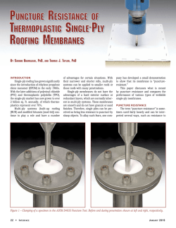
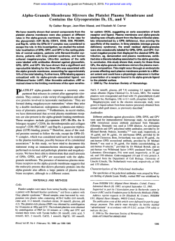
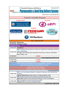
© Copyright 2026