
Increase in Cytosolic Calcium Upregulates the Synthesis of
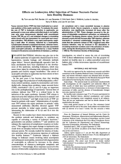
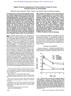
From www.bloodjournal.org by guest on February 6, 2015. For personal use only.
Increase in Cytosolic Calcium Upregulates the Synthesis of Type 1
Plasminogen Activator Inhibitor in the Human Histiocytic Cell Line U937
By Franck Peiretti, Chantal Fossat, Francine Anfosso, Marie-Christine Alessi, Mireille Henry,
Irene Juhan-Vague, and Gilles Nalbone
In the U937 histiocytic cell line, we investigated the effect
of calcium-mobilizingagents with or without tumor necrosis
factor-a (TNF) on the regulation of the synthesis of plasminogen activator inhibitor-type 1 (PAI-l). Cultured U937 cells
were stimulated with ionophore A23187 and thapsigargin
with or without TNF. The response was analyzed in terms
of cytosolic calcium mobilization, PAL1 accumulation in the
medium, and PAI-1 mRNA expression. The study was extended t o urokinase (uPA) secretion and surface expression
of its receptor (uPAR). Using Fluo-3 as a calcium-indicator
dye t o measure cytosolic calcium mobilization, we showed
by flow cytometry that both agents mobilized calcium in
a dose-dependent manner. TNF provoked a slight calcium
mobilization that was also observedby digital imaging microscopy. Association of TNF with the calcium-mobilizing
agents potentiated the calcium mobilization. Both calciummobilizing agents induced at 18 hours a dose-dependent
accumulation of PAL1 in culture medium, whereasuPA was
not affected. TNF alone induced a more marked accumulation of PAI-1than of uPA. Association of TNF with the agents
induced a PAL1 response that was more than additive of
the two, whereas the secretion of uPA was not enhanced.
Membrane expression ofuPAR, measured by flow cytometry, tended t o be slightly augmented by the calcium-mobilizing agents only. All thetreatments resulted in a significant
increase in PAI-1 mRNAlevel at 3 hours after the stimulation,
which was very marked when calcium-mobilizing agents
were present. Incubation of U937 cells in a calcium-free medium totallyprevented both themRNA expression andaccumulation of PAL1 inducedby calcium-mobilizingagents and,
to lesser extent, that induced by TNF. The increase in PAI-1
mRNA expressiondid notrequire de novo protein synthesis,
as cycloheximide did not suppress the increase in PAL1
mRNA induced by calcium-mobilizingagents. It is concluded
that, in U937 cells, calcium triggers a pathway that upregulates PAL1 synthesis andpositively interacts with theTNFinduced pathway that stimulates PAI-1 synthesis. AsuPA
and uPAR were differently affected, it is suggested that an
increase in cytosolic calcium leads to a reduced pericellular
proteolysis.
0 1996 b y The American Society of Hematology.
T
macrophages and smooth muscle cells of human atherosclerotic vessels.” Thus, it now becomes evident that an increase
in PAI-1 not only may
be related to the control of fibrin
degradation at the luminal site of the endothelium or in the
intima but also may affect local matrix proteolysis, thus
controlling the migration of proliferating vascular cells and
angiogenesis” or migratiodrecruitment of monocytes/macrophages.‘”14
Many of the pathophysiologic factors that are released or
are present in the atherosclerotic vessels were demonstrated
in vitro to significantly modify PAI-1 expression of cultured
vascular cells. These include atherogenic
cytokines such as tumor necrosis factor-a (TNF), and interleukin (IL)-l,I9-” and growth factor^.'^.^^ Calcium is believed to play a key role in numerous intracellular function~,’~.’~
some of which (eg, cell proliferation, apoptosis,
angiogenesis, etc) are involved in the development of atherothrombosis.
Some of the pathogenic factors mentioned above that augment PAI-1 synthesis were independently demonstrated also
to alter by different ways the fine tuning of cytosolic calcium
concentration. This is the case with oxidized low-density
lipoproteins (LDL)26and various agonists that bind to tyrosine kinase-linked receptors, such as growth factors or Gprotein-linked receptors such as thrombin.27However, the
role of calcium in the cytokine effect is not clear, and that
of tumor necrosis factor-a (TNF) is still a matter of deIn atherosclerotic vessels, an important feature of
vascular cells is that they are exposed to many signals, and
a cross-talk between the resulting intracellular signalling
pathways may occur. For example, TNF30,31
and calcium
i n f l u ~ ~both
’ . ~ ~activate the mitogen-activated protein (MAP)
kinase pathway. TNF potentiates calcium signals induced by
b r a d ~ k i n i n and
, ~ ~ the calcium ionophore ionomycin synergizes the phorbol ester-induced expression of uPA in a renal
YPE 1 PLASMINOGEN activator inhibitor (PAI-1) is a
serine protease inhibitor that rapidly inhibits the tissuetype plasminogen activator, thereby preventing activation of
plasminogen into plasmin.’ This may result in a slowing
down of fibrin degradation, both in the vasculature and at
its surface. This is of prime interest, because a higher plasma
level of PAI-1 is observed in patients with coronary heart
diseases (CHD) and those presenting with insulin resistance,
a syndrome that is associated with anincreased risk of CHD.’
As demonstrated by several independent groups, PAI-1 is
released by activated platelets and produced in vitro by cultured vascular cells such as endothelial and smooth muscle
cells.3 In vivo, a significant increase in the expression of
PAI-1 mRNA has been observed byin situ hybridization
techniques in human atherosclerotic
By inhibiting
the urokinase-type plasminogen activator (uPA) bound to its
receptor (uPAR), PAL1 is also believed to be involved in
the control of extracellular matrix degradation, which is a
critical step for tissue remodeling, cell migration, and tumor
i n v a ~ i o n . ”Interestingly,
~~
uPAR expression is augmented in
From CJF INSERM 93-12, Laboratoire d’HCmatologie, Faculte‘
de Midecine, Marseille, France.
Submitted April 3, 1995; accepted August 22, 1995.
Supported by funds from INSERM, DRED, and Conseil Giniral
des Bouches-du-RhBne. F.P.isa recipient of Minist2re de 1’Enseignement SupCrieur et de la Recherche.
Address reprint requests to Gilles Nalbone, PhD, CJFINSERM
93-12, Laboratoire d’Himatologie,FacultC de Midecine, 27 Boulevard Jean Moulin, 13385 Marseille Cedex 5, France.
The publication costsof this article were defrayedin part by page
chargepayment. This article must therefore behereby marked
“advertisement” in accordance with 18 U.S.C. section 1734 solely to
indicate this fact.
0 1996 by The American Society of Hematology.
0006-4971/96/8701-0007$3.00/0
162
Blood, Vol 87, No 1 (January l ) , 1996: pp 162-173
From www.bloodjournal.org by guest on February 6, 2015. For personal use only.
CALCIUMMOBILIZATIONAND
PAL1
SYNTHESIS
cell line.3' However, the role of calcium itself or in interaction with other agonists on PAI-1 synthesis has not been
investigated.
Enhanced expression of PAI-1 mRNA was detected in
macrophages present in the atherosclerotic vessels' and those
surrounding the lesion in an experimental model of thrombosis in the rabbit.36 Moreover, it has been demonstrated in
vitro that peripheral blood monocyte^^'^^^ and macrophages
isolated from human atheromatous plaque3' both secrete
PAI- 1. Because the human lymphoma-derived histiocytic
line U937 was shown to synthesize and secrete significant
amounts of PAL2 and PAI-1,'2.40this cell line represents an
interesting model to study the regulation of PM- 1 synthesis
in the context of the role played by monocytedmacrophages
in the development of atherothrombosis. Therefore, we investigated in U937 cells the effect of calcium-mobilizing
agents on PAI-1 and uPA synthesis and uPAR membrane
expression. The molecules studied were ionophore A23 187
and thapsigargin (TG), both of which increase cytosolic calcium in different ways. The former is well known to increase
membrane permeability to calcium and also to discharge
calcium from internal stores. The second irreversibly inhibits
the endoplasmic CaZ+-ATPasepump that drives the ion back
into the internal stores, thus creating a drastic desequilibrium
in favor of a permanent leak of calcium in the cytosolic
compartment:' The effects of ionophore A23187 and TG
were also studied in association with TNF.
Results showed that the agent-induced increase in cytosolic calcium upregulates PAL1 mRNA expression, leading
to a drastic increase in PAL1 accumulation in culture medium. TNF alone stimulated PAL1 synthesis. Association of
TNF and calcium-mobilizing agents potentiated the PAI-1
synthesis, indicating that in U937 cells, calcium positively
regulates the TNF-induced increase in PAI-1 synthesis,
while uPA and uPAR behaved differently.
MATERIALS AND METHODS
Chemicals. Fetal calf serum (FCS) and cell culture reagents were
purchased from Eurobio (Les Ullis, France). A23187, TG, and dimethyl sulfoxide (Me,SO) were purchased from Sigma Chemicals
CO (La Verpillibre, France). Human recombinant TNF-a (specific
activity, 3.8 X lo7U/mg) was from Euromedex (Souffelweyersheim,
(5 X lo7 Wmg) was from
France), and human recombinant L-l@
Boehringer Mannheim (Meylan, France). Fluo-3-acetoxymethyl ester (Nuo3-AM) and 5,5'-dimethyl BAPTA-AM were from Molecular Probes (Eugene, Oregon). Nylon positively charged membranes
and agarose were from Appligene (Illkirch, France). Monoclonal
antibodies specific for PAI-I and cDNA human probes for PAI-l
were a gift from Prof D. Collen, Prof P.J. Declerck, and Dr L. Nelles
(Center for Thrombosis and Vascular Research, Leuven, Belgium).
My0-[2-~H(N)]ionositolwas from NEN-Dupont (Paris, France). The
murine monoclonal antibody (MoAb) directed against uPAR(no.
3936) was from American Diagnostic (Ortho Diagnostic Systems,
Roissy, France). The fluorescein dichlorotriazinyl amino fluorescein
(DTAF)-conjugated F(ab')* fragment goat anti-mouse IgG was from
Immunotech (Marseille, France), and the negative irrelevant mouse
IgGl was from Dako S.A. (Trappes, France).
Cell culture. The U937 histiocytic cell line (provided by Dr
A.M. Benoliel, Marseille, France) was maintained in suspension in
Ham's F12Eagle.s minimal essential medium (MEM; 1/1 vol/vol)
supplemented with 10% heat inactivated FCS, 2 mmovL L-gluta-
163
mine, 100 U/mL penicillin, and 100 pg/mL streptomycin, under 5%
COz in a humidified atmosphere at 37°C. All the experiments, except
video imaging, were performed at a cell density ranging between 1
X 106/mLand 2 X 10⁶/mL.
Assays. Cell viability was assessed by the quantification of lactate dehydrogenase (LDH) release in culture medium. To this end,
the cell suspension was immediately centrifuged at 2,OOOg for 20
minutes at 4°C to sediment cells and cellular debris. Supernatants
collected at 4°C were immediately assayed for the LDH assay using
a routine clinical assay performed on an automatic analyzer (Hitachi
717, Boehringer Mannheim, Mannheim, Germany). Results were
expressed in international units (IU) per IO6 cells. The rest of the
supernatant was stored at -20°C. pending measurements of PAI-l
and uPA accumulation in culture medium. PAL1 and uPA antigen
assays were performed using an enzyme-linked immunosorbent
assay (ELISA) as described by Declerck et al?2,43 Results were
expressed in nanograms of PAI-l or uPA antigen (PAI-1 Ag or uPA
Ag) per lo6 cells.
Measurement of inositol phosphate production. U937 cells (2 X
106/mL)were. first incubated overnight in an inositol-poor medium
(M199) containing my~-[~H]inositol
(4 pCi/mL). Cells were washed
twice and then incubated for 15 minutes with LiCl (40 mmolfl).
They were stimulated either with TNF (100 U/mL) or IL-l (10 U/
mL) for various times, and the stimulation was stopped by addition
of 10 mmolfl ice-cold formic acid. Inositol phosphates were then
extracted and separated on Dowex anion exchange chromatography,
as described by Vigne et al."
Cell stimulation. In the experiments using calcium-mobilizing
agents, cells in 1% FCS-containing medium were stimulated with
the agent (A23187 in pure ethanol and TG in pure MezSO) added
as a single dose at the indicated final concentration and left for 30
minutes. Proper controls with an equivalent volume of ethanol or
MqSO alone were proven not to affect the responses studied. The
stimulated cell suspension was then centrifuged for 5 minutes at
300g, 22"C, and the supernatant was carefully eliminated to remove
the agents. The cellular pellet was resuspended inthe 1% FCS
medium, and the length of incubation from the addition of the agent
depended on the type of analysis performed: 18 hours for PAI-l Ag
and LDH assays in the medium, and 1,3,5, and 18 hours for mRNA
expression. In the studies involving association of agents and TNF,
the cytokine was added as a single dose (final concentration of 100
U/mL) 2 hours before the agent. After the washing procedure, TNF
was added again at the same concentration in the 1% FCS-containing
medium, and incubation continued for indicated times as described
above. The same procedure applied for TNF alone. In the experiments designed to study the effect of calcium-free medium, the 1%
FCS-containing medium was supplemented with EGTA to completely chelate the calcium without any excess of EGTA (calciumfree medium). The measurement of calcium concentration in the
culture medium using a routine clinical assay (Hitachi 717) confirmed that all the calcium was chelated. The cells were incubated
in this medium for 2 hours, then were stimulated with calciummobilizing agents for 30 minutes, and were resuspended in the calcium-free medium. From the onset of stimulation, incubation lasted
3 hours for mRNA analysis and 18 hours for LDH and PAI-I Ag
assays. In experiments using cycloheximide, the inhibitor was added
at a final concentration of 15 pmol/L at 30 minutes before and until
the end of the agent stimulation. Then, cycloheximide was continued
at 3 pmoYL for the next 2.5 hours (&A
expression) or 17.5 hours
(PAL1 Ag secretion). This protocol was selected on the basis of
a minimal cytotoxic effect under our experimental conditions of
stimulation.
Measurement of intracellular calcium mobilization byflow cytometry andfluorescence digital imaging. Intracellular calcium mobilization was measured by flow cytometry using the fluorescent probe
From www.bloodjournal.org by guest on February 6, 2015. For personal use only.
164
Fluo3-AM, which has been shown to be a reliable dye to detect
changes in cytosolic calcium." Fluo3-AM was dispersed in Me,SO
containing 20% pluronic acid. In all the experiments, cells in1%
FCS medium were incubated with 1 pmoVL Ftuo3-AM for 30 minutes, and experiments on dye-loaded cells were performed within
the next 30 to 45 minutes. Indeed, we observed a substantial decay
in basal fluorescence at 1 hour after the end of loading the cells,
which seemed to perturb the fluorescenceresponse in cells stimulated
with the calcium-mobilizing agents. This was not related to a dyeinduced cell membrane disruption, as cell viability measured by flow
cytometry after iodine propidium treatment revealed that at least
95% of the cells were viable at the time of the measurement. This
is perhaps the consequence of a spontaneous leakage of the probe
out of the cell or of an intracellular metabolization into an inactive
probe. In the experiments with a calcium-free medium, cells were
first incubated in this medium for 2 hours, loaded with Flu03-M
as above, and then stimulated with the agents before the fluorescence
was read. To assess the time-dependent persistence of the intracellular calcium-mobilizing effect of the agents after they have been
eliminated, U937 cells in 1% FCS-containing medium were stimulated for 30 minutes, the agents were removed, and the cells were
resuspended in 1% FCS-containing medium. The dye was subsequently loaded at different times after the washing procedure, and
fluorescence was directly read. Analyses were performed on an
EPICS-Profile I1 cytofluorograph (Coulter Electronics Inc. Hialeah,
K) equipped with an air-cooled, 488-nm argon-ion laser that was
run at 15 mW of power. The linearity of the linear amplifier was
performed using four different quantum-24 beads having 4,240,
17,000,38,000, and 77,900 fluorescein equivalents (Flow Cytometry
Standards Corporation, Research Triangle Park, NC) per bead. For
each measurement, 10,OOO events were collected and analyzed at
typical rates of 400 to 600 cells per second. Cells were gated from
debris and putative cell clumps on the basis of forward versus right
angle scatter. Cells excited at 488 nm were detected at 525 nm. The
green fluorescence, which represents the specific binding of cytosolic
calcium, was measured on fluorescence channel 1 (FL 1) and collected after a 525-nm band pass filter. The data were collected in
monoparameter histograms (256 channels) displaying relative log
fluorescence intensity versus cell number by calculating the median
channel number. Data acquisition on the flow cytometer was performed with the EPICS Profile software (Coulter Electronics). Normalized fluorescence is expressed as the ratio of mean fluorescence
over mean basal fluorescence. Fluorescence digital imaging microscopy was also used to analyze calcium mobilization at the singlecell level in cells stimulated with either TNF (100 UlmL) or IL-l
(10 U/mL). This was performed according to the protocol described
by Kaplanski et
Briefly, cells (500,000/mL)were loaded with
Fluo3-AM as described above and examined with an Olympus IMT2
inverted microscope (Olympus, Tokyo, Japan) equipped with a
Reichert heating stage (Reichert, Vienna, Austria). Fluorescence
measurements were performed with a Lhesa 4036 SIT video camera
(Lhesa, Cergy Pontoise, France) mounted on the microscope and
connected to a PC vision + digitizer (Imaging Technology, Wobum,
MA) set in an IBM-compatible desk computer. Changes in fluorescence were recorded every 6 seconds for a period of time up to
IO minutes. Normalized fluorescence in a single responsive cell is
expressed as the ratio of fluorescence over the basal fluorescence.
Flow cyrometry analysis of uPAR expression. Quantitative expression of uPAR on U937 cells under the various conditions of
stimulation was performed by flow cytofluometry, asdescribed."
Briefly, IO6 cells were sedimented and treated for 2 minutes with
0.5 mL of 50 mmol/L glycine-HC1 pH 3.0, 100 mmol/L NaCl; then
neutralized by addition of 0.5 mL 50 mmoVL HEPES pH 7.5, 100
mmoVL NaCl, and immediately centrifuged. The cells were washed
twice with ice-cold phosphate-buffered saline (PBS)/O.1% bovine
PElRETTl ET AL
serum albumin (BSA) containing 0.1% sodium azide and incubated
with the murine MoAb 3936 at a concentration of 2 pg/mL for 30
minutes at 4°C. After extensive washing, cells were incubated for
30 minutes at 4°C with the fluorescein DTAF-conjugated anti-IgG
(dilution, 11200). After extensive washing, the cells were fixed overnight with 2% paraformaldehyde and analyzed by flow cytometry
as described above. Nonspecific binding was assessed by replacing
MoAb 3936 with an irrelevant mouse IgGI.
Preparation of RNA, cRNA probes, and Northern blot analysis.
Total RNA of U937 cells was extracted according to the method
of Chomczynski et
Total RNA (15 pg) was fractionated by
electrophoresis on 1% gel agarose containing 20% formaldehyde.
RNA was transferred under vacuum to a positively charged nylon
membrane. The antisense cRNA probe of PAL1 was transcribed in
vitro from linearized recombinant plasmid (containing the 436-bp
fragment from nucleotide 1045 to 1481 of human PAI-l) using
digoxigenin-l l-uridine triphosphate and SP6 RNA polymerase
(Boehringer Mannheim) according to the protocol described by Melton et al.49 The digoxigenin-labeled human P-actin antisense cRNA
probe was from Boehringer Mannheim Biochemical. Hybridization
was performed with the digoxigenin-labeled antisense cRNA probes
of human PAL1 and p-actin for 16 hours at 68°C under gentle
stirring, as previously described?' Detection of the RNA-cRNA hybrid was performed using a chemiluminescent detection kit as described by the manufacturer (Boehringer Mannheim Biochemical,
no. 1363514). Briefly, after hybridization, blots were incubated with
an antidigoxigenin MoAb conjugated to alkaline phosphatase. The
reaction was initiated by addition of alkaline phosphatase substrate,
and the membrane was exposed to an autoradiography film for a
period of time ranging between 15 and 30 minutes.
RESULTS
Effect of A231 87, TG, TNF, and IL-l on cytosolic calcium
mobilization. A23187 and TG are well known to induce in
various types of cells, each via a different way, an increase
in cytosolic calcium concentration. We analyzed byflow
cytometry the characteristics of cytosolic calcium mobilization induced by these molecules. Cells were stimulated with
A23187 or TG at the indicated concentrations. Fluorescence
was read10 seconds and 60 seconds after addition of A23 187
and TG, respectively, which was determined to be the optimal time for each. With A23187, increasing the dose up
to 1 pmol/L leadto a significant increase in intensity of
fluorescence. Above 1 pmol/L, no further increase in fluorescence was observed (Fig 1A). TG also induced a dosedependent increase in fluorescence, much less markedin
intensity but occurring at a concentration 10 times lower
thanwithA23187 (Fig 1B). The dose response appeared
narrower with TG than with A23187 and reached a plateau
at approximately 0.5 pmoVL and 0.05 pmol/L with A23 187
and TG, respectively. To further confirm the calcium-mobilizing effect of the agents, we analyzed the calcium mobilization in cells preincubated in a calcium-free medium for 2
hours and subsequently stimulated. The fluorescence was
read 10 and 60 seconds after the stimulation with A23187
and TG, respectively. Compared with cells in a normal calcium concentration, we observed a drastic diminution of the
fluorescence signal when cells were incubated in the calcium-free medium and stimulated with the agents (Fig l ,
insets). The diminution of the signal attained 93% and 80%
for A23187 and TG, respectively. Replacing calcium in the
culture medium at a final concentration of 2 mmoVL for I5
From www.bloodjournal.org by guest on February 6, 2015. For personal use only.
CALCIUMMOBILIZATION AND PAI-l SYNTHESIS
165
minutesalloweddetection of a fluorescencesignalwhen
or TG, although for both
cells were stimulated with A23 187
the signals were slightly less intense than under normal conditions of stimulation (Fig 1, insets). We then investigated
the calcium mobilizationin cells stimulatedby TNF with or
without the agents. Cells were treated for
2 hours with TNF,
loaded with the dye, and then stimulated with either A23187
or TG,andthefluorescencewasread
10 seconds and 60
secondsafterthestimulation,respectively.Resultswere
comparedwithcellsstimulatedwithcalcium-mobilizing
15
2
0
t
Q
V
c
Q
V
2
0
3
-c
7
a
EV
-c
2
9
E
Q
A
11
m
m
A
T
13
T
l
Il-
5
3
1
0
t
Fig 2. Calcium mobilization in U937 cells by TNF, A23187, TG, or
agents in association with TNF, as measured by flow
cytometry. Cells
were incubated for1.5 hours in l % FCS with or withoutTNF (100U/
mL1, then incubatedwith 1 pmol/L Fluo3-AM, and stimulated ornot
with A23187 l1 pmollLI orTG (0.1 pmol/Ll. Normalizedfluorescence
values calculated as in Fig 1 are the means (+SDI of two separate
experiments, each performed in duplicate.
Q
V
c
Q
V
01
E0
zl
E
c
Q
m
to
EV
-c
I
0.0
1.o
0.5
1.5
I
'
2.0
2.5
A23187 (wM)
"I
2.0
1.2
,
1.0
0.0
0.1
,l,
1.Q
0.3
0.4
fhapsigargin (PM)
0.2
,
(
0.5
-I
0.6
Fig l.Dosedependent effect of calcium-mobilizing agentson cytosolic calcium mobilization in U937 cells as measured by flow cy.
tometry. Cells in 1% FCS medium were loaded with Fluo3-AM for
30 minutes and then stimulated with the calcium-mobilizing
agent.
Fluorescence was read before and then10 seconds and 60 seconds
after the addition A23187
of
(A) and TG (B),respeetively. Normalized
fluorescence values (ratio of the
means after stimu1ation:before stimulation) are the means (+SDI of two separate experiments, each
performed in duplicate. Insets Effect of extracellular EGTA on calcium mobilization in cells stimulated bylpmol/L A23187 (A) or 0.1
pmol/L TG (B).Control cells, solid bars; plus EGTA, open bars; reintroduction of calcium for 20 minutes, shaded bars. Values are the
means (+SDI of two separate experiments, each performed in duplicate.
agents alone or TNF alone. As shown in Fig 2, TNF alone
induced a modest increasein fluorescence within the minutes
that followed stimulation. Similar observations were made
with IL-l at 10 UlmL (not shown).A potentiation of calcium
mobilization induced by the association of TNF with either
A23187 or TG was noticed, as more than an additive effect
was observed. The weak fluorescent signal induced by TNF
maybe inherenttoflowcytometrymeasurements,which
may average a heterogenous response within a cell population. To further explore the calcium mobilization induced
by TNF, we analyzed the response aatsingle-cell level using
digitalimagingmicroscopy.Resultswereconsistentwith
flow cytometry observations, as about 40% of the cells responded to TNF within the first 10 minutes and displayed a
in Fig 3. In our experimenheterogenous response, as shown
tal protocol of stimulation designed to study the regulation
of PAL 1 synthesis, cells were stimulated for 30 minutes, the
in 1%
agentwasremoved,andincubationwascontinued
FCS (see below). Therefore, we investigated by flow cytometry how long the increase in the level of cytosolic calcium
persisted in cells after removal of the agent. As shown in
Table 1, A23 187-stimulatedcells did not sustain an elevated
cytosolic calcium level, as 30 minutes after the removal of
A23187,thefluorescencenearlyreturnedtothelevelof
control cells.In contrast, in TG-stimulated cells, the fluorescenceremainedat a constantlevelcomparablewiththat
read at the initial measurement (compare with Fig 1). This
indicates that the effect of TG on intracellular calcium,
in
contrast with that of A23 187, persisted after its removal from
the medium.
We investigated if the weak calcium mobilization induced
by cytokineswasmediated by inositolphosphate-3(IP3)
release. After labeling the cells with rny~[~H]inositol,
they
were stimulated with either TNF (100UlmL) or IL-I (10 U/
mL) for 0,2,5, 10, and 15 minutes. Any significant increase
From www.bloodjournal.org by guest on February 6, 2015. For personal use only.
166
PElRElTl ET AL
f
V
.
0.5
0
1.0
l.S
2.0
A23187 (PM)
0
30
60
120
90
Time
150
180
(S)
Fig 3. Patterns of TNF-induced calcium mobilization at a singlecall level, as measured by digital imaging microscopy. U937 cells
were incubated with 1 pmol/L Fluo3-AM for 30 minutes and stimulated with TNF (100 UlmL). Fluorercence was recorded every
6 seconds as described in Materials and Methods. Five typical curves are
shown.
in IPS above the control level was detected in these conditions (data not shown).
Effects of A23187, TG, and cytokines on PAI-I accumulation in the culture medium. Cells in 1% FCS medium were
stimulated for 30 minutes with increasing doses of A23187
or TG. As showninFig 4, both agents induceda dosedependent effect on PAI-1 accumulation, which was more
pronounced with TG than with A23187. The response was
linearly related to a dose up to 1 pmol/L and 0.1 p m o K
for A23187andTG,
respectively, andthendeclined
for
A23187 and plateaued for TG. Evaluation of cell viability,
Table 1. Measurement by Flow Cytometry of the Penistence of
Calcium Mobilization in U937 Cells PreviouslyStimulated With
A23187 or TG
Time After Removal of the Calcium-Mobilizing Agent (min)
Agent
A23107
TG
30
60
90
1.3 ? 0.16
1.94 ? 0.26
1.05 2 0.11
2.55 2 0.21
1.05 2 0.10
2.18 2 0.45
Cells in 1% FCS medium were stimulated for 30 minutes with
A23187 (1 pmol/L) or TG (0.1 pmol/L), washed, and resuspended in
1% FCS medium. Cells were loaded with Fluo3-AM for 30 minutes,
and then fluorescence was measured at the indicated time. Detailed
experimental conditions of flow cytometry
analysis and data processing are described in Materials and Methods. Values (ratio of fluorescence in stimulated cel1s:fluorescence in unstimulated cells) are
indicated as fold-increases in fluorescence and are the means ( S D )
of two separate experiments, each performed in duplicate.
00.2
0.1
0.3
Thepsigargin
0.4
0.5
0.6
(PM)
Fig 4. Dose-dapendent affectof cakium-mobilizing agents on PAI1 Ag accumulation in medium from U937 cells. Celtsin 1% FCS medium were stimulated for 30 minutes with A23187 (A) or TG (B).
Then, cells were washed and resuspended in 1%FCS medium as
indicated in Materials and Methods. Media ware collected 18hours
after the addition of the agents. Unstimulated control calls were
similarly handled. Valuea are
the means SD of three separate experiments, each performed in triplicate.
*
performed by thetrypanblue dye exclusion test,didnot
reveal significantly highercell injury, except for at the highest dose of A23187. This is confirmed by the measurement
of LDH in culture medium, which showed for this dose an
increased release in the culture medium (X4) that represents
about 30% of the total intracellular LDH that can be released
(data not shown). Therefore, the decrease in PAI-1 accumulation at 2 pmoVL A23 187 could be related to a cytotoxic
effect of the molecule at this concentration. Combining the
results of the dose-dependent calcium mobilization (Fig 1)
and PAI- 1 accumulation (Fig
4) enabled us to set the experimental concentrations of A23187 and TG in all subsequent
studies at 1 pmoYL and 0.1 pmoVL, respectively. The doses
of 100 U/mL of TNF and 10 U/mL of IL- 1optimally induced
over control cells about 22 times and five times more secreted PAL1 Ag, respectively. TNF at 100 U/mL was, thus,
selected inthesubsequent studies. When TNF-stimulated
From www.bloodjournal.org by guest on February 6, 2015. For personal use only.
CALCIUMMOBILIZATIONAND
PAL1 SYNTHESIS
Control
1
167
TNF
A23187
2
3
TG
TNFtA23187
4
5
TNFtTG
6
Fig 5. Effect of TNF, A23187, TG, or agents in association with TNF on PAI-1 synthesis. (A) Effect on PAL1 accumulation. Stimulation with
A23187 alone (1pmollL1 or TG alone (0.1 pmol/L) was performed as in Fig 4. In the experiments with TNF alone, TNF was added as a single
dose 1100 UlmL) and left for 2.5 hours. Cells were then washed, and TNF (100 U/mL) was added again as a single dose. In the experiments
associating TNF and agent, the agent was added 2 hours after initial addition ofTNF and left for 30 minutes, and the experiment continued
as indicated above. Media were recovered 18 hours after agent addition (ie, 20 hours after initial TNF addition). There was no significant
difference in the accumulationof PAL1 whether the agent was added at the same time as TNF (not shown) or 2 hours after. Values are the
means t SD of t w o separate experiments, each performed in triplicate. (B) Effect on PAL1 mRNA expression. RNA was extracted at a fixed
time (3 hours after agent addition). Additions are, from left t o right, in the same order as in panel A. Scanning densitometry of the PAL1
mRNA signal relativeto mRNA p-actin signal shows the following values from leftt o right: 0, 0.28, 1.8, 2.6,2.2, and 2.8. This Northern blot is
representative oft w o separate experiments.
cells were incubated for 30 minutes with A23187 ( I pmol/
L) or TG (0.I pmol/L), the accumulation of PAI- 1 was more
than additive of the two separately (Fig S). In all the situations, the protein synthesis inhibitor cycloheximide drastically abolished the increase in PAL I accumulation, indicating that neither agents nor TNF induced the release of a
preformed pool of PAI-I. To confirm the role of calcium
in PAI-I release, cells were preincubated in a calcium-free
medium 2 hours before the stimulation and throughout the
experiment. Results were compared with those treated under
the same conditions at normal calcium concentration. Results
are listed in Table 2. Incubation in the calcium-free medium
almost totally preventedthe A23187- or TG-induced increase in PAI-l. Incubation in a calcium-free medium was
not cytotoxic as judged by LDH release. However, those
stimulated with A23 187 in the calcium-free medium released
slightly more LDH (+40%). Deprivation of extracellular calcium reduced by about 47% the TNF-induced increase in
the release of PAI-I. Insets in Fig I show that even after a
preincubation with EGTA, calcium is still mobilizable by
agents, although weakly. This minor pool may account for
the remaining 53% of PAI-I in EGTA-treated cells stimulated with TNF. Therefore, we used the intracellular calcium
chelator BAPTA-AM at a concentration of 1 pmol/L, which
was shown not to be
cytotoxic at this concentration. Cells
were preincubated with EGTA for 2 hours and then incubatedwithBAPTA-AM for 30 minutes.Wefirstchecked
byflow cytometry that the stimulation of these calciumchelated cells by A23 187 did notinduce any greater increase
in fluorescence, attesting for the complete absence of mobiTable 2. Effect of EGTA and BAPTA-AM on PAL1 Accumulation in
Culture Medium From U937 Cells
Conditions
Normal calcium
EGTA
BAPTA-AM
EGTA + BAPTA-AMC
A23187
TG
TNF
I1 umollLl (0.1 umolR) 1100 UIrnLl Unstimulated
100
1.2
ND
ND
100
100
302.8
ND
ND
100
53.1
184.8
61.9
350
ND
Cells were preincubated for 2 hours with EGTA before stimulation
and left throughout the experiment. BAPTA-AM was added 30 minutes before stimulation and left throughout the experiment. Values,
expressed as percent of the PAL1 Ag release observed with normal
calcium concentration (standard calcium), are the means of two separate experiments, each performed in triplicate. For each treatment,
the 100% ranged in the values shown in Fig 5.
Abbreviation: ND, not determined.
The so-called "calcium-chelated" cells.
From www.bloodjournal.org by guest on February 6, 2015. For personal use only.
PElRElTl ET AL
168
Table 3. Effect of Various Treatments on uPA Accumulation in the
Medium and uPAR Membrane Expression in U937 Cells
uPAR (specific mean fluorescence)
uPA
(ng11O6cells).
18 h
Treatment
Control
TNF
A23187
TG
TNF + A23187
TNF + TG
1.37
4.47
1.10
0.90
2.86
3.39
ir 0.17
10 h
5h
18 h
0.45 ir 0.04 0.63 ir 0.07
0.60 t 0.2
2 0.89 0.44 ir 0.04 0.42 2 0.07
0.42
ir 0.39
2 0.83
ir 1.34
%
0.47
0.41
0.39
0.39
ir 0.05 0.89
ir 0.11 0.64
t 0.05 0.86
t 0.10 0.60
t
ir
t
ir
0.60
0.21 0.99
0.15 0.73
0.17 0.80
0.014 0.91
ir 0.25
2 0.32
ir 0.25
ir 0.22
ir 0.51
U937 cells were treated as described in Fig
5. Details of assay conditions are described in Materials and Methods. Fluorescencevalues
are expressed as the mean of fluorescence and were corrected for
fluorescence measuredwith irrelevant IgG as a negative control. Values are the means -c SD oftwo separate experiments,each performed
in duplicate.
lizable calcium. These calcium-chelated cells were stimulated by TNF for 18 hours. Table 2 shows that TNF-induced
PAI-I release from calcium-chelated cells was similar to
EGTA-treated cells. This indicates that the remaining intracellular calcium after EGTA preincubation is not responsible
for the TNF-induced PAI-I secretion from EGTA-treated
cells. Curiously, TNF-treated cells incubated with BAPTAAM released about twice more PAI-I than cells treated with
TNF alone. In unstimulated control cells, deprivation of extracellular calcium reduced the basal accumulation of PAI1 by 70%, whereas BAPTA-AM increased the basal accumulation by about a factor of 3.
Effect of A23187, TG, and TNF on uPA accumulation in
culture medium and uPAR membrane expression. The
same experimental conditions were applied to analyze the
synthesis of uPA and its receptor (Table 3). Calcium-mobilizing agents alone didnot induce significant alteration in
uPA Ag accumulation in the culture medium, whereas TNF
produced about a threefold increase over the control cells.
Association of TNF with either A23187 or TG tended to
decrease the TNF-induced increase in uPA Ag. Flow cytometry analysis ofuPAR showed that its membrane surface
expression was not significantly altered by TNF at 5, 10,
and18hours.
However, calcium-mobilizing agents either
alone or in association with TNF tended to increase the
surface expression of uPAR mainly at 18 hours when com-
pared with unstimulated cells. Although statistically not significant, the effects appeared slightly moremarkedat
10
hours and 18 hours with A23 187( + 4 1 8 and +65%, respectively) thanwith TG ( + 2 1 8 at 18 hours). As a positive
control of uPAR surface expression, U937 cells were treated
twice with 5 pmol/L of phorbol myristate acetate (PMA) at
24-hour intervals. Approximately a 10-fold increasedsurface
expression of uPAR was detected in PMA-treated cells over
control cells (data not shown).
Effect ?f A23I87, TG, and TNF on PAI-I mRNA level.
To see if the augmentation of PAI-I release in medium
induced by calcium-mobilizing agents waslinked to its
mRNA expression, we analyzed by Northern blot the PAI1 mRNA expression at different times after stimulation with
calcium-mobilizing agents. As shown in Fig 6, PAI- I mRNA
(3.2 and 2.3 kb transcripts) expression was strongly induced
3 hours after the onset of agent stimulation and then declined
at 5 hours both for A23 187 andTG. At I8 hours, expression
almost disappeared with A23187, whereas the signal remained quite stable with TG. TNF alone or in association
with the agents was then studied. TNF alone ( 1 0 0 U/mL;
Fig 7A and B) induced a slight PAI-I mRNA expression 3
hours after the onset of stimulation. The signal remained the
same at 5 hours and disappeared at 7 hours. When calciummobilizing agents were added in association with TNF (Fig
7A and B), the profile of the time-dependent PAI-I mRNA
expression was comparable with that observed when the
agents were added alone (compare with Fig 6); ie, with TG,
the expression of PAI-I mRNAremained quite constant
from 7 hours up to 18 hours, whereas with A23187, it declined. To see the effect of the association of TNF with
calcium-mobilizing agents on PAI-I mRNA, we compared
its expression at a fixed time (3 hours after the addition of
the agent, ie, 5 hours after initial addition of TNF) under
different conditions: control, 1% FCS; TNF (100 U/mL);
A23 187 (1 pmol/L); TG (0.1 pmol/L); TNF + A23 187; and
TNF + TG. As shown in Fig 5, the effects of TNF associated
either with A23187 or TG appear additive of the two. In all
situations, @-actin takenas a control showed slight modifications that were unrelatedto the drastic modifications obtained
with PAI-I mRNA. To examine if the observed increase in
PAI-I mRNA required a de novo protein synthesis,cells were
treated with cycloheximide. Treatment with the protein synthesis inhibitor did not suppress agent-induced PAI-I mRNA
A
R
J-
PAI-1
p-ACTIN
[
1
2
3
4
1
2
3
4
Fig 6. Time-dependent effect of calcium-mobilizingagents on PAL1 mRNA expression in U937 cells. Cells were stimulated as indicated in
Fig 4, and RNA was extracted at l hour (lane l),
3 hours (lane 21, 5 hours (lane 3).and 18 hours (lane 4) after the onset of the stimulation by
A23187 (A) or TG (B). This Northern blot is representative of three separate experiments.
From www.bloodjournal.org by guest on February 6, 2015. For personal use only.
CALCIUM MOBILIZATION AND PAI-1 SYNTHESIS
169
A
I
i
PAL1
~~
Fig 7. Time-dependent effect
of TNF alone or in association
with either A23187 or TG on PAI1 mRNA expression in U937
cells. The protocol of stimulation
is the same as in Fig 5. CO and
C20, control cells in 1% FCS at
times 0 and 20 hours, respectively. Cells were
stimulated
with TNF alone (-1 or in association with agents (+) A23187 (A)
and TG (B).Total RNA was extracted 3,5,7.and 20 hours after
the initial TNF addition (ie, 1, 3,
5, and 18 hours when considering the addition of the agent).
These Northern blots are representative oftwo separate experiments.
CO
~
c20
,-
+l
+I
j-
L
+I
1-
I
B
[
13.2 kb
~
CO
c20
1-
+I
j
-
+I
c
expression, which was even higher for the 3.2-kb form (Fig
8). PAI-I mRNA expression of cycloheximide-treated cells
was identical to that of control cells (data not shown). The
effect of calcium-free medium on PAI-I mRNA expression
in cells stimulated with A23 187
or TG was investigated. Interestingly, the A23187- or TG-induced increase in mRNA normally observed 3 hours after the beginning of stimulation in
standard calcium concentrationwas now completely abolished
by deprivation of extracellular calcium (Fig 8).
DISCUSSION
We investigated the role of calcium-mobilizing agents (ionophore A23187 and TG) alone or in association with TNF
in the regulation of PAI-I synthesis and extended the study
to include uPA and uPAR. Both calcium-mobilizing agents
induced a drastic increase in PAI-I accumulation in the cul-
2
3
+,
I 2.3 kb
-
,t
t 7 t 5 t 3
1
+I
t 20
t 7 t 5 t 3
PAI-1
-
3.2 kb
2.3 kb
+I
20
ture medium. Three arguments are in favor of a calciummediated effect. (1) The two structurally unrelated molecules
dramatically stimulated PAI-I accumulation, thus ruling out
a possible side effect unrelated to calcium. (2) A comparable
shape of the dose-dependence curves of PAI-I release and
calcium mobilization was observed with either A23187 or
TG. (3) The complete chelation of extracellular calcium drastically prevented the agent-induced increase in PAI-Irelease. This is reflected by flow cytometry analysis, revealing
that in cells cultured for 2 hours in a calcium-free medium,
calcium mobilization was strongly reduced. Therefore,
depletion of theextracellular calcium 2 hours beforestimulation practically emptied the agent-sensitive intracellular
stores by a progressive efflux of calcium from these stores
to the outside of the cell. This is in agreement with studies
clearly showing that a calcium-free mediumresulted in a
significant loss of calcium from intracellular
How-
4
5
6
Fig 8. Effect of calcium-free medium and cycloheximide onPAL1 mRNA expression in U937 cells stimulated with eithercalcium mobilizing
agent. Cells in 1% FCS were stimulated with A23187 (A) or TG (B)for 30 minutes, and total RNA was extracted 3 hours after the additionof
the agent. Experimental conditions with calcium-free medium were thesame as in Table 1, except the experiment lasted3 hours. Protocol
for cycloheximide is detailed
in Materials and Methods. A23187 alone, lane 1; A23187 + cycloheximide, lane 2;A23187 in calcium-free medium,
lane 3; TG alone, lane 4; TG + cycloheximide, lane 5; TG in calcium-free medium, lane 6. This Northern blot isrepresentative of two separate
experiments.
From www.bloodjournal.org by guest on February 6, 2015. For personal use only.
170
ever, chelating extracellular calcium did not irreversibly affect the ability of cells to maintain calcium homeostasis.
The upregulation of PAL 1 accumulation induced by elevated
levels of cytosolic calcium is not due to the secretion of a
preformed pool of PAI-1. First, cycloheximide suppressed
the agent-induced secretion of PAI-l. Secondly, the PAL1
mRNA expression was drastically increased after stimulation
by either A23 187 or TG. The effect of TG on PAI-1 mRNA
expression persisted for a longer time than that of A23187.
This may be the result of the irreversible inactivation of the
Ca2’-ATPase pump by TG?’ which prolonged the increase
in cytosolic calcium for a longer period than with A23187.
Indeed, we showed that the calcium-mobilizing effect of TG
remained at its initial value 90 minutes after the agent had
been eliminated, whereas it disappeared rapidly
with
A23187. Whether this higher level of mRNA is due to an
enhanced transcription rate of the gene andor a higher stabilization of PAL1 mRNA has not been investigated here.
Because the basal level of PAI-1 mRNA in quiescent cells
is undetectable, it is likely that the markedly elevated level
of PAI-l mRNA after stimulation mainly results from an
enhanced transcription process. However, a stabilizing effect
on PAI-l mRNA cannot be ruled out, as it has been demonstrated with A23187 on IL-3 ~ I R N A . ’A~ calcium-free medium totally prevented the increase inmRNA expression
after stimulation by the calcium-mobilizing agent, which
again reinforces the role of calcium as an important intracellular messenger involved in the regulation of PAI-I gene
expression. The calcium effect does not require a de novo
protein synthesis, because treatment of cells with cycloheximidedidnot
suppress A23187- and TG-induced PAI-I
mRNA expression. A slight overincrease in the 3.2-kb transcript expression was observed, a phenomenon explained by
a stabilizing effect of cycloheximide on this mRNA.s4.55
The interaction of calcium-mobilizing agents with TNF is
of interest. TNF upregulates PAL1 production and mRNA
expression in U937 cells, which has already been reported
in various tissues” and other types of cells.57The role of
calcium mobilization in signal transduction by TNF is still
controver~ial.*~*~~
Some of the discrepancies reported can be
explained by the calcium-buffering capacities of the fluorescent probe used” and/or by the different sensitivity of cells
to TNF. Using Fluo3, we found in U937 cells that TNF
alone weakly mobilized calcium. In other types of cells, the
use of F ~ r a 2 ’ did
~ , ~not allow detection of any significant
calcium increase after stimulation by TNF even at very high
doses:’ whereas in human neutrophils stimulated with TNF
at 1,000 U/mL, calcium oscillations were observed with
Flu03 in some of the responsive cells.62The modest increase
we observed with TNF did not appear to be the result of a
calcium release from IP3-sensitive stores, as we could not
measureany significant IP production. This is consistent
with previous results6’ and with our digital imaging microscopy observations that showed a heterogeneous slow calcium
increase in about 40% of cells. IP3 is known to be rapidly
produced and toinduce within minutes a transient significant
increase in calcium. However, the possibility that a secondary, very late, and low sustained increase in IP3 occurs cannot be ruled out.@It is evident from our results that calcium
PEIRElTI ET AL
is not indispensable but does contribute to the TNF-induced
pathway of PAI-1 synthesis. In support of a contributive
role, calcium-mobilizing agents and EGTA have an opposite
effect on PAI-1 Ag production triggered by TNF. On the
other hand, calcium is not indispensable for TNF action.
because in experiments in which bothextracellular and intracellular calcium have been buffered, TNF still induced about
60% of PAI-1 release. The individual opposite effects of
BAPTA-AM and EGTA on PAL1 synthesis in TNF-stimulated cells deserve some comments. By preventing the refilling of intracellular stores, BAPTA-AM may secondarily induce a calcium influx, in a comparable manner to the effect
of TG.6’-hs Inthe presence of TNF and EGTA, BAPTA-AM
did not further lower the reduced PAI-1 response provoked
by EGTA, suggesting that chelation of extracellular calcium
was a determinant to reduce the TNF action on PAL I synthesis. Collectively, these data suggest that calcium influx plays
an important role in the upregulation of PAL1 synthesis
triggered by TNF. Recently, it was found in adherent human
fibroblasts that extracellular calcium predominates in IL- I stimulated calcium
Thus, we can hypothesize that a
calcium influx induced by either TNF or calcium-mobilizing
agents primes at a certain step a common pathway thatregulates PAI-1 expression. The tyrosine kinase pathwayis a
possible candidate. Recently, it has been shown in human
endothelial cells that TNF-induced increase in PAI-1 gene
transcription follows a genistein-sensitive pathway involving
a tyrosine kinase phosphorylation ~ t e p . ~Interestingly,
”
in
human lymphocytes, the influx of calcium controlled by the
filling state of intracellular calcium pools is regulated by the
tyrosine kinase activity.” Also, TNF”,” and calcium
activate the MAP kinase pathway, and TNF potentiates calcium signals induced by brad~kinin.’~
The potentiating effect
of agents on calcium mobilization and PAI-l synthesis in
TNF-stimulated cells could not be observed at the level of
PAL1 mRNA. The stability and export of mRNA and the
translation efficiency are intermediate control mechanisms
that may be significantly altered by the different treatments,
thus preventing establishment of a rigorous relationship between mRNA expression and its corresponding protein released in the culture medium. Therefore, whether this effect
acts at a transcriptional, posttranscriptional, or translational
level cannot be determined on the basis of the present data.
The triad PAI-l/uPA/uPAR is believed to regulate pericellular proteolysis, which conditions cell rnigrati~n.~~’’
We,
therefore, investigated uPA and uPAR expression. We found
that uPAR membrane expression was not significantly altered by TNF and was weakly altered by calcium-mobilizing
agents 18 hours after the stimulation. However, uPAR surface expression may be transient. First, uPAR can be internalized by the low-density lipoprotein receptor-related protein (LRP) together with uPA/PAI-I and recycled back to
the membrane.72Second, uPAR can bereleased from the
membrane into the medium.’’ Thus, we investigated the possibility that we missed a more marked transient uPAR expression before 18 hours.This does not appear to be the case,
because no significantly higher surface expression could be
observed over control cells at 5 hours and 10 hours after the
Stimulation. Thus, it is reasonable to assume that within the
From www.bloodjournal.org by guest on February 6, 2015. For personal use only.
CALCIUM MOBILIZATION AND PAL1 SYNTHESIS
duration of the experiment, only calcium-mobilizing agents
tended to induce a weak uPAR surface expression. Under our
experimental conditions (ie, 18 hours after the stimulation),
A23187 or TG alone did not increase uPA accumulation in
the medium, which is in accordance with previous data.35
TNF increased uPA release as already
although
to a much lesser extent than PAI-1. Interestingly, calciummobilizing agents did not potentiate TNF-induced uPA release, indicating that the upregulation exerted by the calcium
agents either themselves or in interaction with TNF appears
specific for PAI-1 within the proteins regulating the surface
proteolysis. It may be hypothesized that in monocytes the
calcium-mediated signal that specifically upregulates PAI- 1
production shifts the pericellular proteolytic potential toward
hypoproteolysis or hypofibrinolysis.
In conclusion, our results demonstrate the role of calcium
as being an intracellular messenger able to upregulate the
expression of PAI-l in U937 cells and also to interact with
TNF by potentiating its effect. This process may be of prime
importance inthe migration andrecruitment of activated
monocytes/macrophages during atherosclerosis, as recently
sugge~ted,'~.'~
orin the focal accumulation of PAI-1 in a
lesion contributing to fibrin deposition.
ACKNOWLEDGMENT
We are indebted to R. Lijnen (Louvain, Belgium) for uPA assays,
A.M. Benoliel (INSERM, Marseille, France) for digital imaging
microscopy, P. Vigne (CNRS, Sophia-Antipolis, France) for the IP3
assay, and Dr H. Lu (INSERM, Paris, France) for his suggestions.
We are grateful to N. Fernandez for excellent technical assistance
(flow cytometry); J. Ansaldi and M. Luchi (PAI-I Ag assay); the
Laboratoire de Biochimie, CHU-Timone (LDH assay); and S. Debroas and A. Olivi for technical help.
REFERENCES
1. Hekman CM, Loskutoff DJ: Kinetic analysis of the interactions
between plasminogen activator inhibitor 1 and both urokinase and
tissue plasminogen activator. Arch Biochem Biophys 262:199, 1988
2. Juhan-Vague I, Alessi MC: Plasminogen activator inhibitor 1
and atherothrombosis. Thromb Haemost 70:138, 1993
3. Andreasen PA, Georg B, Lund LR, Riccio A, Stacey SN:
Plasminogen activator inhibitors: Hormonally regulated serpins. Mol
Cell Endocrinol 68:1, 1990
4. Schneidermann J, Sawdey MS, Keeton MR, Bordin GM, Bernstein EF, Dilley RB, Loskutoff DJ: Increased type 1 plasminogen
activator inhibitor gene expression in atherosclerotic human arteries.
Proc Natl Acad Sci USA 89:6998, 1992
5. Lupu F, Bergonzelli GE, Hein DA, Cousin E, Genton CY,
Bachman F, Kruithof EKO: Localization and production of plasminogen activator inhibitor- 1 in human healthy and atherosclerotic arteries. Arterioscler Thromb 13:1090, 1993
6. Chomiki N, Henry M, Alessi MC, Anfosso F, Juhan-Vague I:
Plasminogen activator inhibitor-] expression in human liver and
healthy or atherosclerotic vessel walls. Thromb Haemost 72:44, 1994
7. Grimaldi G, Di Fiore P, Locatelli EK, Falco J, Blasi F Modulation of urokinase plasminogen activator gene expression during the
transition from quiescent to proliferative state in normal mouse cells.
EMBO J 5:855, 1986
8. Pepper MS, Sappino AP, Montesano R, Orci L, Vassali JD:
Plasminogen activator inhibitor-l is induced in migrating endothelial
cells. J Cell Physiol 153:129, 1992
9. Schneiderman J, Sawdey M, Craig H, Thinnes T, Bordin G,
171
Loskutoff DJ: Type 1 plasminogen activator inhibitor gene expression following partial hepatectomy. Am J Pathol 143:753, 1993
10. Thornton AJ, Bruzdzinski CJ, Raper SE, Gelehrter TD: Plasminogen activator inhibitor-] is an immediate early response gene
in regenerating rat liver. Cancer Res 54:1337, 1994
11. Noda-Heiny H, Daugherty A, Sobel BE: Augmented urokinase receptor expression in atheroma. Arterioscler Thromb Vasc
Biol 15:37, 1995
12. van Hinsbergh VWM, Kooistra T, Emeis JJ, Koolwijk P
Regulation of plasminogen activator production by endothelial cells:
Role in fibrinolysis and local proteolysis. Int J Radiat Biol 60:261,
1991
13. Lundgren CH, Sawa H, Sobel BE, Fujii S: Modulation of
expression of monocyte/macrophage plasminogen activator activity
and its implications for attenuation of vasculopathy. Circulation
901927, 1994
14. Gyetko MR, Todd RF, Wilkinson CC, Sitrin RC: The urokinase receptor is required for human monocyte chemotaxis in vivo.
J Clin Invest 93:1380, 1994
15. Stiko-Rahm A, Wiman B, Hamsten A, Nilsson J: Secretion
of plasminogen activator inhibitor- 1 from cultured human veinendothelial cells is induced by very low density lipoproteins. Arteriosclerosis 10:1067, 1990
16. Latron Y, Chautan M, Anfosso F, Alessi MC, Nalbone G,
Lafont H, Juhan-Vague I: Stimulating effect of oxidized low density
lipoproteins on plasminogen activator inhibitor 1 synthesis by endothelial cells. Arterioscler Thromb 11:1821, 1991
17. Mussoni L, Manucci L, Sirtori M, Camera M, Maderna P,
Sironi L, Tremoli E: Hypertriglyceridemia and regulation of fibrinolytic activity. Arterioscler Thromb 12:19, 1992
18. Kugiyama K, Sakamoto T, Misumi I, Sugiyama S, Ohgushi
M, Ogawa H, Horiguchi M, Yasue H: Transferable lipids in oxidized
low density lipoproteins stimulate plasminogen activator inhibitor1 and inhibit tissue-type plasminogen activator release from endothelial cells. Circ Res 73:335, 1993
19. Sawdey M, Podor TJ, Loskutoff DJ: Regulation of type 1
plasminogen activator inhibitor gene expression in cultured bovine
aortic endothelial cells. Induction by transforming growth factoralpha. J Biol Chem 264:10396, 1989
20. van Hinsbergh VWM, Kooistra T, van den Berg EA, Princen
HMG, Fiers W, Emeis JJ: Tumor necrosis factor increases the production of plasminogen activator inhibitor in human endothelial cells
in vitro and in rats in vivo. Blood 72:1467, 1988
21. van den Berg EA, Springers ED, Jaye M, Burgess W, Maciag
T, van Hinsbergh VWM: Regulation of plasminogen activator inhibitor-l mRNA in human endothelial cells. Thromb Haemost 60:63,
1988
22. Emeis JJ, Kooistra T: Interleukin 1 and lipopolysaccharide
induce an inhibitor of tissue-type plasminogen activator in vivo and
in cultured endothelial cells. J Exp Med 163:1260, 1986
23. Pepper MS, Belin D, Montesano R, Orci L, Vassalli JD:
Transforming growth factor PI modulates basic fibroblast growth
factor-induced proteolytic and angiogenic properties of endothelial
cells in vitro. J Cell Biol l 1 1:743, 1990
24. Scharff 0, Foder B: Regulation of cytosolic calcium in blood
cells. Physiol Rev 73:547, 1993
25. Clapham DE: Calcium signaling. Cell 80:259, 1995
26. Nkgre-Salvayre A, Fitoussi G, Reaud G, Pierraggi M, Thiers
JC, Salvayre R: A delayed and sustained rise of cytosolic calcium
is elicited by oxidized LDL in cultured bovine endothelial cells.
FEBS Lett 299:60, 1992
27. Bemdge MJ: Inositol triphosphate and calcium signalling.
Nature 361:315, 1993
28. Camussi G, Albano E, Tetta C, Bussolino F: The molecular
action of tumor necrosis factor-alpha. Eur J Biochem 202:3, 1991
From www.bloodjournal.org by guest on February 6, 2015. For personal use only.
172
29. Beyaert R, Fiers W: Molecular mechanisms of tumor necrosis
factor-induced cytotoxicity. What we do understand and what we
do not. FEBS Lett 340:9, 1994
30. Winston BW, Lange-Carter C, Gardner AM, Johnson GL,
Riches DWH: Tumor necrosis factor a rapidly inactivates the mitogen-activated protein kinase (MAPK) cascade in a MAPK kinase
kinase-dependent, c-Raf-l -independent fashion in mouse macrophages. Proc Natl Acad Sci USA 92:1614, 1995
Lee JK, L u n g CC, Raffin TA: TNF-a induces
31.RafieeP,
tyrosine phosphorylation of mitogen-activated protein kinase in adherent human neutrophils. J Immunol 154:4785, 1995
32. Franklin RA, Tordai A, Mazer B, Terada N, Lucas J, Gelfand
EW: Activation of MA=-kinase in B lymphocytes by calcium ionophores. J Immunol 153:4890, 1994
33. Rosen LB, Ginty DD, Weber MJ, Greenberg ME: Membrane
depolarization and calcium influx stimulate MEK and MAP kinase
via activation of Ras. Neuron 12:1207, 1994
34. Amrani Y, Martinet N, Bronner C: Potentiation by tumor
necrosis factor-a of calcium signals induced by bradykinin and carbachol in humantracheal smooth muscle cells. Br J Pharmacol 1 14:4,
1995
35. Naganamine Y, Ziegler A, Kiefer B: Modulation of uPA gene
expression by novel signalling pathways. Fibrinolysis 6:121, 1992
(SUPPI 4)
36. Sawa H, Fuji S, Sobel BE: Augmented arterial wall expression of type-l plasminogen activator inhibitor induced by thrombosis. Arterioscler Thromb 12:1507, 1992
37. Castellote JC, Grau E, Linde MA, Pujol-Moix N, Rutllan ML:
Detection of both type 1 and type 2 plasminogen activator inhibitors
in human monocytes. Thromb Haemost 63:67, 1990
38. Ritchie HM, Jamieson A, Booth NA: Stimulation of monocytes with thrombin causes increased plasminogen activator inhibitor
2 (PAI-2) synthesis. Fibrinolysis 8:119, 1994 (abstr)
39. Tipping PG, Davenport P, Gallicchio M, Filonzi EL, Apostolopoulos J, Wotja J: Atheromatous plaque macrophages produce
plasminogen activator inhibitor type 1 and stimulate its production
by endothelial cells and vascular smooth muscle cells. Am J Pathol
143:875, 1993
40. Wohlwend A, Belin D, Vassalli JD: Plasminogen activatorspecific inhibitors produced by human monocyteshnacrophages. J
Exp Med 165:320, 1987
41. Thastrup 0,Cullen PJ, Drobak BK, Hanley MR, Dawson AP:
Thapsigargin, a tumor promoter, discharges intracellular Ca2+ stores
by specific inhibition of the endoplasmic reticulum Ca2f-ATPase.
Proc Natl Acad Sci USA 87:2466, 1990
42. Declerck PJ, Alessi MC, Verstreken M, Kruithof EKO, JuhanVague I, Collen D: Measurement of plasminogen activator inhibitor
1 (PAI-l) in biological fluids with a murine monoclonal antibody,
based on enzyme-linked immunoadsorbant assay. Blood 71:220,
1988
43. Declerck PJ, Van Keer L, Verstreken M, Collen D: An enzyme-linked immunoabsorbent assay for urokinase-type plasminogen activator (U-PA)and mutants and chimeras containing the serine
protease domain of U-PA. Thromb Haemost 67:95, 1992
44. Vigne P, Marsault R, Breittmayer JP, Frelin C, Lazdsunski
M: Endothelin stimulates phosphatidylinositol hydrolysis and DNA
synthesis in brain capillary endothelial cells. Biochem J 266:415,
1990
45. Minta A, Kao JPY, Tsien RY: Fluorescent indicators for cytosolic calcium based on rhodamin and fluorescein. J Biol Chem
264:8171, 1989
46. Kaplanski G, Famarier C, Benoliel AM, Foa C, Kaplanski S,
Bongrand P: A novel role for E- and P-selectins: Shape control of
endothelial cell monolayers. J Cell Sci 107:2449, 1994
47. Wilhelm 0, Weidle U, Hohl S, Rettenberger P, Schmitt M,
PElRETTl ET AL
Graeff H: Recombinant soluble urokinase receptor as a scavenger
for urokinase-type plasminogen activator (uPA). FEBS Lett 337:131,
1994
48. Chomczynski P, Sacchi N: Single step method of RNA isolation by acid guanidium-thiocyanate-phenol-chloroformextraction.
Anal Biochem 162:156, 1987
49. Melton DA, Krieg PA, Rebagliati MR, Maniatis T, Zinn K,
Green MR: Efficient in vitro synthesis of biologically active RNA
and RNA hybridization probes from plasmids containing a bacteriophage SP6 promoter. Nucleic Acids Res 12:7035, 1984
50. Anfosso F, Alessi MC, Nalbone G, Chomiki N,HenryM,
Juhan-Vague I: Up-regulated expression of plasminogen activator
inhibitor-l in Hep G2 cells: Interrelationships between insulin and
insulin-like growth factor 1. Thromb Haemost 73:268, 1995
5 1. Chandra S, Fewtrell C, Millard PJ, Sandison DR, Webb WW,
Morisson GH: Imaging of total intracellular calcium and calcium
influxand efflux in individual resting and stimulated tumor mast
cells using ion microscopy. J Biol Chem 269: 15186, 1994
52. Hoth M, Penner R: Depletion of intracellular calcium stores
activates a calcium current in mast cells. Nature 355:353, 1992
53. Wodnar-Filipowitcz A, Moroni C: Regulation of interleukin
3 mRNA expression in mast cells occurs at the posttranscriptional
level and is mediated by calcium ions. Proc NatlAcad Sci USA
87:777, 1990
54. Heaton JH, Kathju S, Gelehrter TD: Transcriptional and posttranscriptional regulation of type 1 plasminogen activator inhibitor
and tissue-type plasminogen activator gene expression in HTC rat
hepatoma cells by glucocorticoids and cyclic nucleotides. Mol Endocrinol 653, 1992
55. Fattal PG, Schneider DJ, Sobel BE, Billadello JJ: Post-transcriptional regulation of expression of plasminogen inhibitor type
1 mRNA by insulin and insulin-like growth factor. J BiolChem
267:12412, 1992
56. Sawdey MS, Loskutoff DJ: Regulation of murine type 1 plasminogen activator inhibitor gene expression in vivo. J Clin Invest
88:1346, 1991
57. Medcalf RL, Kruithof EKO, Schleuning WD: Plasminogen
activator inhibitor 1 and 2 are tumor necrosis factorkachectin-responsive genes. J Exp Med 168:751, 1988
58. Menitt JE, McCarthy SA, Davies MPA, Moores KE: Use of
fluo-3 to measure cytosolic Ca2+ in platelets and neutrophils. Biochem J 269513, 1990
59. Richter J, Anderson T, Olsson I: Effect of tumor necrosis
factor and granulocytehacrophage colony-stimulating factor on
neutrophil degranulation. J Immunol 142:3199, 1989
60. Yamato K, El-Hajjaoui Z, Kuo JF, Koeffler P: Granulocytemacrophage colony-stimulating factor: Signals for its mRNA accumulation. Blood 74:1314, 1989
61. Schiitze S, Berkovic D, Tomsing 0, Unger C, Kronke M:
Tumor necrosis factor induces rapid production of I '2'-diacylglycerol by a phosphatidylcholine-specific phospholipase C. J Exp Med
174:975, 1991
62. Schumann MA, Gardner P, Raffin TA: Recombinant human
tumor necrosis alpha induces calcium oscillation and calcium-activated chloride current in human neutrophils. J Biol Chem 268:2134,
1993
63. Laudanna C , Miron S, Berton G, Rossi F Tumor necrosis
factor activates the 02-generating system of human neutrophils independently of the hydrolysis of phosphoinositides and the release of
arachidonic acid. Biochem Biophys Res Commun 166:308, 1990
64. Beyaert R, Heyninck K, De Valck D, Boeykens F, Van Roy
F, Fiers W: Enhancement of tumor necrosis factor cytotoxicity by
lithium chloride is associated with increased inositol phosphate accumulation. J Immunol 151:291, 1993
65. Kwan CY, Takemura H, Obie JF, Thastrup 0, Putney JW
From www.bloodjournal.org by guest on February 6, 2015. For personal use only.
CALCIUMMOBILIZATION AND PAL1 SYNTHESIS
Jr: Effect of MeCh, thapsigargin, and La3+ on plasmalemmal and
intracellular Ca2+ transport in lacrimal acinar cells. Am J Physiol
258:C1006, 1990
66. Ely JA, Ambroz C, Baukal AJ, Christensen BS, Balla T,
Catt W :Relationship between agonist- and thapsigargin-sensitive
calcium pools in adrenal glomerulosa cells. J Biol Chem 266:18635,
1991
67. Dolor W,Hunvitz LM, Mirza 2, Strauss HC, Whorton R:
Regulation of extracellular calcium entry in endothelial cells: Role
of intracellular calcium pool. Am J Physiol 262:C171, 1992
68. Robinson M,Cheek TR, Burgoyne R D : Ca2+ influx induced
by the Ca2+-ATPase inhibitors 2,5-di-(t-butyl)l,4-benzohydroquinone and thapsigargin in bovine adrenal chromaffin cells. Biochem
J 288:457, 1992
69. Arora PD, Ma J, Min W, Cruz T, McCulloch AG: Interleukinl-induced calcium flux in human fibroblasts is mediated through
focal adhesions. J Biol Chem 270:6042, 1995
70. van Hinsbergh VWM, Vermeer M, Koolwijk P, Grimbergen
173
J, Kooistra T: Genistein reduces tumor necrosis factor alpha-induced
plasminogen activator inhibitor-l transcription but not urokinase expression in human endothelial cells. Blood 84:2984, 1994
71. Tepel M, Kiihnapfel S , Theimeier G, Teupe C, Schlotmann
R, Zidek W: Filling state of intracellular Ca2+ pools triggers transplasma membrane Na+ and Ca2+ influx by a tyrosine kinase-dependent pathway. J Biol Chem 269:26239, 1994
72. Herz J, Clouthier DE, Hammer RE: LDL receptor-related
protein internalizes and degrades u-PA:PAI-l complexes and is essential for embryo implantation. Cell 71:411, 1992
73. Sitrin RG, Todd RF, Mizukami IF, Gross TJ, Shollenberger
SB, Gyetko MR: Cytokine-specific regulation of urokinase receptor
(CD87) expression by U937 mononuclear phagocytes. Blood
84:1268, 1994
74. Gyetko MR, Schollenberger S , Sitrin RG: Urokinase expression in mononuclear phagocytes: Cytokine-specific modulation by
interferon-y and tumor necrosis factor-cr. J Leukoc Biol 51:256,
1992
From www.bloodjournal.org by guest on February 6, 2015. For personal use only.
1996 87: 162-173
Increase in cytosolic calcium upregulates the synthesis of type 1
plasminogen activator inhibitor in the human histiocytic cell line
U937
F Peiretti, C Fossat, F Anfosso, MC Alessi, M Henry, I Juhan-Vague and G Nalbone
Updated information and services can be found at:
http://www.bloodjournal.org/content/87/1/162.full.html
Articles on similar topics can be found in the following Blood collections
Information about reproducing this article in parts or in its entirety may be found online at:
http://www.bloodjournal.org/site/misc/rights.xhtml#repub_requests
Information about ordering reprints may be found online at:
http://www.bloodjournal.org/site/misc/rights.xhtml#reprints
Information about subscriptions and ASH membership may be found online at:
http://www.bloodjournal.org/site/subscriptions/index.xhtml
Blood (print ISSN 0006-4971, online ISSN 1528-0020), is published weekly by the American
Society of Hematology, 2021 L St, NW, Suite 900, Washington DC 20036.
Copyright 2011 by The American Society of Hematology; all rights reserved.

© Copyright 2026