
Signal Pathway Regulation of Interleukin-8-Induced Actin
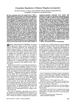
From www.bloodjournal.org by guest on February 6, 2015. For personal use only.
Signal Pathway Regulation of Interleukin-8-Induced Actin
Polymerization in Neutrophils
By Ronald L. Sham, Pradyumna D. Phatak, Trenton P. Ihne, Camille N. Abboud, and Charles H. Packman
Interleukin-8 (IL-8). a recently described peptide cytokine,
is a neutrophil chemoattractant and activator that exerts
effects similar t o fMLP, yet their receptors and their roles
in pathophysiology differ. The effect of IL-8 on the neutrophil cytoskeleton has not been well studied; therefore, we
compared and contrasted the effects of IL-8 and fMLP on
neutrophil actin conformation and on the signal pathway
regulation of actin responses. IL-8 caused a rapid, dosedependent increase in neutrophil F-actin content within 30
seconds. The maximum increase was twofold. These
changes were accompanied by the development of F-actin-rich pseudopods, as noted with fluorescence microscopy and scanning electron microscopy. Selected biochemical inhibitors were used to study the regulation of
the IL-8-induced actin changes. Incubation of neutrophils
with 2 pg/mL pertussis toxin resulted in a 67%inhibition of
the IL-8-induced F-actin increase. The protein kinase C
(PKC) inhibitors, staurosporine and H7, did not inhibit the
increase in F-actin caused by IL-8. IL-8 caused a rapid increase in neutrophil intracellular calcium that could be
completely inhibited by the chelating agent 1,2-bis(oaminophen0xy)ethane-N,N-”,”-tetraacetic
acid
(BAPTA). However, BAPTA-treated neutrophils retained
the ability to increase F-actin in response to IL-8. Similar
results were seen with fMLP, indicating that, similar to
fMLP, the IL-8-induced actin response is mediated
through pertussis-toxin-sensitive G-proteins but is neither
dependent on PKC nor increases in cytosolic calcium.
Thus, although IL-8 and fMLP exert their effects on neutrophils through different receptors, the signal transduction
pathways used and the effects on actin conformation and
pseudopod formation are similar.
0 1993 by The American Society of Hematology.
N
ing the chemoattractant effect on neutrophils to decrease
unwanted tissue damage, we investigated the regulation of
IL-8-induced actin polymerization. We found that IL-8
causes an increase in neutrophil F-actin that is dependent
on a pertussis-toxin-sensitive G-protein, is independent of
PKC, and can occur in the absence of an increase in intracellular calcium.
EUTROPHIL MIGRATION to sites of inflammation
occurs in a variety of pathologic states. The regulation of neutrophil migration involves the coordinated action of surface receptors, second messengers, and the cytoskeleton.’,’ Actin is a key component of the neutrophil
cytoskeleton, and its reversible polymerization is central to
neutrophil m i g r a t i ~ n . The
~ , ~ signal pathway regulation of
actin polymerization is an area of intense study, as is the
structure and function of many physiologic neutrophil activators. One such activator, interleukin-8 (IL-8), is a cytokine produced by a variety of cell types including monocytes, T lymphocytes, fibroblasts, and endothelial cells.’
IL-8 induces many responses in neutrophils similar to those
seen with fMLP including chemotaxis, neutrophil degranulation, and respiratory
Recently, IL-8 was shown to
increase F-actin in neutrophil^,^.'^ a finding that correlates
with the known effect of IL-8 on chemotaxis, and parallels
observations with fMLP. The signal-pathway regulation of
fMLP-induced actin responses has been studied in great detail. However, the role of calcium, protein kinase C (PKC),
and G proteins in IL-8-induced actin responses remains to
be determined. Given the proinflammatory effects of IL-8
in a variety ofclinical settings and the potential for modulatFrom the University ofRochester School of Medicine and Dentistry; the Department of Medicine, Rochester General Hospital:
and the Department of Medicine, University of Rochester Medical
Center, Rochester, NY.
Submitted March 11. 1993; accepted June 21, 1993.
Supported in part by the Department ofMedicine, Rochester General Hospital and Public Health Services Grant No. POI HL1820818.
Address reprint requests to Ronald L. Sham, MD, Hematology
Unit, Rochester General Hospital, 1425 Portland Ave, Rochester,
NY 14621.
The publication costx of this article were dejrayed in part by page
charge payment. This article must therefore be hereby marked
“advertisement” in accordance with 18 U.S.C.section I734 solely to
indicate this fact.
0 1993 by The American Society of Hematology.
0006-4971/93/8208-0003$3.00/0
2546
MATERIALS AND METHODS
Preparationofneutrophils. Neutrophils were isolated from citrated blood obtained from normal volunteers using dextran sedimentation followed by Ficoll-Hypaque centrifugation. Saline lysis was
2.0
1
i
0
2
4
6
8
10
TIME (min)
Fig 1. Effect of IL-8 on F-actin content in neutrophils. Doses of
IL-8 ranging from 1.2 nmol/L to 1 2 nmol/L (10 ng/mL to 1 0 0 ng/
mL) were applied to neutrophils and samples were taken at the
indicated times, and F-actin content was determined by flow cytometry. The relative F-actin content is plotted against time. A
dose-dependent increase was observed, with the 12 nmol/L dose
resulting in a 1.85-fold increase. There was some lowering of F-actin content within 5 to 10 minutes after the initial response, but it
did not return to the baseline. Results shown are the mean of four
experiments 2 SE.
Blood, Vol 82, No 8 (October 15). 1993: pp 2546-255 1
From www.bloodjournal.org by guest on February 6, 2015. For personal use only.
NEUTROPHIL ACTIN AND INTERLEUKIN-8
2547
Fig 2. Effect of IL-8 on F-actin distribution and morphology of neutrophils. Neutrophils were fixed before (t = 0 )and after the addition of
12 nmol/L IL-8 (t = 1, 3,and 10 minutes). The cells were stained with rhodamine phalloidin and prepared for fluorescence microscopy as
described. The photographs show that t h e cells have undergone shape change, actin redistribution, and the development of F-actin-rich
pseudopods, which occurred over time: (A), 0 minutes; (E), 1 minute; (C), 3 minutes; and (D), 10 minutes.
used to removeerythrocytes. Neutrophils were resuspended in phosphate-buffered saline (PRS)at a Concentration of S X 106/mL for
Use.
Activution of ncwtroplri1.v. FMLP (Sigma Chemical Co. St
Louis. MO). 1.2-bis(o-aminophenoxy)ethane-N.N-N',N-tetraacetic acid (RAPTA). and staurosporine (Calbiochem. San Diego. CA)
were stored in dimethyl sulfoxide (DMSO) at -2OOC. For use. they
were thawed and diluted in RPMI/O.Ir;s bovine serum albumin
(RSA) and added to I mL of cell suspension to achieve the desired
final concentration. l~S-isoquinolinylsulfonyl~3-mcthylpiperizinc (H7; Seikagaku America Corp. St Petcrsburg. FL) and IL-8
(Calbiochem. San Diego, CA) were refrigerated in distilled water
and used directly from the stock solution. Pertussis toxin (Calbiochem) was refrigerated and used directly from stock solution. For
many experiments. cells were incubated with inhibitors before the
addition ofcell activators. All incubations were performed at 37°C
as follows: H7 for IS minutes. staurosporine for 5 minutes. pertussis toxin for 180 minutes, and RAPTA for 30 minutes. All experiments were performed at room temperature. For each experiment.
aliquots of the cell suspension containing I X IO6 cclls/200 pL were
removed before and at appropriate times after the addition of activating agents and inhibitors. placed into I mL icecold 3.2% paraformaldehyde, and refrigcrated for 48 hours to permeabilize the
cells for subsequent analysis.
Flow cytomcvry. Cells were prepared for flow cytometry using
previously descrihed methods.' Cells were washed with PRS containingO.l% RSA and incubated in thedark with theF-actin-specific prohe. 7-nitrobenz-3-oxadiazole(NRD) phallacidin (0.6 pmol/
L) (Molecular Probes Inc. Eugene. OR), for 45 minutes. The cells
were rewashed and filtered through 53-pm nylon mesh. The final
volume of 0.5 mL contained about I x lo6cells.
The F-actin content of cells was measured on an EPICS Profile
flow cytometer (Coulter Corp. Hialeah. FL). A IS-mw laser was
used for fluorescence excitation at 488 nm. and green fluorescence
was measured at 525 nm as was forward-angle light scatter. The log
green fluorescence was converted to a relative linear scale and plotted against time. A total of 10.000 cells were collected for each
measurement.
F/rcorcwcnt mic*ro.scopr. A total of IO pL rhodamine phalloidin
(0.22 pmol/L Molecular Probes Inc) was added to I SO pL aliquots
of permeabilized cells and incubated in the dark for 45 minutes.
The cells were then washed with PRS/0. 1% RSA and cylospin preparations were made. The cell preparations were studied with fluorescent microscopy. and representative cells were photographed.
Scunnin,q c k t r o n micmwopy. Aliquots of fresh cell samples
taken at appropriate time points were pipetted into 1% glutaraldehyde in Sorcnsen's phosphate buffer and fixed for I hour. Cellswcre
then rinsed with Sorensen's buffer and attached to poly-L-lysinecoated mica chips. They were post-fixed in 1 % osmium tetroxide
for I hour and rinsed again. Samples were dehydrated in a graded
ethanol series and critical point dried. Cells were coated with gold
palladium and ohserved with a JEOL T330A scanning electron
microscope (JEOL USA Inc. Peabody, MA).
C'alcirtm depletion of cc1l.v and nwusrtrcmcnt of cytoplusmicjiw
culcirtm. Neutrophil intracellular calcium measurements were
made before and after cell activation and in the presence and a h
From www.bloodjournal.org by guest on February 6, 2015. For personal use only.
SHAM ET AL
2548
Fig 3. Effect of IL-8 on pseudopod formation, morphology, and membrane ruffling. Neutrophils were fixed before (t = 0) and after the
addition of 12 nmol/L IL-8 (t = 1, 3. and 10 minutes). The cells were prepared for scanning electron microscopy as described. The
micrographs show that the neutrophils developed membrane ruffles and pseudopods over time: (A), 0 minutes; (8). 1 minute; (C), 3
minutes; and (D), 10 minutes.
scncc of the calcium chelator RAPTA. Ancr cell separation. neutrophils were rcsuspcnded in RPMI/O. Irk RSA at a concentration of 1
X IO6 cells/mL. RAPTA was added to ccll suspcnsions during vortexing so that a linal Concentration of 25 pmol/L was achieved.
DMSO was added to control cells. Samples were then incubated for
30 minutes at 37°C. pclleted. and resuspcndcd in loading huffcr
containing Hanks Ralanccd Salt Solution (HRSS). I mmol/L Ca".
I mmol/l. Mg". and 15' RSA.
Neutrophils were then loaded with the calcium-sensitive fluom
cent prolx fura-2-AM (fun-2-tetraacetomcthoxy ester: Molecular
Prohes Inc) using previously described methods." A suspension of
5 x 10'cells/mL in HRSS was incubated at 37OC for 10 minutes in
a shaking water bath. The cells were diluted sixfold and incubated
for 20 minutes. This suspcnsion was further diluted fivefold. ccntrifuged at 2 5 0 , for
~ 10 minutes. and muspcndcd at 2 X 10"cclls/mL
in I IRSS containing I mmol/L calcium and I mmol/L magnesium
for thccalcium determinations. I-un-2 fluorescence was monitored
continuously with a SPEX Fluorolog photofluonmeter (SPEX Industries, Edison. N J ) using dual exitation monochromators. The
intracellular ionized free calcium Concentration (Caf') was determined as follows:
R is the ratio ofemission intensities at 505 nm on excitation at 340
and 380 nm. R,, was obtained hv inducing cell lysis with 0.1";.
Triton. which exposcd the fun-2 to 1 mmol/L external calcium.
and R,,, was determined at 7ero calcium on addition of 6.25
mmol/L EGTA to the lysed cellular suspcnsion at ptl 8.5. K is the
product. kd X (FdFs). where kd is the effcctivediswciation constant
of f u n 2 for calcium (224 nmol/L). fyo is the 380-nm excitation
signal in the absence of calcium. and I-, is the 380-nm excitation
signal in the prescncc of calcium.
RESULTS
I:'/rct of 1 1 A on F-actiti contmt and ccdl sliap>it1 ncittrophi1.r. Neutrophils treated with IL-8 showed a dosedependent increase in F-actin content which occured within 30 to
60 seconds of IL-8 addition (Fig I). The doses used ranged
from 0.12 nmol/L to I2 nmol/L ( I ng/mL to IO0 ng/mL).
The lowest dose resulted in essentially no change. and the
maximum increase was twofold. The magnitude and time
course of the responses were similar to those seen with comparable doses of ~TVILP..',~
Fluorescence microscopy using
rhodamine phalloidin staining showed actin redistribution
and the development of F-actin-rich pseudopods after IL-8
treatment (Fig 2). Scanning electron microscopy showed
pseudopod formation and membrane rutling that correlated with the fluorescence microscopy (Fig 3).
Rolc (?Sititracdliilart.alc*iiimin II,-X-indiiccvl F-actin in-
From www.bloodjournal.org by guest on February 6, 2015. For personal use only.
2549
NEUTROPHIL ACTIN AND INTERLEUKIN-8
250
Q-z
200
-
-
* FMLP (BAPTA)
-
FMLP (Buffer)
---b 11-8 (BAPTA)
11-8 (Buffer)
Buffer (BAPTA)
Buffer
0
za
I-
6
y8
150-
100-
"
0
0
2
4
6
8
TIME (min)
Fig 4. Effect of IL-8 on intracellular calcium and the effect of
BAPTA. Neutrophils were treated with IL-8, in the presence or absence of BAPTA, and intracellular calcium was determined spectrophotometrically as described. After obtaining a baselinetracing for
60 seconds, 12 nmol/L IL-8 was added (at t = 1 minute) and
caused a rapid increase in intracellular calcium, from 50 nmol/L to
170 nmol/L within 30 seconds (A).Cells pretreated with the intracellular calcium chelator BAPTA, 25 Mmol/L, had no increase in calcium after IL-8 application. Control cells and those exposed to
BAPTA alone had no change in intracellular calcium. Parallel experiments were performed with 10 nmol/L fMLP; intracellular calcium
increased to 200 nmol/L ( 0 ) and was completely blocked by
BAPTA. Results shown are the mean of five experiments k SE.
creases in neutrophils. Intracellular calcium increased
from 50 nmol/L to 170 nmol/L within 30 seconds of addition of 12 nmol/L I L 8 to resting neutrophils (Fig 4). Intracellular calcium increased from 50 nmol/L to 200 nmol/L
within 30 seconds of addition of 10 nmol/L FMLP (Fig 4).
The intracellular calcium chelator BAPTA completely
blocked the increase in intracellular calcium caused by IL-8
and FMLP (Fig 4). BAPTA-treated neutrophils from the
same sets of experiments were also used to determine the
effect of calcium depletion on IL-8- and FMLP-induced
actin polymerization. Although the cells pretreated with
BAPTA did not increase their F-actin content to the extent
of that seen in the control cells, IL-8 and FMLP still caused
1S5-fold increases in F-actin content in the absence of any
rise in intracellular calcium (Fig 5). Fluorescence microscopy was used to evaluate neutrophils from the BAPTA
experiments. IL-8 and FMLP caused F-actin-rich pseudopods to form in the absence of an increase in intracellular
calcium. These pseudopods appeared similar to cells not
treated with BAPTA; however, their baseline appearance (at
t = 0 minutes) was not as spheroidal as the control neutrophils.
Effect of pertussis toxin on IL-&induced F-actin increases in neutrophils. The IL-8 receptor is G-proteinlinked, yet separate and distinct from the fMLP receptor.
Pertussis toxin inhibits fMLP-induced actin polymerization in neutrophils,12and, therefore, the effect on IL-8 responses was studied. Neutrophils were incubated with 1 pg/
mL and 2 pg/mL pertussis toxin for 180 minutes and then
treated with 12 nmol/L IL-8. Pertussis toxin inhibited, in a
dose-dependent fashion, the previously shown increase in
F-actin seen after IL-8 treatment (Fig 6). The 2 pg/mL dose
resulted in a 67% inhibition of the F-actin increase. This
dose of pertussis toxin resulted in a 55% inhibition of the
F-actin increase in neutrophils treated with 10 nmol/L
FMLP.
Effect of PKC inhibitors on IL-&induced F-actin increases in neutrophils. To evaluate the potential role of
PKC in IL-8-induced neutrophil actin responses, the PKC
inhibitorsH7 and staurosporinewere used. Exposure of neutrophils to 100 pmol/L H7 for 15 minutes or 100 nmol/L
staurosporine for 5 minutes before the addition of 12 nmol/
L IL-8 did not inhibit the previously shown increase in F-actin content (Fig 7). Both H-7 and staurosporine inhibited
the F-actin increase induced by 10 nmol/L 12-0-tetradecanoyl phorbol-13-acetate (TPA). This was essentially a total
inhibition at 0.5 and 1.O minutes with a subsequentincrease
to 1.2- to 1.4-fold above the baseline F-actin content at 5 to
10 minutes.
DISCUSSION
Studying the signal pathway regulation of cytokine-mediated responses in neutrophils will enhance our understanding of inflammation, and the pharmacologic modulation of unwanted inflammation may hold promise in the
BAPTA+IL-8
Buffer+IL-8
BAPTA + FMLP
----D-- Buffer + FMLP
-0- Control
--t
----t
---*--
1.8 1.6 -
1.4
-
0.8
0
2
4
6
8
10
TIME (min)
Fig 5. Effect of calcium chelation on IL-%induced F-actin responses in neutrophils. BAPTA-pretreated neutrophils were used
to study the effect of calcium chelation on IL-Ginduced actin responses. In these experiments, control cells displayed a 1S f o l d
increase in F-actin in response to 12 nmol/L IL-8, and BAPTA-pretreated cells had a 1.55-fold increase. The BAPTA-treated cells
had no increase in intracellular calcium, whereas those not treated
with BAPTA had a calcium increasefrom 50 nmol/L to 170 nmol/L.
Although the magnitude of the actin response is not as great in
BAPTA-treatedcells, significant increasesin F-actin content of neutrophils occurs in response to IL-8 in the absence of any increase in
intracellular calcium. Parallel experiments were performed using
10 nmol/L fMLP and showed similar respoges. with an F-actin
increase of 1.55-fold in the absence of any increase in intracellular
calcium. Results shown are the mean of five experiments 2 SE.
From www.bloodjournal.org by guest on February 6, 2015. For personal use only.
SHAM ET AL
2550
clinical management of patients with inflammatory states.
IL-8 is somewhat unique among cytokines in that it does
not prime neutrophils for physiologic actions but directly
causes many responses in neutrophils, including chemotaxis. Neutrophil chemotaxis is dependent on the coordinated polymerization of actin, the regulation of which is
best understood in experimental systems using fMLP.' Our
studies show that the regulation of IL-&induced neutrophil-actin polymerization is via a signal transduction pathway similar to that described for fMLP.
The signals generated after fMLP-receptor occupancy include a pertussis-toxin-sensitive G-protein step and activation of the phosphotidylinositol pathway with subsequent
PKC activation and calcium mobili~ation.'~
The roles of
these two second messenger systems in fMLP-induced actin-polymerizationresponses have been studied extensively.
Despite evidence of PKC activation and calcium mobilization, neither pathway can account for the observed actin
response^.'^
The IL-8 receptor and the M L P receptor are both G-protein-linked.'~~However, in human leukocytes, there is minimal sequence homology between the two re~ept0rs.I~
Pertussis toxin inhibits many effects of fMLP on neutrophils,
such as degranulation and actin polymerization.I6Pertussis
toxin also inhibits IL-&mediated events such as respiratory
burst, exocytosis and shape change.6Shape changes in neutrophils are mediated by changes in actin conformation and
other cytoskeletal proteins. Our studies show that pertussis
toxin inhibits IL-8-induced actin polymerization, further
supporting both the shape-change observations and the
functional similarity with fMLP.
22
-
EI-
Control
PT 2 u g " + 11-8
I
PT1 ugml +I14
Bulfer+lL-8
o
9
W
L
+
a
T
A
w
U
0.8
I
0
'
i
I
2
4
6
8
10
TIME (min)
Fig 6. Effect of pertussis toxin on IL-8-induced F-actin
changes. Neutrophils were incubated with pertussis toxin for 1 8 0
minutes before the addition of 1 2 nmol/L IL-8. Two doses of pertussis toxin were used (1 pg/mL and 2 pg/mL). A dose-dependent inhibition of IL-8-induced actin responses was seen only with F-actin,
increasing 1.25-fold with the higher dose of pertussis toxin. This
represents a 67% inhibition compared with control cells. Results
shown are the mean of three experiments k SE. Similar results
were obtained with fMLP.
5
241
2.2
Control
H 7 +IL-8
Staurosporine + IL-8
Buffer + IL-8
20
1.8
1.6
1.4
1.2
1.o
0
2
4
6
8
10
TIME (min)
Fig 7. Effect of PKC inhibitors on IL-8-induced F-actin changes.
Neutrophils were incubated with the PKC inhibitors staurosporine
(100 nmol/L) and H-7 (100 pmol/L) before the addition of 1 2 nmol/L
IL-8. Neither agent inhibited the peak F-actin response. However,
H-7-treated cells appeared to have a more rapid returnto the baseline. Both H-7 and staurosporine inhibited a TPA-induced increase
in F-actin (data not shown). Results shown are the mean of three
experiments 2 SE.
The role of calcium in fMLP-induced neutrophil-actin
polymerization has been studied extensively. In neutrophils, calcium ionophores cause increases in F-actin, yet
fMLP-induced actin polymerization still occurs when intracellular calcium is held table.".'^ It is generally accepted
that increases in intracellular calcium are not necessary to
initiate actin polymerization. Our data confirm that IL-8
causes an increase in intracellular c a l c i ~ m ~and
~ ~show
,'~
that a calcium increase is not necessary for IL-&induced
actin polymerization in neutrophils. In these experiments,
the F-actin response in BAPTA-treated cells was 65%ofthat
in control cells, suggesting some inhibitory effect of calcium
chelation. However, it is notable that a 1.55-foldincrease in
F-actin content occurred despite the complete inhibition of
a calcium spike, indicating that an increase in intracellular
calcium is not required for IL-&induced actin polymerization.
The role of PKC in neutrophil-actin polymerization has
also been studied.20,2L,22
Although direct PKC activation
with phorbol esters results in increases in F-actin and although surface receptor activators such as fMLP cause actin
polymerization and PKC-mediated phosphorylation, the
relationship between PKC and chemotactic peptide-receptor-mediated events is not entirely understood. When the
effects of PKC activators and fMLP on neutrophils are compared, differences in pho~phorylation~~
and cytoplasmic
streaming are ~bserved?~
which may account for differences in the effects on actin conformation. The PKC inhibitor H7 has no effect on fMLP-induced actin polymerization
in neutrophil^,^^ and our current work shows this to be true
for IL-&mediated actin responses.
In vivo, tissue destruction occurs following neutrophil re-
From www.bloodjournal.org by guest on February 6, 2015. For personal use only.
NEUTROPHIL ACTIN AND INTERLEUKIN-8
cruitment and activation, events that are triggered by IL-8
and are dependent on the cytoskeleton. The proinflammatory effects of IL-8 have been implicated in a variety of
conditions, including idiopathic pulmonary fibrosis:6 rheumatoid
ps0riasis,2~ulcerative colitis,30and reperfusion i n j ~ r y .To
~ ’ date, there are few studies linking the
in vitro alteration of neutrophil behavior with clinical benefit; however, studies with drugs such as pentoxyphyline3*
suggest that the signal transduction mechanism may be an
important target for future therapy. The studies described
here increase the understanding of IL-%mediated changes
in neutrophils and may help focus attempts to pharmacologically modulate these changes.
ACKNOWLEDGMENT
The authors thank Kelly Belanger, Rachel Goss, Maureen
Kempski, and Caroline Braggins for expert technical assistance.
REFERENCES
1. Stossel T: From signal to pseudopod. How cells control cytoplasmic actin assembly. J Biol Chem 264: 18261, 1989
2. Snyderman R, Smith CD, Verghese, M W Model for leukocyte regulation by chemoattractant receptors: Roles of a guanine
nucleotide regulatory protein and polyphosphoinositide metabolism. J Leukoc Biol40:785, 1986
3. Howard TH, Meyer WH: Chemotactic peptide modulation of
actin assembly and locomotion in neutrophils. J Cell Biol98: 1265,
1984
4. Wallace PJ, Wersto RP, Packman CH, Lichtman, MA: Chemotactic peptide-induced changes in neutrophil actin conformation. J Cell Biol 99:1060, 1984
5. Baggiolini M, Walz A, Kunkel S L Neutrophil-activatingpeptide-l/interleukin 8, a novel cytokine that activates neutrophils. J
Clin Invest 84:1045, 1989
6. Thelen M, Peveri P, Kernen P, von Tschamer V, Walz A,
Baggiolini M: Mechanism of neutrophil activation by NAF, a novel
monocyte-derived peptide agonist. FASEB J 2:2702, 1988
7. Huber AR, Kunkel SL, Todd RF, Weiss SJ: Regulation of
transendothelial neutorphil migration by endogenous interleukin8. Science 254:99, 1991
8. Peveri P, Waltz A, DeWald B, Baggiolini M: A novel neutrophil-activating factor produced by human mononuclear phagocytes. J Exp Med 167:1547, 1988
9. Detmers PA, Powell DE, Walz A, Clark-Lewis I, Baggiolini
M, Cohn ZA: Differential effects of neutrophil-activatingpeptide
1/IL-8 and its homologues on leukocyte adhesion and phagocytosis. J Immunol 147:4211, 1991
10. Westlin WF, Kiely JM, Gimbrone MA: Interleukin-8 induces changes in human neutrophil actin conformation and distribution: Relationship to inhibition of adhesion to cytokine-activated endothelium. J Leukoc Biol 52:43, 1992
11. Cobbold PH, Rink TJ: Fluorescence and bioluminescence
measurement of cytoplasmic free calcium. Biochem J 48:3 13,1987
12. Shefcyk J, Yassin R, Volpi M, Molski TFP, Naccache PH,
Munoz JJ, Becker EL, Feinstein MB, Sha’afi RI: Pertussis toxin but
not cholera toxin inhibits the stimulated increase in actin association with the cytoskelaton in rabbit neutrophils: Role of “G proteins” in stimulus-response coupling. Biochem Biophys Res Commun 126:1174, 1985
13. Bemdge MJ: Inositol triphosphates and diacylglycerol: Two
interacting second messengers. Annu Rev Biochem 56:159, 1987
2551
14. Naccache PH: Signals for actin polymerization in neutrophils. Biomed Pharmacother 41:297, 1987
15. Holmes WE, Lee J. Kuang WJ, Rice GC,Wood WI: Structure and functional expression of a human interleukin-8 receptor.
Science 253:1278, 1991
16. Verghase MW, Smith CD, Snyderman R Potential role for a
guanine nucleotide regulatory protein in chemoattractant receptor
mediated polyphosphoinositide metabolism. Ca++ mobilization
and cellular responses by leukocytes. Biochem Biophys Res Commun 127:450, 1985
17. Sha’afi RI, Shefcyk J, Yassin R, Molski TFP, Volpi M, Naccache PH, White JR, Feinstein MB, Becker E L Is a rise in intracellular concentration of free calcium necessary or sufficientfor stimulated cytoskeltal-associatedactin? J Cell Biol 102:1459, 1986
18. Downey GP, Chan CK, Trudel S, Grinstein S: Actin assembly in electropermiabilized neutrophils: Role of intracellular calcium. J Cell Biol 1101975, 1990.
19. Walz A, Meloni F, Clark-LewisI, von Tschamer V, Baggioh i M. [Ca2+],changes and respiratory burst in human neutrophils
and monocytes induced by NAP- l/interleukin-8, NAP-2, and pro/
MGSA. J Leukoc Biol50:279, 1991
20. Howard TH, Wang D Calcium ionophore, phorbol ester,
and chemotactic peptide-induced cytoskeleton reorganization in
human neutrophils. J Clin Invest 79: 1359, 1987
2 I. Rao KMK Phorbol esters and retinoids induce actin polymerization in human leukocytes. Cancer Lett 28:253, 1985
22. Downey GP, Chan CK, Lea P, Takai A, Grinstein S:Phorbol
ester-induced actin assembly in neutrophils: Role of protein kinase
C. J Cell Biol 116:695, 1992
23. Pontremoli S, Melloni E, Salamino F, Sparatore B, Michetti
M, Sacco 0, Horecker BL: Phosphorylation of proteins in human
neutrophils activated with phorbol myristate acetate or with chemotactic factor. Arch Biochem Biophys 250:23, 1986
24. Roos FJ, Zimmerman A, Keller H U Effect of phorbol myristate acetate and the chemotactic peptide fNLPNTL on shape and
movement of human neutrophils. J Cell Sci 88:399, 1987
25. Sham RL, Packman CH, Abboud CN, Lichtman MA: Signal
transduction and the regulation of actin conformation during myeloid maturation: Studies in HL60 cells. Blood 77:363, 1991
26. Carre PC, Mortenson RL, King TE Jr, Noble PW, Sable CL,
Riches DW: Increased expression of the interleukin-8 gene by alveovar macrophages in idiopathic pulmonary fibrosis. A potential
mechanism for the recruitment and activation of neutrophils in
lung fibrosis. J Clin Invest 88:1802, 1991
27. Koch AE, Kunkel SL, Burrows JC, Evanoff HL, Haines GK,
Pope RM, Strieter RM: Synvovial tissue macrophage as a source of
the chemotactic cytokine IL-8. J Immunol 147:2187, 1991
28. Seitz M, Dewald B, Gerber N, Baggiolini M: Enhanced production of neutrophil-activating peptide-I/interleukin-S in rheumatoid arthritis. J Clin Invest 87:463, 1991
29. Nickoloff BJ, Karabin GD, Barker JN, Griffiths CE, Sarma
V, Mitra RS, Elder JT, Kunkel SL, Dixit VM: Cellular localization
of interleukin-8 and its inducer, tumor necrosis factor-alphain psoriasis. Am J Pathol 138:129, I99 1
30. Izzo RS, Witkon K, Chen AI, Hadjiyane C, Weinstein MI,
Pellecchia C: Interleukin-8 and neutrophil markers in colonic mucosa from patients with ulcerative colitis. Am J Gastroenterol
87:1447, 1992
31. Metinko AP, Kunkel SL, Standiford TJ, Strieter RM: Anoxia-hyperoxia induces monocyte-derived interleukin-8. J Clin Invest 90:791, 1992
32. McDonald RJ: Pentoxyfylline reduces injury to isolated
lungs perfused with human neutrophils. Am Rev Respir Dis
144:1347, 1991
From www.bloodjournal.org by guest on February 6, 2015. For personal use only.
1993 82: 2546-2551
Signal pathway regulation of interleukin-8-induced actin
polymerization in neutrophils
RL Sham, PD Phatak, TP Ihne, CN Abboud and CH Packman
Updated information and services can be found at:
http://www.bloodjournal.org/content/82/8/2546.full.html
Articles on similar topics can be found in the following Blood collections
Information about reproducing this article in parts or in its entirety may be found online at:
http://www.bloodjournal.org/site/misc/rights.xhtml#repub_requests
Information about ordering reprints may be found online at:
http://www.bloodjournal.org/site/misc/rights.xhtml#reprints
Information about subscriptions and ASH membership may be found online at:
http://www.bloodjournal.org/site/subscriptions/index.xhtml
Blood (print ISSN 0006-4971, online ISSN 1528-0020), is published weekly by the American
Society of Hematology, 2021 L St, NW, Suite 900, Washington DC 20036.
Copyright 2011 by The American Society of Hematology; all rights reserved.


© Copyright 2026