
Calcium Ionophore, A23187, Induces Commitment to
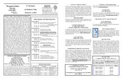
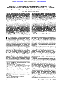
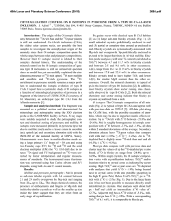
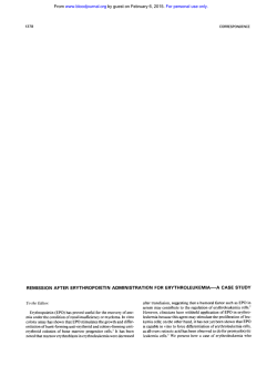
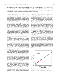
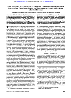
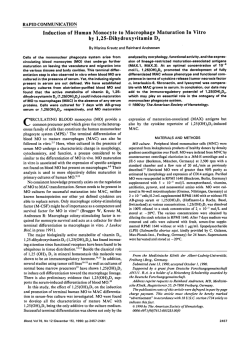
From www.bloodjournal.org by guest on February 6, 2015. For personal use only.
Calcium Ionophore, A23187, Induces Commitment to Differentiation But Inhibits
the Subsequent Expression of Erythroid Genes in Murine Erythroleukemia Cells
By Jack 0. Hensold, George Dubyak, and David E. Housman
Murine erythroleukemia (MEL) cells are a useful model for
studying the processes that regulate erythroid differentiation because exposure of these cells to a variety of chemical
inducing agents results in expression of erythroid-specific
genes and the resultant loss of cellular immortality. Previously it has been suggested that the calcium ionophore,
A23187, has effects on the early cellular events that lead to
the commitment of these cells to differentiation, but was not
in itself sufficient to induce differentiation. We demonstrate
here that A23187, as well as another calcium ionophore,
ionomycin, are capable of inducing commitmentto differentiation. Unlike other inducing agents, continual exposure to
A23187 inhibits transcription of the erythroid-specific genes,
pglobin and Band 3. This effect is not attributable to an
increase in cytosolic calcium concentration, because cells
induced by ionomycin produce normal amounts of hemoglobin. These effects of A23187 on MEL cells confirm that
commitment to differentiation is a distinct event from the
subsequent transcriptional activation of erythroidgenes. The
ability of both ionophores to induce commitment to differentiation suggests that an increase in cytosolic calcium can
trigger commitment to differentiation. These agents should
prove useful in investigatingthe cellular processes that are
responsiblefor commitment to differentiation.
0 1991 by The American Society of Hematology.
F
necessary for the subsequent occurrence of ~ommitment.3.~
Although these early changes are rever~ible,~.~
commitment
results in the irreversible loss of cellular immortality and
the concomitant expression of the erythroid phenotype.
Changes in cytosolic calcium concentration have been
suggested to play a role in producing these early changes6-’
In cells induced by DMSO, both EGTA and procaine block
commitment to differentiation, and this block is reversed by
the addition of excess calcium to the medium.”8 Further, a
brief (1 hour) exposure of MEL cells to the calcium
ionophore, A23187, was shown to abolish the latent period
that occurred with subsequent exposure to
More
recently though, direct measurements have shown that
cytosolic calcium concentration decreases slightly when
cells are exposed to DMSO.” In contrast, for MEL cells
induced to differentiate by the P-chain of the peptide
hormone inhibin, an increase in cytosolic calcium concentration was measured.’’ Thus, the significance of these changes
for erythroid differentiation remains uncertain.
Although A23187 induces cells to traverse the latent
period, this agent has not been reported to be capable by
itself of inducing commitment to differentiation. We demonstrate here that A23187, as well as a chemically distinct
calcium ionophore, ionomycin, can induce commitment to
differentiation. However, these agents have different effects
on the expression of the erythroid phenotype. Whereas the
committed colonies that arise from A23187-treated cells
are fully hemoglobinized, cells grown continually in this
agent do not express elevated levels of @-globinor Band 3
transcripts. Rather, despite increasing numbers of cells
committed to differentiation, these transcripts only begin to
accumulate when the cells are placed in medium free of the
ionophore. A23187 also blocks hemoglobinization of cells
that have previously become committed to differentiate in
DMSO. Thus, A23187 triggers commitment to differentiation while inhibiting the subsequent expression of the
differentiated erythroid phenotype. This effect is not seen
with ionomycin, which induces commitment and erythroid
gene expression in a manner similar to previously described
inducing agents. The similar effect of these ionophores on
commitment suggests that an increase in intracellular
calcium concentration can trigger differentiation of MEL
cells. The differing effect of these agents on erythroid gene
RIEND VIRUS-TRANSFORMED murine erythroleukemia (MEL) cells are a useful model system for
studying growth and differentiation in the erythroid lineage. These malignant pronormoblasts grow indefinitely in
culture, but when treated with dimethyl sulfoxide (DMSO)
or a variety of other inducing agents (eg, hexamethylene
bisacetamide, hypoxanthine, X-irradiation) undergo terminal differentiation with full expression of the erythroid
phenotype, as well as a limited capacity for growth.’,’
Inducing agents act to trigger the normal pathways of
differentiation, because continual exposure to an inducer is
not necessary for complete expression of the erythroid
p h e n ~ t y p eCells
. ~ that have undergone this triggering event
are said to be “committed” to differentiation. Committed
cells can be detected in cultures by subcloning into semisolid medium without inducers and determining the phenotype of the resulting colonies after 4 days of growth.
Colonies arising from committed cells are fully hemoglobinized and less than 32 cells in size (reflecting a limited
potential for growth), while cells that have not committed
have an unlimited growth capacity (colonies greater than 64
cells in size) with an erythroblastic m~rphology.~
Following inducer exposure, a latent period of 8 to 12
hours occurs before the cells begin to commit to differentiate. During this time, changes occur in the cells that are
From the Center for Cancer Research, Massachusetts Institute of
Technology, Cambridge; and the The R. Livingston Ireland Cancer
Center and the Department of Medicine, and the Department of
Physiology and Biophysics, Case Westem Reserve University Medical
School, Cleveland, OH.
Submitted February 9, 1990; accepted November 8, 1990.
Supported by Public Health Service Grants DK-01392 and P30-CA43703toJ.O.H., CA-l7575toD.E.H.,andGM-36387to G.D. G.D. is
an Established Investigator of the American Heart Association.
Address reprint requests to Jack 0. Hensold, MD, Ireland Cancer
Center, University Hospitals of Cleveland, 2074 Abington Rd, Cleveland, OH 44106.
The publication costs of this article were defrayed in part by page
charge payment. This article must therefore be hereby marked
“advertisement” in accordance with 18 U.S.C. section 1734 solely to
indicate this fact.
0 1991 by The American Society of Hematology.
OOO6-4971/91/7706-OOO3$3.
0010
1362
Blood, Vol77, N o 6 (March 15). 1991: pp 1362-1370
From www.bloodjournal.org by guest on February 6, 2015. For personal use only.
1363
A231874NDUCED COMMITMENT OF MEL CELLS
expression suggests that A23187 inhibits these latter processes through a mechanism other than an increase in
cytosolic calcium concentration.
MATERIALS AND METHODS
MEL cells were subclones of 745-PC4-Bl-2A- (15, 17, 17f, 15,
17i, and 19) that had been selected for rapid inducibility in DMSO.
These subclones responded similarly to each other when exposed
to inducers. Cells were grown in Dulbecco’s modified Eagle’s
medium (DMEM) supplemented with either 12% or 15% fetal
bovine serum (FBS; Armour Pharmaceutical Co,Kankakee, IL)
and 2 mmom L-glutamine, at densities to maintain log phase of
growth (0.5 to 10 x lo5cellslml). Cell growth and differentiation
in DMSO (1.5% volhrol) were similar under these conditions.
Because of significant binding of the ionophores to serum proteins,
concentrations of these agents were standardized for serum concentration as indicated in the text. A23187 and 4-bromo-A23187 were
dissolved in DMSO at concentrations of 1 mg/mL. Ionomycin was
dissolved in DMSO at a concentration of 1 mmol/L. Experiments
demonstrated that at these final solvent concentrations (<0.15%),
DMSO had no effect on MELgrowth or differentiation.
Commitment to differentiation was assessed as described by
Gusella et al.) Briefly, following exposure to inducer, aliquots of
cells were washed and plated in inducer-free plasma clot culture at
a clonal density of 2000 cells/mL. After 4 days of growth, the clots
were fixed with glutaraldehyde, stained with benzidene, and
counterstained with hematoxylin. The percentage of colonies that
reacted with benzidene (an indication of hemoglobin accumulation) was determined on triplicate samples of 0.1-mL plasma clot
cultures. Cloning efficiencies were calculated by dividing the
number of colonies in each clot by 200 (the number of cells plated
per 0.1-mL plasma clot) and multiplying by 100.
For determination of growth rate, cells were maintained at
concentrations that ensured log phase growth of control cells (ie,
0.5 to 10 X 10S/mL). Cells were diluted as necessary with prewarmed medium to maintain these densities. Cell counts were
performed with a Coulter Counter (Model Z, Coulter Electronics,
Hialeah, FL) and absolute cell number calculated by considering
the previous dilutions.
Cell cycle profiles were determined as described by Crissman
and Steinkamp.” Briefly, cells in log phase of growth were exposed
to inducer and at various times following inducer exposure,
aliquots of cells were removed from culture, washed with ice-cold
phosphate-buffered saline (PBS), and fixed with ice-cold 70%
ethanol. Before analysis, cells were treated with RNAse A (200
&mL) to reduce fluorescence from RNA, then stained with
propidium iodide (10 pg/mL). DNA-bound propidium iodide was
excited at a wavelength of 480 nm in a Cytofluorograf System 50
(Ortho Diagnostic Systems, Raritan, NJ) and cellular DNA content was determined by fluorescence at a wavelength of 630 nm.
Cell cycle distribution was calculated using an Ortho 2150 System
computer and the 2150 Program Constant method.
Relative rates of DNA synthesis were determined by pulselabeling with tritiated thymidine (’H-TdR). 3H-TdR (10 pCi/mL)
was added to cultures of cells for 30 minutes. Incorporation into
DNA was terminated by the addition of 5 vol of ice-cold PBS
followed by two washes in PBS. Cells were lysed and DNA
precipitated by the addition of ice-cold 10% trichloroacetic (TCA)
acid. Lysed cell pellets were incubated for 30 minutes on ice before
collection by filtration on Whatman (Maidstone, England) GF/C
glass fiber filters. The filters were washed extensively with 5%
TCA, with a final wash in ice-cold 70% ethanol. Incorporated
radioactivity was determined by scintillation counting in a Packard
(Downers Grove, IL) Tri-carb Liquid Scintillation Spectrometer.
For determination of mRNA levels, cytoplasmic RNA was
extracted from cells by the method of Favaloro et al,” except that
cell lysis was performed in 5 mmol/L Tris (pH 7.4), 2.5 mmol/L
MgCI,, 1.5 m m o m KCI, 1% Triton X-100 (Sigma, St Louis, MO),
and 0.5% deoxycholate and the nuclei pelletted in Eppendorf
centrifuge tubes by a 60-second spin at 10,OOOg. Extracted RNA
was quantitated by absorbance at 260 nm and separated by
electrophoresis in 1.2% agarose/20 mmol/L 3-[N-Morpholino]
propanesulfonic acid (MOPS)/2.2 m o m formaldehyde gels. Following electrophoresis, the RNA was transferred to nitrocellulose
membranes (Schleicher and Schuell, Keene, NH) by Northern
blotting. Transferred RNA was hybridized at 42°C in 5X SSPE, 5X
Denhardt’s solution, 50% formamide, and sheared salmon sperm
DNA (100 pg/mL) with the indicated cloned fragments of DNA
that had been labeled with [”PI-dmP in the manner described by
Feinberg and V0ge1stein.l~The cloned DNA fragments used in
these experiments and the nuclear run-off experiments (see below)
were a 2.2-kb fragment of the mouse p-globin gene,” a 600-bp
cDNA encoding mouse actin16 and a 3.4-kb cDNA fragment
encoding the murine erythrocyte Band 3 protein.” The Northern
blot hybridization results were standardized for ribosomal RNA
loaded by rehybridizing all the filters with a cloned fragment of the
human 18.5 rRNA gene.”
Nuclear run-off transcriptions were performed as described by
Greenberg and Ziff.I9 An excess amount of linearized plasmid
DNA was bound to nitrocellulose and hybridized for 48 hours with
standardized amounts of 32P-UTP-labeled run-off transcripts. In
addition to the cloned DNA fragments indicated above plasmids
containing a human hsp90 cDNA,” a hamster GRP78 CDNA;~and
a murine hsc7O cDNA:* were also used in these experiments.
Relative transcription rates were internally standardized to levels
of actin transcripts because previous experiments had demonstrated that the relative mRNA abundance and transcription rate
of this gene was unaltered during MEL cell differentiation.
Free cytosolic calcium concentrations were determined as previously described?’ Briefly, cells were removed from growth medium,
washed, and resuspended in buffer containing 5 mmol/L glucose
and 12% FBS. The serum was added to ensure that measurements
were performed at similar effective concentrations of ionomycin
and 4-bromo-A23187 as those present in the growth medium. Cells
were loaded with fura2-AM ester (Molecular Probes, Eugene, OR)
and fura2 fluorescence (339-nm excitationB00-nm emission) determined as previously described?’ Calcium-dependent fura2 fluorescence was calibrated using standard techniquesxBzsafter lysis of the
cells with digitonin. To determine the effect of prolonged incubations on cytosolic calcium, cells were exposed to inducers for the
times described in the text, then pelletted and resuspended in
one-half of the supernatant medium and loaded with fura2
acetoxymethyl ester (AM) for 30 minutes at 37°C. The cells were
then pelletted and resuspended in the remaining half volume of
medium and cytosolic calcium determined as above.
RESULTS
A23187 induces commitment to differentiation of MEL
cells. Previous work in this laboratory had demonstrated
that prior exposure of MEL cells to the calcium ionophore,
A23187 (1 pg/mL), abolished the normally occurring latent
period of 10 to 12 hours that precedes the onset of
commitment to differentiation in DMSO-exposed cells: To
further evaluate the cellular changes that occurred during
this time preceding differentiation, we sought to determine
the changes in gene expression that took place in MEL cells
following A23187 exposure. However, in initial experiments
From www.bloodjournal.org by guest on February 6, 2015. For personal use only.
1364
HENSOLD, DUBYAK, AND HOUSMAN
it was noted that, following overnight exposure of the cells
to A23187 (1 pg/mL, 15% FBS), committed colonies began
to appear. Because this result suggested that the ionophore
not only shortened the latent period but was capable of
inducing commitment to differentiation, this effect was
examined more closely. MEL cells were exposed to either
DMSO or A23187 (1 pg/mL) for up to 48 hours, then
subcloned into inducer-free plasma clot cultures and commitment determined as previously described’ The results
of this experiment (Fig 1A) demonstrated that DMSO and
A23187 induce commitment to differentiation with similar
kinetics. However, relative to DMSO, A23187 produced a
slightly higher percentage of committed colonies at early
times of exposure (by 9 hours, 2.4% v 1.5%), and a lower
percentage of committed colonies with prolonged exposure
times (by 48 hours, 71% v 87%). This data demonstrated
that A23187 was at least as efficient at inducing the
metabolic changes that occur during the latent period as
DMSO and that the ionophore was sufficient by itself to
induce commitment of MEL cells to differentiation.
To determine the optimal dose of A23187 for inducing
commitment to differentiation, MEL cells were exposed to
varying concentrations of A23187 for 48 hours, and the
percentage of committed cells was determined. The results
of this experiment are indicated in Fig 1B. Concentrations
below 0.5 pg/mL had no detectable effect on growth or
differentiation. As ionophore concentration was increased,
up to 1 pg/mL, an increased percentage of committed cells
was observed. For the dose ranges and times examined here
toxicity was not evident, as demonstrated by the cloning
efficiencies indicated at the bottom of Fig 1. However,
B
r
ar
20
OO
40
A23187 CONCENTRATION
(j~glmll
INDUCER EXPOSURE
I hours)
ODMSO
r
4-
0
3.5
9
I2
17
EXPOSURE (hour.)
24
48
Fig 1. (A) A23187 and DMSO induce commitmentto differentiation
with similar kinetics. MEL cells were exposed to either DMSO (1.5%
vol/vol) ( 0 )or A23187 (1.0 pg/mL) ( 0 )for the times indicated. The
cells were then washed, and seeded at clonal density in inducer-free
plasma clot culture. After 4 days of growth the percentage of
committed cells was determined by staining the resulting colonies
with benzidene. (B) A23187 dose-response curve. MEL cells were
exposed for 48 hours to A23187 at the concentrations indicated.
Commitment to differentiation was determined as above. Cloning
efficiencies are indicated in the bar graphs at the bottom of the figure.
concentrations of over 1 pg/mL were associated with
toxicity, and this was occasionally evidenced at a concentration of 1 pg/mL, particularly with prolonged incubations.
Thus, this agent is similar to previously described inducing
agents in having a maximal effect on differentiation just
below the concentration where toxicity is manifested.’ In
subsequent experiments, an A23187 concentration of 0.9
pg/mL, in 15% FBS, or a comparable dose of 0.72 pg/mL in
12% FBS was used.
A23187and DMSO have different effects on growth ofMEL
cells. While these experiments demonstrated that the
ionophore functioned as a complete inducer of differentiation, differences were noted in cells grown in A23187
compared with those grown in DMSO. While the growth
rate in DMSO was equivalent to that of control cells for up
to 2 to 3 days, cells cultured with A23187 rapidly ceased to
grow. In addition, despite high percentages of committed
cells in cultures induced with either DMSO or A23187,
hemoglobin synthesis appeared to be minimal in A23187exposed cells as judged by the color of the cell pellets.
Therefore, we investigated these differences more carefully.
Growth rates were compared in cells treated with DMSO
or A23187 and in untreated cells. As the growth curves in
Fig 2A demonstrate, normal growth rates persisted in all
cells for the first 12 hours of inducer exposure. After this,
cell number in the A23187-treated cultures did not change,
while cells grown in DMSO continued to grow at rates
identical to control for up to 72 hours. As previously shown,
with longer DMSO exposures a gradual slowing of the
growth rate occurs (not shown).
Exposure of MEL cells to DMSO results in a transient
Previous
arrest of cells in the GI phase of the cell cy~le.’~~~’
experiments had demonstrated that for these clones, the G,
arrest occurred at approximately 7 to 9 hours of DMSO
exposure.’‘ To determine if the ionophore’s effect on
growth rate was to prolong this normally occurring G,
arrest, cell-cycle profiles were determined on MEL cells
following increasing time of exposure to A23187. As demonstrated in Fig 2B, a small decrease in the percentage of cells
in S phase occurred following exposure to A23187. However, significant alterations in the cell-cycle distribution did
not occur until 49 hours, a time when a majority of the cells
had become committed to differentiation (see Fig 1). This
late accumulation of cells in G, has also been observed in
DMSO-exposed cells as
It was evident from this data
though that the change in growth rate that occurred after 12
hours of A23187 exposure was not caused by an arrest in a
particular phase of the cell cycle. Therefore, it is likely that
the transit time through all phases of the cell cycle must be
slowed. To confirm that the rate of DNA synthesis at 24
hours was indeed slowed despite a large percentage of cells
still in S phase, MEL cells were pulse-labeled with 3H-TdR
and the incorporation of label into trichloroactic acidinsoluble material in control and A23187-exposed cells was
determined. This experiment (Fig 2C) demonstrated an
87% reduction in thymidine incorporation following 24
hours of A23187 exposure. This finding confirmed that
transit through S phase of the cell cycle was indeed slowed
From www.bloodjournal.org by guest on February 6, 2015. For personal use only.
1365
A23187-INDUCED COMMITMENT OF MEL CELLS
B
10
0
20
30
40
CELL CYCLE ANALYSIS
50
Hours
C
THYMIDINE INCORPORATION
(per 105cells)
3or
24 HRS
Fig 2. A23187 produces cell cycle slowing of MEL cells. (A) Growth rate of MEL cells exposed to either DMSO or A23187. Cells were grown in
medium with DMSO (1.5% vol/vol) (O), A23187 (0.9 pg/mL) ( W ) or without inducers (0)and diluted with prewarmed medium to maintain
densities between 1 and 10 x 1Q and the dilutions figured in the final calculation of cell number. Cell counts were determined using a Coulter
Counter (model&),( 8 )Cell-cycle profilesfollowing exposure toA23187. DNA content of MEL cells exposedto A23187for the times indicated was
determined by fluorescence of propidium bound to DNA. The left-most peak in the figure represents (GO G1 cells (2 N amount of DNA), the
right-most peak represents6 2 M cells (4 N amount of DNA), and the area between the two peaks represents cells in S phase (between 2 N and 4
N amount of DNA). The percentage of cells in these cell-cycle phases was determined as described in Materials and Methods. (C) DNA synthesis
rates in control cells and in cells exposed to A23187. The relative rate of DNA synthesis in MEL cells exposed to A23187 was determined by
labelingthe cells for 30 minutes with PHI-TdR. and determining the incorporationof labelled PHI-TdR into TCA acid-insoluble material. Following
the labeling period, cells were lysed in ice-cold 10% TCA and the precipitatescollected on Whatman GF/C filters. The results were standardized
for cell number.
+
by exposure to A23187. Thus, in contrast to cells induced in
DMSO, A23187 slows MEL cell growth rate and the transit
time through all stages of the cell cycle is affected.
A23187 inhibits expression of P-globin and band 3 genes in
committed MEL cells. As noted above, cells induced to
differentiate in A23187 did not synthesize significant
amounts of hemoglobin (as assessed by lack of red color of
the cell pellet). Because the rate of commitment to differentiation was similar in DMSO and A23187-exposed cells, and
because both inducers produced committed colonies which
reacted with benzidene (after 4 days of growth in inducerfree medium) this suggested that continual exposure to
A23187 had an inhibitory effect on accumulation of hemoglobin. A decrease in protein synthetic rate occurs in MEL
cells following exposure to inducers of differentiation?'
Because this effect is more pronounced in A23187-exposed
cells (data not shown), the failure to accumulate significant
amounts of hemoglobin might reflect a decrease in translation of globin mRNA in A23187-induced cells. In such a
case, globin mRNA would increase in parallel with commit-
+
ment, but the cells would remain poorly hemoglobinized.
Therefore, levels of @-globinmessage were determined in
cells exposed to DMSO and A23187 by hybridization of
Northern-blotted RNA with a "P-labeled P-globin DNA
fragment. Preliminary experiments demonstrated that
P-globin mRNA did not increase in cells treated with
A23187 for up to 60 hours, a time when a significant
increase in this message had occurred in DMSO-treated
cells (see text below and Fig 3A). A similar effect was also
seen for accumulation of Band 3 mRNA (data not shown).
These results implied that, despite inducing commitment to
differentiation, A23187 inhibited erythroid gene expression. To determine if this was the case, MEL cells were
exposed to either DMSO or A23187 for 54 hours to induce
commitment in a high percentage of the cells. Cell cultures
were then split and maintained in the presence or absence
of inducer for an additional 18 hours. P-globin mRNA
levels were determined as above. As shown in Fig 3A, in the
continued presence of A23187, P-globin mRNA level does
not significantly increase relative to control cells. However,
From www.bloodjournal.org by guest on February 6, 2015. For personal use only.
1366
HENSOLD, DUBYAK, AND HOUSMAN
B
.
r
P-GLOBIN
P-GLOBIN
18s
r
n
D/A
by 18 hours after withdrawal of the ionophore, this message
has accumulated nearly threefold. In contrast, cells induccd
in DMSO accumulated high levels of f3-globin message in
the continual presence of the inducer. Thc extent of this
increasc could not be quantitated by densitometry of these
gels, bccausc at exposure times sufficient for detection of
p-globin mRNA in control cclls, thc signal from p-globin
mRNA from the DMSO-exposed cells had saturated thc
film. The inhibitory cffccts of the ionophore on p-globin
mRNA accumulation also extended to cclls that had bccn
induced in DMSO. As shown in Fig 3B, cclls that had been
induced by exposure to DMSO for 26 hours and thcn were
incubated in both A23187 and DMSO accumulated significantly less p-globin mRNA than those maintained exclusivcly in DMSO. This cffcct was not because of a toxic cffcct
of coincubation with both agents, because cell growth
following rcmoval of the agcnts was similar for cclls
exposed to either DMSO or DMSO and A23187 (data not
shown).
Thc high level of p-globin mRNA accumulation in cells
induced with DMSO is the result of an increase in the rate
of transcription of the gene,”-’* as well as the stability of the
p-globin mcssage.” To determine if the inhibitory effects of
the ionophorc on exprcssion of p-globin and Band 3
occurred at the transcriptional or posttranscriptional Icvel,
the relative transcription rates for these two gencs were
determined by nuclcar run-off assay. Transcription rates
were determined following 60 hours of cxposurc to DMSO
or A23187, a time when significant accumulation of both
p-globin and Band 3 messagc had occurrcd in DMSO-
Fig 3. A23187 i
nhim acalmul.tion ol p-gkMn
mRNA In co”W MEL calk. (A) BQloMn mRNA
only accumulates in MEL cells Induced to dHlerentiate in A23187 when removed from the inducer. MEL
cells were exposed to either A23187 (0.9 pglmL) or
DMSO (1.5% vollvol) for 52 hours (A52 and D52). The
cultures were then split and for the following 18
hours maintained in medium elther with inducer (A70
and 070) or without inducer (A52/70 and M2/70).
Cytoplasmic RNA was extracted from the cells at the
indicated timer and p-globin mRNA levels determined by blot hybridizationwith a [UP~labeledmolns
p-globin gene fragment. pglobin mRNA Ieveh were
quantitated by densitometry and internally standardized to the amount of 18s rRNA loaded/lane and is
indicated in the graph below the blots. Ouantitation
of signal in the DMSO lanes was not attempted
because of saturation of the film. (9)A23187 inhibtts
accumulation of (+globin mRNA in cells indueed to
differentiate In DMSO. MEL cells were induced in
DMSO (l.Soh vol/vol) for 26 houn, then split and
grown in either DMSO alone, or in both DMSO (1.5%)
and A23187 (0.9 pg/mL) for the ensuing 18 hours.
p-globin mRNA levels were determined and quantitated as above.
induced cells. These results arc shown in Fig 4. Following
60 hours of DMSO exposurc. the rate of p-globin transcription had incrcascd 15-fold. At similar times of A23187
exposure, p-globin transcripts had not changcd relative to
control cells. A similar effect was dctccted for Band 3 gene
transcription. This cffcct on the transcription rates of
p-globin and Band 3 genes was spccific, bccause the
transcription of actin, GRW8, HSP90, and hsc70 transcripts persisted in cells cxposed to A23187. These experiments dcmonstratcd that, although A23 187 induccscommitmcnt of MEL cells, it can block the subsequent
transcriptional activation of at least two gcncs whose
expression is characteristic of terminal erythroid differentiation.
Ionomycin induces both commitment and &globin gene
eqmssion in MEL cells. The ability of A23187 to induce
commitment to differentiation suggcsted that a decrease in
cytosoliccalcium (previously reportcd toaccur with DMSO)
was not necessary for commitmcnt. However, bccausc the
effccts of A23187 on cation pcrmcability are not selcctive
for calcium, it is possible that A23187-induced commitment
is mediated by other cffccts of this agent. Thercforc. we
investigated the effects of ionomycin on MEL cell commitment and erythroid gene cxprcssion. This agent has a more
selectivc effect on membrane permeability to calcium than
docs A23187.”The ability of this agent to induce diffcrcntiation was dctcrmined by a 48-hour cxposurc to a rangc of
concentrations as previously donc for ,423187. As dcmonstrated in Fig SA, within the same molar range of conccntralions as A23187 (0.7 pg/mL A23187 is 1.36 pmol/L),
From www.bloodjournal.org by guest on February 6, 2015. For personal use only.
A23187-INDUCED COMMITMENT OF MEL CELLS
1367
DMSO A23187
60h
CON- - 6 0 hFig4. &gkbln.ndh n d 3 gumsam nattr8maip
tiona~.ctivateddespiteco"imwnttodifferentlation of MEL cells in A23187. Relative transcription
mea were determined by labeling of " o f f transcripts with tYpl-UTP as described by Omenberg and
Ztff." Tim- of expoaure (e0houn) to either DMSO
(D) or A23187 (A) More labeling of " o f f transcripts are indkated abow each blot The labeled
tramaipta were hybridized with an exceaa at linearized plaamld DNA that had been previously bound to
nitrocelluloae filters. The plaamid DNAa uaed in theae
experiments are deacribed in Materials and Methods
and include DNA fragmenta encoding mwae actin,
Band 3, (+globin, hsc70. harnater GRP78, and human
HSPSO. pUCl9 plnmid DNA was uaed as a negative
control. The filtera were developed by autoradiography.
GRP78
hsc70
hsp90
Actin
pUCI9
Band 3
p- globin
ionomycin induced commitment to differentiation in a
doxdcpendcnt manncr. However, in contrast to A23187,
cells induced to diffcrcntiate in ionomycin accumulate
p-globin mRNA at a ratc approximatcly equal to cells
induced in DMSO (Fig 5B). Thus, ionomycin is capable of
inducing commitment to diffcrcntiation and concomitant
crythroid gcnc cxprcssion in a manncr similar to DMSO
and othcr, prcviously described inducing agents.
Thc cffccts of ionomycin on commitmcnt and erythroid
gcnc expression suggcstcd that a dccrcase in cytosolic
calcium is ncither ncccssary for the occurrence of commitment to diffcrcntiation (as previously suggcstcd
for DMSO),'a'' nor rcsponsiblc for inhibiting erythroid
gene cxprcssion (as in cells cxposcd to A23187). To bc
ccrtain that the ionophorc concentrations uscd in thcsc
cxpcrimcnts were sufficicnt to incrcasc cytosolic calcium,
this paramctcr was mcasured in cells cxposcd to thcse
agcnts using fura2 fluorcsccncc. Because cytosolic calcium
conccntration cannot be dctcrmincd in cclls cxposcd to
A23187 (thc ionophorc has a fluorcscent spcctra similar to
fura2), thc 4-bromo-analog of A23187 was used in these
studics. This agent has bccn shown to have similar cffccts
on calcium pcrmcability, but has a diffcrcnt fluorescent
spectrum than thc parental compound." At a conccntration
of 4-bromo-A27187of 0.8 pg/mL (cquimolar to 0.72 pg/mL
A23187). cytosolic calcium increascd from 155 nmol/L to
475 nmol/L (scc Tablc 1). A similar cffcct on final cytosolic
calcium conccntration was also sccn at a 4-bromo-A23187
conccntration of 0.4 pg/mL, although the time rcquircd to
rcach this lcvcl was longer. Ionomycin at concentrations
ranging from 0.5 to 1.5 pmol/L similarly increased cytosolic
calcium, up to 435 nmol/L at thc latter concentration.
Consistent with prcvious findings, DMSO produccd a small
dccrcasc in cytosolic calcium concentration (data not
shown).
Thcse results demonstrated that acute exposure to these
ionophorcs resulted in an incrcase in cytosolic calcium.
Howcvcr, it is possible that thc cclls rapidly adapt to this
increase and, hence, cytosoliccalcium concentrationsmight
not be increased by the time the cells began to commit to
diffcrcntiatc. Therefore, wc investigated whether cytosolic
calcium concentration remained clcvatcd in cells cxposed
to thc ionophorcs. To obtain thcsc mcasurcmcnts, cclls
werc grown for either 13 or 48 hours in ionomycin and
loaded with fura2 in the same medium. cytosolic calcium
concentration was dctcrmincd with thc cells in their normal
growth medium. with ionomycin added. Thcsc studies
dcmonstratcd that at 13 and 48 hours of ionomycin cxposurc cytosolic calcium remaincd clcvatcd, at 399 nmol/L
and 416 nmol/L, respectively. cytosolic calcium conccntration in control cells measured undcr idcntical conditions
was 262 nm. Thus, calcium ionophorcs induce commitmcnt
to diffcrcntiation whilc producing a sustained incrcasc in
cytosoliccalcium conccntration.
DISCUSSION
The data presented in this paper demonstrates that
exposure of MEL cclls to A23187 is sufficient to induce
commitment to diffcrcntiation. However. this inducer differs from those previously described because continued
cxposurc to the ionophorc, whilc inducing commitmcnt to
diffcrcntiation, inhibits cxprcssion of at lcast wogenes that
arc charactcristic of diffcrcntiated erythroid cells. Thcse
findings confirm that commitmcnt to differentiation is a
distinct evcnt from the ultimatc expression of the erythroid
phcnotype in inducerexposed MEL cells. Thc dcmonstration that ionomycin can also inducc diffcrcntiation of MEL
cclls, hut docs not inhibit f3-globin gcne cxprcssion. suggcsts that an incrcasc in cytosolic calcium can triggcr
commitmcnt and that this incrcasc is not responsible for the
inhibition of erythroid gcne expression in cclls cxposed to
A232 187.
It has been previously demonstrated that a 1-hour
exposure to A23187 eradicates the latcnt period of diffcrentiation in MEL cells subscqucntlyexposed to DMSO.".'Thc
From www.bloodjournal.org by guest on February 6, 2015. For personal use only.
HENSOLD, DUEYAK, AND HOUSMAN
1360
ability of A23187 to induce commitment to differentiation
was not detected in these studics. The reason for these
differences may bc thc intrinsic propcrtics of A23187 that
influence its potency. A23187 is a hydrophobic molecule
that binds to albumin and partitions into ccll mcmbrancs."
hOphore
Control
4-bromo-A23107
lonomycin
A
Concentration
0.4 pmoVL
0.0 pmoVL
0.5 pmoVL
1.O pmoUL
1.5 pmoVL
Cytowliic Cakium
155 nmoVL
475nmolR
475 nmoVL
300 nmoVL
390 nmoUL
435 nmoVL
Cells were loadedwith furs2 AM in buffered solution containing 12%
FCS. Ionophore was added 81 the concentration indicated and cytosolic
calcium concentration determined as described in Materials and Methods.
-
0 1.0 125 1.5
IONOMKIN
CONCENTRATION ()I M)
B
p-GLOBlN
Fig 5. Th. u k h ionopt", ionomydn, induces both commit-
ment to dlfferemtiatlon and hemoglablnlzatlon of MEL cells. (A)
lonomydn i n d m co"imHnt to differentiation of MEL cells. MEL
cells were incubated for 48 hours with ionomycin at the concontrations lndkated below tho graph. Commitment to differentiation was
detennlned as described in tho text. Cloning .(fkiemies for the
ionomycin treated cells (0.5 pg/mL 94%. 1.0 pg/mL 71%. 1.5 pg/mL
100%) ware similar to control cells (77%). (E) lonomydn Induces
p-globln mRNA accumulati6n in MEL cells. MEL cells were exposed to
DMSO (1.5% vol/vol) or ionomycin (1.5 rmol/L) for 65 hours and
cytoplasmic RNA extmcted as described in the text. Ten micrograms
of RNA was anakzed for pglobin mRNA by Northem blotting and
hybridization with a p)-labeled fragment of the m w w pglobin
gene.
Because its cffcctivc concentration is rclativc to the conccntration of ccll mcmbrancs in the medium. at low ccll
conccntrations A23 187 has greater cffccts as an ionophore
than at higher ccll conccntrations. Thus, both scrum
conccntration and ccll concentration must bc standardized
in determining the optimal dosc. In thc previous cxpcrimcnts only brief exposures to A23187 wcrc cvaluatcd
bccause toxicity was notcd with prolongcd cxposurcs. It is
likely that thcse ohscrvations wcrc made undcr diffcrcnt
conditions than thosc uscd in the cxpcrimcnts prcscntcd
hcrc. Thc obscrvation of commitment in our carly cxpcrimcnts likely rcsultcd from the scrcndipitous occurrcncc of
conditions that produced an cffcctivc dosc of A23187 with
limitcd toxicity. Using this initial observation as a starting
point, optimal inducing conditions wcrc thcn established as
dcscribcd hcrc.
Previous cxpcrimcnts havc dcmonstratcd that inhibition
of hcmoglobin accumulation by imidazolc docs not affcct
commitmcnt to diffcrcntiation of MEL cells simultaneously
cxposcd to DMSO."Our results confirm thosc ohscrvations
and cxtcnd them to includc Band 3 gcnc cxprcssion, but
differ in that the ability to induce commitmcnt and the
ability to block hcmoglobinization arc shared properties of
a single agcnt. A27187 inhibits transcriptional activation of
both the p-globin and Band 3 genes, as dcmonstratcd in thc
nuclcar run-off cxpcrimcnts. Thus, commitment to diffcrcntiation of these cclls is most closely linkcd to their loss of
immortality and rcprcscnts a distinct cvcnt that can tic
separated from the subscqucnt transcriptional activation of
lincagc-spccificgenes.
Thc cffccts of A23187 on transcription of erythroid genes
cannot bc simply attributed to an increase in cytosolic
calcium concentration. Exposure of MEL cells to an cquimolar concentration of ionomycin, which produces a similar
affcct on cytoplasmic calcium conccntration (1.5 pmol/L
ionomycid435 nmol/L [Ca" I, 1.4 pmol/L4-bromo-A23187/
475 nmol/L [Ca2*]),induccs commitment but docs not
inhibit p-globin gcnc expression. Thc continued transcrip
tion of thc actin, hsc70. hsp90, and GRP78 gcncs suggests
that the transcriptional activation of newly induced genes is
most sensitive to inhibition by this agcnt. Our data do not
allow us to draw a conclusion as to the mechanism of this
cffcct. However, the ability of ionomycin to inducc commitmcnt without inhibiting erythroid gene cxprcssion suggcsts
From www.bloodjournal.org by guest on February 6, 2015. For personal use only.
1369
A23187-INDUCED COMMITMENT OF MEL CELLS
that the difference in selectivity for cations of these two
ionophores may underlie their differing effects on gene
expression.
The mechanisms by which agents as diverse as DMSO,
hypoxanthine, X-irradiation, and actinomycin D act to
induce commitment to differentiation of MEL cells remains
unknown. The ability of the calcium ionophores, ionomycin
and A23187, to induce commitment to differentiation
suggests that an increase in cytosolic calcium concentration
may trigger commitment. Although our data does not
exclude that the ionophores have a synergistic effect with
the low levels of DMSO used to solubilize them, this
interpretation would still suggest that calcium has a role in
triggering commitment. Early experiments suggested that
an increase in cytosolic calcium concentration played a
critical role in inducing ~ommitment.6-~.~
More recently,
Shibata et al" have also demonstrated that MEL cells
induced to differentiate by the @-subunitof the hormone
inhibin increase their cytosolic calcium. In contrast, Arrow
and Macara have demonstrated that DMSO decreases
cellular calcium levels in MEL cells.'o Thus, the role of
changes in calcium concentration in triggering commitment
is uncertain. However, it seems likely that an increase in
cytosolic calcium concentration is one possible mechanism
that may be used by inducing agents to trigger differentiation.
ACKNOWLEDGMENT
We acknowledge Bruce Spiegelman for the cloned mouse actin
cDNA, Seth Alper for the mouse Band 3 cDNA, Lutz Giebel for
the hsc7O cDNA, Amy Lee for the GRP78 cDNA, and Neil Rebbe
for the HSP90 cDNA used in these experiments.
REFERENCES
1. Housman D, Volloch V, Tsiftsoglou A, Levenson R, Gusella
J, Kernan J, Mitrani A Analysis of the molecular basis of
commitment in murine erythroleukemia (MEL), in Rossi G (ed):
In Vivo and In Vitro Erythropoiesis: The Friend System. Amsterdam, The Netherlands, ElsevierNorth Holland Biomedical, 1980,
p 273
2. Marks P, Rifkind R: Erythroleukemic differentiation. Annu
Rev Biochem 47:419,1978
3. Gusella J, Geller R, Clarke B, Weeks V, Housman D:
Commitment to erythroid differentiation by Friend erythroleukemia cells: A stochastic analysis. Cell 9:221, 1976
4. Levenson R, Housman D: Memory of MEL cells to a previous
exposure to inducer. Cell 17:485,1979
5. Levenson R, Housman D: Developmental program of murine
erythroleukemia cells. Effect of the inhibition of protein synthesis.
Dev Biol82:715,1979
6. Bridges K, Levenson R, Housman D, Cantley L Calcium
regulates the commitment of murine erythroleukemia cells to
terminal differentiation. J Cell Biol90:542,1981
7. Chapman L: Effect of calcium on differentiation of Friend
leukemia cells. Dev Biol79:243, 1980
8. Tsiftsoglou A, Mitrani A, Housman D: Procaine inhibits the
erythroid differentiation of MEL cells by blocking commitment:
Possible involvement of calcium metabolism. J Cell Physiol 108:
327,1981
9. Levenson R, Macara I, Smith R, Cantley L, Housman D: Role
of mitochondrial membrane potential in the regulation of murine
erythroleukemia cell differentiation. Cell 28:855,1982
10. Faletto D, Macara I: The role of Ca2+in dimethyl sulfoxideinduced differentiation of Friend erythroleukemia cells. J Biol
Chem 260:4884,1985
11. Shibata H, Ogata E, Etoh Y, Shibai H, Kojima I: Erythroid
differentiation factor stimulates hydrolysis of polyphoinositide in
Friend erythroleukemia cells. Biochem Biophys Res Comm 146:
187,1987
12. Crissman H, Steinkamp J: Rapid simultaneous measurement of DNA, protein and cell volume in single cells from large
mammalian populations. J Cell Biol59:766,1973
13. Favaloro J, Treisman R, Kamen R: Transcription maps of
polyoma virus-specific R N A Analysis by two dimensional S1 gel
mapping. Methods Enzymol65:718,1980
14. Feinberg A, Vogelstein B: A technique for radiolabelling
DNA restriction endonuclease fragments to high specific activity.
Anal Biochem 132:6,1983
15. Tilghman S, Tiemeier D, Polsky F, Edge11 M, Seidman J,
Leder A, Enquist L, Norman B, Leder P: Cloning specific segments
of the mamalian genome: Bacteriophage lamdba containing mouse
globin and surrounding gene sequences. Proc Natl Acad Sci USA
74:4406,1977
16. Spiegelman B, Frank M, Green H: Molecular cloning of
messenger RNA for glycerophosphate dehydrogenase and other
differentiation-dependent proteins during adipocyte development.
J Biol Chem 258:10083,1983
17. Kopito R, Lodish H: Primary structure and transmembrane
orientation of the murine anion exchange protein. Nature 316:234,
1985
18. Wilson G, Hollar B, Waterson J, Schmeckel R: Molecular
analysis of cloned human 18s ribosomal DNA segments. Proc Natl
Acad Sci USA 755367,1978
19. Greenberg M, Ziff E: Stimulation of 3T3 cells induces
transcription of the c-fos proto-oncogene. Nature 311:433,1984
20. Rebbe N, Ware J, Bertina R, Modrich P, Stafford D:
Nucleotide sequence of a cDNA member of the human 90 kDa
heat-shock protein family. Gene 53:235,1987
21. Lee A, Delegeane A, ScharE D: Highly conserved glucoseregulated protein in hamster and chicken cells: Preliminary characterization of its cDNA clone. Proc Natl Acad Sci USA 78:4922,
1981
22. Giebel L, Dworniczak B, Bautz E Developmental regulation of a constitutively expressed mouse mRNA encoding a 72 kDa
heat shock-like protein. Dev Biol 125:200,1988
23. Dubyak G, Cowen D, Mueller L Activation of inositol
phospholipid breakdown in HL60 cells by P2-purinergic receptors
for extracellular ATP. J Biol Chem 263:18108,1988
24. Dubyak G: Extracellular ATP activates polyphosphoinositide breakdown and calcium mobilization in Ehrlich ascites tumor
cells. Arch Biochem Biophys 245:84,1986
25. Grynkiewicz G, Poenie M, Tsien R: A new generation of
Ca2+indicators with greatly improved fluorescence properties. J
Biol Chem 260:3440,1985
26. Friedman E, Schildkraut C Lengthening of the G, phase is
not strictly correlated with differentiation in Friend erythroleukemia cells. Proc Natl Acad Sci USA 75:3813,1978
27. Terada M, Fried J, Nude1 U, Rifkind R, Marks P Transient
inhibition of initiation of S-phase associated with dimethyl sulfoxide induction of murine erythroleukemia cells to erythroid differentiation. Proc Natl Acad Sci USA 74:248, 1977
28. Hensold J, Housman D: Decreased expression of the stress
From www.bloodjournal.org by guest on February 6, 2015. For personal use only.
1370
protein HSP70 is an early event in murine erythroleukemia cell
differentiation. Mol Cell Biol8:2219,1988
29. Friedman E, Schildkraut C: Terminal differentiation in
cultured Friend erythroleukemia cells. Cell 12:901,1977
30. Sherton C, Kabat D: Changes in RNA and protein metabolism preceding onset of hemoglobin synthesis in cultured Friend
leukemia cells. Dev Biol48:118,1976
31. Gilmour R, Harrison P, Windess J, M a r a N, Paul J: Globin
messenger RNA synthesis and processing during hemoglobin
induction in Friend cells: 1. Evidence for increased transcription
during erythroid differentiation in clone M2. Cell Differentiation
3:9,1974
32. Orkin S, Swerdlow P: Globin RNA synthesis in vitro by
isolated erythroleukemic cell nuclei: Direct evidence for increased
transcription during erythroid differentiation. Proc Natl Acad Sci
USA 74:2475,1977
33. Volloch V, Housman D: Stability of globin mRNA in
HENSOLD, DUBYAK, AND HOUSMAN
terminally differentiating murine erythroleukemic cells. Cell 23:
509,1981
34. Liu C, Herman T: Characterization of ionomycin as a
calcium ionophore. J Biol Chem 253:5892,1978
35. Faletto D, Arrow A, Macara I: An early decrease in
phosphatidylinositol tumover occurs on induction of Friend cell
differentiation and precedes the decrease in c-myc expression. Cell
43:315,1985
36. Deber C, tom-Kun J, Mack E, Grinstein S: Bromo-A23187
A nonfluorescent calcium ionophore for use with fluorescent
probes. Anal Biochem 146:349,1985
37. Vidaver G, Lee J: Rapid stoping of A23187 action by
phophatidylcholine. FEBS Lett 155:117,1983
38. Gusella J, Tsiftsoglou A, Volloch V, Weil S, Neumann J,
Housman D: Dissociation of hemoglobin accumulation and commitment during murine erythroleukemia cell differentiation by treatment with imidazole. J Cell Physiol113:179,1982
From www.bloodjournal.org by guest on February 6, 2015. For personal use only.
1991 77: 1362-1370
Calcium ionophore, A23187, induces commitment to differentiation
but inhibits the subsequent expression of erythroid genes in murine
erythroleukemia cells
JO Hensold, G Dubyak and DE Housman
Updated information and services can be found at:
http://www.bloodjournal.org/content/77/6/1362.full.html
Articles on similar topics can be found in the following Blood collections
Information about reproducing this article in parts or in its entirety may be found online at:
http://www.bloodjournal.org/site/misc/rights.xhtml#repub_requests
Information about ordering reprints may be found online at:
http://www.bloodjournal.org/site/misc/rights.xhtml#reprints
Information about subscriptions and ASH membership may be found online at:
http://www.bloodjournal.org/site/subscriptions/index.xhtml
Blood (print ISSN 0006-4971, online ISSN 1528-0020), is published weekly by the American
Society of Hematology, 2021 L St, NW, Suite 900, Washington DC 20036.
Copyright 2011 by The American Society of Hematology; all rights reserved.
© Copyright 2026