
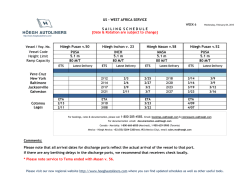
Parietal and intravascular innate mechanisms of vascular inflammation
Ramirez et al. Arthritis Research & Therapy (2015) 17:16
DOI 10.1186/s13075-015-0528-2
REVIEW
Open Access
Parietal and intravascular innate mechanisms of
vascular inflammation
Giuseppe A Ramirez1,2*, Patrizia Rovere-Querini1,2, Maria Grazia Sabbadini1,2 and Angelo A Manfredi1,2
Abstract
Sustained inflammation of the vessel walls occurs in a
large number of systemic diseases (ranging from
atherosclerosis to systemic vasculitides, thrombotic
microangiopathies and connective tissue diseases),
which are ultimately characterized by ischemia and
end-organ failure. Cellular and humoral innate immunity
contribute to a common pathogenic background and
comprise several potential targets for therapeutic
intervention. Here we discuss some recent advances
in the effector and regulatory action of neutrophils
and in the outcome of their interaction with circulating
platelets. In parallel, we discuss novel insights into
the role of humoral innate immunity in vascular
inflammation. All these topics are discussed in light of
potential clinical and therapeutic implications in the
near future.
The clinical spectrum of vascular inflammation
Blood vessels act as tissue integrators by granting the
diffusion of oxygen, nutrients and particulate signals
throughout the body. The immune function emerges during evolution as a tool to defend the circulatory system
from threats to its integrity. Each static player (that is, the
vessel walls) or dynamic player (that is, blood components) of the circulatory system rapidly shifts towards a
defensive, inflamed state and cooperates with evolutionary
more recent adaptive immune responses. Vessels might
thus represent the archetypical scenario for the very early
initiation of the inflammatory response.
Under physiological conditions, self-limiting inflammatory processes occur in the circulating blood that
necessarily involve the vessel walls, when the immune
system effectively copes with microbial and nonmicrobial threats, eliminating the original noxa and guiding
* Correspondence: [email protected]
1
IRCCS Ospedale San Raffaele, via Olgettina 60, 20132 Milan, Italy
2
Università Vita Salute San Raffaele, via Olgettina 58, 20132 Milan, Italy
vessel regeneration and eventual healing. Threats that
cannot be removed or persistent deregulated immune responses directed against endogenous vascular constituents
in turn underlie vascular diseases.
Atherosclerosis and its complications represent the
leading cause of mortality in westernized countries and
the most frequent clinical manifestations of the effects of
persisting vessel inflammation. The priming event in vascular inflammation in atherosclerosis is exquisitely metabolic, since the origin of the disease is associated with the
accumulation of lipoproteins endowed with oxidative
potential in the intimal layer with ensuing lipidogenic persistent inflammation. The characteristic atherosclerotic lesion (that is, the atheromasic plaque) typically develops
assuming an eccentric shape.
In addition to these consolidated data, novel evidence
is progressively emerging about the implications of persistent vascular inflammation for a large number of systemic
diseases; in particular, those diseases in which autoimmunity plays a crucial role such as systemic sclerosis (SSc), systemic lupus erythematosus (SLE), dermatomyositis and
other connective tissue diseases, thrombotic microangiopathies (TMAs) and systemic vasculitides. Some of these
diseases have received more significant attention in recent years and could serve as clinical and pathophysiological paradigms.
SSc is an autoimmune disease of unknown etiology,
characterized by widespread organ dysfunction, peripheral
ischemia and fibrotic substitution. Vascular immunemediated injury of small arteries and capillaries is an early
event in the natural history of the disease and often takes
place before fibrosis is established. Endothelial activation
and apoptosis are thought to constitute the priming
process in the progression of vascular injury. Recent studies provide evidence for a role of neutrophil-dependent
interleukin (IL)-6 signaling in mediating the early phase of
vascular injury in SSc [1]. Vessel remodeling and intimal
proliferation in turn could arise as a response to endothelial dysfunction and rheologic disturbances [2,3]. Endothelial cells and myofibroblasts could both be involved in
© 2015 Ramirez et al.; licensee BioMed Central. This is an Open Access article distributed under the terms of the Creative
Commons Attribution License (http://creativecommons.org/licenses/by/4.0), which permits unrestricted use, distribution, and
reproduction in any medium, provided the original work is properly credited. The Creative Commons Public Domain
Dedication waiver (http://creativecommons.org/publicdomain/zero/1.0/) applies to the data made available in this article,
unless otherwise stated.
Ramirez et al. Arthritis Research & Therapy (2015) 17:16
neointima formation in SSc. The latter cell subset can
derive from resident pericytes, transdifferentiating cells
or bone marrow-derived precursors [4]. Lung involvement comprises interstitial lung disease and pulmonary
arterial hypertension and represents a major issue in the
management of SSc, given the high mortality rate and
the poor efficacy of available treatments. Conventional
immunosuppressive treatments are only partially or not
at all effective in controlling and reversing vascular
events (for example, pulmonary arterial hypertension)
whose pathogenesis is only partially defined [2].
TMAs such as thrombotic thrombocytopenic purpura,
hemolytic uremic syndrome (HUS) and pre-eclampsia are
characterized by widespread endothelial injury and expression of thrombogenic stimuli such as von Willebrand
factor (vWF), due to the release of endotheliotropic toxins
(characteristic of typical HUS), impaired inhibition of the
complement system (atypical HUS) or other noncharacterized stimuli, possibly in the setting of jeopardized
ADAMTS-13 activity (thrombotic thrombocytopenic purpura) [5]. Endothelial injury/activation in turn reflects on
platelets and the coagulation system, with microvascular
thrombosis and end-organ ischemia.
Systemic vasculitides comprise heterogeneous diseases,
characterized by persistent inflammatory damage of the
vessel walls [6]. According to the Chapel Hill Consensus
Conference, there are seven classes of systemic vasculitides:
large vessel vasculitides (including giant cell arteritis (GCA)
and Takayasu’s arteritis), small vessel vasculitides (including anti-neutrophil cytoplasmic antibody-associated
vasculitides (AAV), IgA vasculitis and cryoglobulinemic
vasculitis), medium vessel vasculitides (including Kawasaki’s
disease and polyarteritis nodosa), variable vessel vasculitides
(including Behçet’s disease), single organ vasculitides, vasculitides associated with systemic disease and vasculitides
associated with probable etiology. Although the pathogenetic mechanisms and clinical scenarios differ, the diseases
share the inflammatory involvement of vessels as the primary event in the disease natural history and the associated
multiorgan systemic involvement.
Blood vessel checkpoints: role of vessel-residing
cells in the initiation of the inflammatory response
Circulating leukocytes interact with cells that resides
within the vessel walls as well as with other circulating
cells that interact with blood vessels in order to gain information about ongoing damage in surrounding tissues
and eventually to extravasate. To this purpose, either cells
located in the lumen of blood vessels or cells located at
the periphery of the vessel wall are able to productively
interact with circulating and extravasating leukocytes and
drive their subsequent effector responses. Cells that define
the internal wall of blood vessels, such as the endothelium
or the platelets recruited at sites of vessel injury to
Page 2 of 12
surrogate the function of the endothelium, indeed sense
potential threats to the integrity of vessels and surrounding tissues through an array of pattern recognition receptors (PRR) [7,8]. After rapid mobilization of intracellular
stores, endothelial cells and platelets expose a large
array of signaling molecules such as P-selectin (from the
endothelial and platelet side), vWF (from endothelial
Weibel–Palade bodies) and inflammatory signals such
as the high mobility group box 1 protein, the soluble
form of CD40 ligand, leukotrienes LTA4 and LTB4 and
tissue factor [7]. Later responses involve the transcriptionally regulated synthesis of E-selectin, vascular cell
adhesion molecule 1 and intercellular adhesion molecules (Figure 1B1).
Rolling/crawling neutrophils, monocytes and innatelike lymphocytes, besides being directly activated by the
original inflammatory stimuli, recognize the changes on
the vessel cell surface, and in turn generate signals that expand the sensitivity of blood vessels [6]. The recognition
of potentially harmful agents in the context of blood vessels thus prompts the development of a tripartite crosstalk
involving the endothelium, platelets and leukocytes [7].
During migration towards target tissues, leukocytes (at
least of the myeloid lineage) receive additional information
after interacting with vascular pericytes (Figure 1B2,B3).
These poorly defined cells, wrapped around the endothelium, play a role that has only recently gained attention as
a secondary pre-tissue checkpoint. Venular neuron–glial
antigen 2-negative/alpha-smooth muscle actin-positive
(NG2− α-SMA+) pericytes directly provide navigation support during extravasation, modifying their shape in response to inflammatory signals forming gaps and thus
providing preferential exit routes to neutrophils through
the venular wall [9] (Figure 1B2). By contrast, capillary or
arteriolar neuron–glial antigen 2-positive/alpha-smooth
muscle actin-positive (NG2+ α-SMA+) pericytes recruit
myeloid leukocytes after completion of diapedesis (even
from relatively distant sites) and enhance their survival as
well as the speed and the linearity of their migration in the
perivascular interstitial space (Figure 1B3). As such, this
latter pericyte subset might be specifically involved in
the maintenance of inflammation associated with small
arteries and capillaries [10]. Pericytes contribute to the
remodeling of vessels and of surrounding tissues under
conditions of hypoxia [4,11] and regulate the vascular
tone, possibly by acquiring vascular smooth muscle celllike features [11].
Spreading of inflammation through large arterial vessel
walls involves unique pathophysiological pathways. In fact,
large arteries are themselves vascularized, as they are
served by a specific set of small vessels in the adventitial
layer, called the vasa vasorum. Adventitial dendritic cells
are thought to coordinate the recruitment of activated
CD4+ T lymphocytes from the vasa vasorum and to
Ramirez et al. Arthritis Research & Therapy (2015) 17:16
Page 3 of 12
Figure 1 Checkpoints on leukocyte migration through inflamed vessels and tissues. In the setting of inflamed large arterial vessels,
leukocytes adhere to the walls of the vasa vasorum and eventually extravasate through them. After accessing the adventitial layer of large
arteries, leukocytes interact with vessel-residing dendritic cells that induce the generation of a follicle-like structure during chronic inflammation
(A). In general, circulating leukocytes that approach an inflamed tissue (B) interact with signaling molecules expressed by activated endothelial
cells (and platelets), which promote their adhesion to the vessel walls and eventually their migration through endothelial cells and perivascular
connective tissue (1). This process mainly occurs at the level of postcapillary venules, where leukocytes are assisted by a first subset of neuron–glial
antigen 2-negative/alpha-smooth muscle actin-positive (NG2− α-SMA+) pericytes (2). Both luminal and pericyte-derived signals enhance leukocyte
survival and activation. As an example, neutrophils exposed to vascular cell adhesion molecule 1 (VCAM1) live longer: notably VCAM1 is required for
the full development of anti-neutrophil cytoplasmic antibody-associated vasculitides glomerulonephritis, while its soluble and endothelial surface
expression in rheumatoid arthritis correlates with joint damage and late-stage vascular injury, respectively. After diapedesis, leukocytes move through
the interstitial space, mainly following slow and nonlinear routes. Interactions with a second subset of NG2+α-SMA+ arteriolar/capillary pericytes prompt
leukocytes to progress faster and more linearly towards target tissues and might be involved in perpetuating vascular inflammation (3). CAM, cell
adhesion molecules; vWF, von Willebrand factor.
influence the deployment of T-helper (Th)1/Th17-driven
immune responses in the underlying vessel layers [12]
(Figure 1A). Interactions between vessel-residing dendritic
cells and T cells have been extensively studied in GCA
and are required for full establishment of the disease [13].
However, evidence that similar phenomena also occur in
atherosclerosis is progressively being acquired [14]. In the
specific setting of GCA, two cytokine clusters apparently
drive the inflammatory process: (a) the steroid-sensible
IL-6/IL-17 cluster, which would be sustained by Th17
cells; and (b) the IL-12/interferon gamma cluster due to
the persistent activity of Th1 cells, responsible for the progression or refractoriness of the disease despite corticosteroid or anti-IL-6 drugs [13].
Downstream of the activation of vessel-residing dendritic cells and extravasated lymphocytes, stromal cells are
thought to participate in vessel inflammation by providing
quantitative and qualitative alterations of the extracellular
matrix. (1) Quantitative expansion of the extracellular
matrix, together with the proliferation of stromal cells and
infiltrating leukocytes, is responsible for vessel thickening
and eventually occlusion. (2) In addition, the disruption of
Ramirez et al. Arthritis Research & Therapy (2015) 17:16
the histological architecture of the vessel wall is accompanied by changes in the signal that the extracellular space
provides to residing and infiltrating cells. In particular, recent evidence suggests that imbalances in the immuneregulatory functions of the stromal microenvironment (for
example, signaling through galectin 1A, fibronectin and
syndecan) could be responsible for the constitutional activation of dendritic cells in large vessel vasculitides [13].
Endothelial dysfunction constitutes one of the pathogenic
hallmarks of atherosclerosis, which is also characterized
by a centrifugal development of vessel inflammation from
the luminal side towards the medial and adventitial layers.
By contrast, large vessel vasculitides are thought to be
characterized by a centripetal pattern of inflammation.
Nonetheless, signs of endothelial activation are also detectable in patients with GCA [15,16] and recent studies
support a role of anti-endothelial antibodies in causing
vascular injury in this context [17].
Innate players in vascular inflammation
Neutrophil effector functions
Myeloid cells and neutrophils in particular undergo an
acute burst of activation during very early phases of atherothrombosis [18] and play a pivotal role in the initiation
and perpetuation of vascular and tissue inflammation in
small and medium vessel vasculitides [19], SLE [20], SSc
[1,21] and other inflammatory illnesses. During acute inflammatory responses, regardless of whether in response
to microbial or sterile inciting stimuli, these cells are
recruited from the blood to sites of inflammation. After
recognition of endothelial-derived, platelet-derived and
eventually pericyte-derived signals, activated neutrophils:
(a) firmly adhere to the vessel walls and eventually extravasate, while granule contents (myeloperoxidase (MPO), proteinase 3 (PR3), the targets of anti-neutrophil cytoplasmic
antibodies in AAV – and pentraxin 3 (PTX3) in particular;
see below) migrate to the cell surface and are eventually released; (b) undergo an oxidative burst and generate reactive
oxygen species; (c) become resistant to programmed cell
death and as such prolong their survival; and (d) in certain
conditions generate neutrophil extracellular traps (NETs)
[22] – that is, extracellular grids of decondensed and extensively modified (for example, by citrullination) chromatin
that enhance the host response to pathogens by providing
locally high concentrations of microbicidal moieties and by
promoting immunothrombosis [23].
Neutrophils also interact directly with adaptive immunity
by recruiting Th1 and Th17 at sites of inflammation and by
supporting B-cell survival and maturation [23,24]. Furthermore, neutrophils influence vessel permeability and might
influence the regulation of the coagulation cascade by
prompting thrombosis, an event that has been proposed recently to exert a protective action by ensnaring intravascular microbes (immunothrombosis). Neutrophils themselves
Page 4 of 12
constitute a circulating source of autoantigens and inflammatory signals, which become exposed to the immune
system during neutrophil activation and/or NETosis and
might in certain conditions contribute to autoimmunity
[24,25]. In physiology, NETs contribute to the host response to invading microbes and, when generated at high
neutrophil density, aggregate and favor the termination of
the inflammatory response by degrading cytokines and
interfering with the further activation and recruitment of
neutrophils [26]. However, neutrophils also contribute to
the establishment of a vicious circle sustaining inflammation in SLE [27-29], rheumatoid arthritis (RA) [30] and
AAV [31] (Figure 2).
NETting neutrophils expose an alarming burden of
autoantigens such as DNA, citrullinated histones, MPO
and PR3 [28,30,31]. Upon cross-presentation, they prompt
the generation of autoantibodies recognizing DNA (antiDNA), RNP (anti-ribonuclear protein (aRNP)), LL37
peptide, citrullinated moieties (anti-citrullinated peptides
(ACPA)) as well as MPO and PR3 (anti-neutrophil cytoplasmic antibody (ANCA)) [28,30,31]. Antibodies in turn
promote neutrophil activation, NETosis and persistence of
NETs through: (a) the direct engagement of surface molecules, such as self-antigens like LL37 peptide, MPO and
PR3 [32], and immunoglobulin receptors (FcγR); or (b)
the impairment of regulation mechanisms such as enzymes involved in the degradation of NETs (for example,
DNAse) [27] or interference with the function of cells involved in the phagocytic clearance of neutrophil antigens
(that is, macrophages). Under physiological conditions,
NET clearance is indeed a relatively uneventful process,
which shares with the clearance of early apoptotic cells
the lack of production of inflammatory signals, such as
IL-1β, IL-6 and tumor necrosis factor (TNF) [33]. Disturbance in the process could conversely result in amplification of bystander inflammatory events [33].
The events initiating the loop are poorly understood.
An immature inflammatory phenotype has been shown
to characterize circulating neutrophils in SLE [20,34].
This particular subset of low-density granulocytes is
characterized by an enhanced bactericidal signature that
results in an increased tendency to NETosis. NETting lowdensity granulocytes induced endothelial injury in vitro
and were associated with the development of vasculitis in
patients with SLE [20]. Neutrophils from RA patients easily
undergo NETosis and in this process generate a panel of
circulating moieties of possible diagnostic interest [35]. In
a similar way, neutrophils from patients with SSc showed
an enhanced response to inflammatory stimuli even in the
absence of priming [21]. On the other hand, patients with
AAV and RA have enhanced, genetically determined expression of PR3 on the surface of neutrophils [36]. Enhanced spontaneous, platelet-induced or antibody-induced
neutrophil activation, possibly through NETosis, results in
Ramirez et al. Arthritis Research & Therapy (2015) 17:16
Page 5 of 12
Figure 2 Self-sustaining and amplifying feedback loops of NETosis and subsequent humoral response. Enhanced neutrophil activation
and the accumulation of neutrophil extracellular traps (NETs) characterize several autoimmune diseases. NETs are a source of diverse antigens that
include myeloperoxidase, proteinase 3, human neutrophil peptide (HNP), the chemotactic peptide LL37, ribonucleoproteins, citrullinated residues,
various nuclear proteins as well as DNA itself. Owing to the adjuvant effect of NET-associated signals such as the high mobility group box 1
protein, autoantigens are productively processed by dendritic cells (DC) and presented to T cells that undergo productive activation and favor
the production of autoantibodies upon clonal expansion, proliferation and differentiation of antigen-specific B lymphocytes. Recognition of
nucleic acids by plasmacytoid dendritic cells (pDC) induces the release of interferon alpha (IFNα), which in turn promotes NET generation.
Autoantibodies generated against neutrophil nuclear or cytosolic components such as anti-citrullinated peptides (ACPA), anti-ribonuclear protein
(aRNP) or anti-neutrophil cytoplasmic antibody (ANCA) antibodies as well as anti-LL37, anti-HNP, anti-DNA and anti-DNAse antibodies promote
neutrophil activation and NETosis: (a) by direct interaction with immunoglobulin receptors (FcγR) on the cell membrane, (b) by recognizing
their target antigens on the surface of neutrophils and (c) through inhibition of the clearance of NET by macrophages and hindrance with their
enzymatic degradation by enzymes such as DNAse. Locally produced soluble pattern recognition receptors, such as pentraxin (PTX)3, could
be implicated at various levels. On the other hand, NET-derived nuclear components recognized by macrophage promote the assembly of
inflammasomes with eventual extensive release of cytokines such as interleukin (IL)-1β and IL-18. These in turn promote neutrophil activation and
the synthesis of innate humoral mediators such as PTX3, which can further affect NET clearance. AAV, ANCA-associated vasculitides; Ab, antibody;
Ag, antigen; N, neutrophil; Mϕ, macrophage; RA, rheumatoid arthritis; SLE, systemic lupus erythematosus.
a fallout of potentially dangerous events, such as endothelial injury and thrombosis [37].
In particular, immunothrombosis is emerging as a
novel intravascular effector of innate immunity [25]. In
contrast to hemostasis, immunothrombosis occurs in intact blood vessels, results from the activation of multiple
innate processes involved in antimicrobial responses and
could thus cooperate with other neutrophil effector functions, such as NETosis, in the development of sepsis [22].
NETosis plays a fundamental role in promoting thrombosis since NETs neutralize regulatory agents of the coagulation cascade such as tissue factor pathway inhibitor
and activating factor XII, and employ captured vWF and
histone proteins to enhance platelet recruitment and activation [25,34]. On the other hand, the expression of
intravascular tissue factor by activated platelets and immune cells contributes to the development of immunothrombosis through the activation of the coagulation
cascade [25]. The activation of thrombin can in turn potentiate the activation of the endothelium and of circulating platelets through protease-activated receptors [7].
Neutrophil anti-inflammatory responses
Myeloid-derived suppressor cells comprise heterogeneous
immature myeloid cells with immunosuppressive properties, arising from the bone marrow when systemic perturbation in the cytokine network occurs during cancer or
inflammation [24]. It could be attractive to speculate
about a possible application of these cells in a setting of
adoptive immuno(suppressive) therapy. However, the
Ramirez et al. Arthritis Research & Therapy (2015) 17:16
milieu required for generating myeloid-derived suppressor
cells is poorly characterized [38]. Pillay and colleagues
have recently identified a specific CD16brightCD62Ldim
CD11bbrightCD11cbright neutrophil subset, elicited by systemic inflammation, that showed reduced expression of
adhesion molecules and impaired extravasation and suppressed T-cell proliferation [38]. Suppressor neutrophils
are detectable in patients with GCA and their systemic
expansion is apparently modulated by steroid therapy.
Moreover, they could be induced ex vivo by high concentrations of IL-17 and IL-6 [39]. The source of the functional instruction required to promote the differentiation
of suppressor neutrophils has not yet been identified, and
the nature of their involvement in the natural history of
large vessel vasculitis, whether causative or epiphenomenal, remains to be defined.
Platelets and their interactions with leukocytes
Enhanced platelet activation and deregulated interactions with circulating leukocytes occur in the setting of
small vessel vasculitides [40] and large vessel vasculitides
[7], as well as in SSc [41], transfusion-related acute lung
injury [37,42] and other inflammatory conditions. Platelet–leukocyte aggregation reflects interactions physiologically occurring after acute vessel injury: during this
process, neutrophils undergo an early burst of activation,
characterized by degranulation and tissue factor expression. Extensive adhesion culminates in platelet phagocytosis and eventual neutrophil anergy (Figure 3) [41].
Platelet–leukocyte heterotypic aggregates occur in several
diseases in which vessel involvement is prominent, ranging from atherothrombosis [43] to vasculitis [7]. The outcomes of platelet–leukocyte cross-talk widely differ and
are thought to significantly affect the global ischemic risk.
In particular, a failure in the homeostatic control of the reciprocal platelet and neutrophil activation could acutely
cause thrombosis, associated with accelerated neutrophil
extravasation and bystander vessel/tissue damage. Chronically, it might jeopardize vessel and/or perivascular tissue
healing [3]. Significant efforts have been spent in recent
years on the quest for safer and more efficacious antiplatelet agents. Besides an undisputed protective action
in patients with cardiovascular and thromboembolic
diseases, some agents might exert additional effects on
vascular inflammation that could be valuable in the context of systemic vasculitis and SSc (Table 1) [44].
The complement system: an old dog learning new tricks
The complement system comprises an arsenal of plasma
proteins sequentially activated by diverse stimuli to converge towards the generation of opsonins, anaphylotoxins
and a terminal complement complex with prominent cytolytic functions [45] (Figure 4). Activation of the complement system, of the endothelium and of neutrophils and
Page 6 of 12
platelets is intermingled in vivo [45]. Recognition of complement metabolites (such as the anaphylotoxins C3a and
C5a or of noncytolytic forms of terminal complement
complex) by the endothelial layer enforces a feed-forward
loop, with enhanced surface expression of adhesion molecules, tissue factor and vWF [45], which in turn favors the
activation of blood neutrophils and platelets. The generation of anaphylotoxins directly impacts on neutrophil
activation, while platelet activation facilitates the further
activation of the complement cascade, which in turn amplifies thrombin-dependent platelet aggregation. Humoral
immunity triggers complement activation and genetically
determined or acquired immune defects influence the risk
of developing vascular inflammation (Table 2) [45,46]. On
the other hand, surface molecules (for example, CD46,
CD55, CD59) and soluble molecules (for example, factor
H, factor I, vitronectin, clusterin) that quench the complement activation play a crucial role in the protection of vessels and of the perivascular tissues [45].
Defective control and/or enhanced activation of the
complement system are key pathogenic factors in TMAs
[5,47]. TMA-like features are also detectable in other
autoimmune diseases such as SSc, small vessel vasculitis,
SLE and anti-phospholipid syndrome, which possibly
share a deregulated activation of the alternative pathway
of complement activation [48]. Intravascular assembly of
terminal complement complex is detectable in the early
phases of atherosclerosis and precedes monocyte infiltration and foam cell formation [45], while the deficiency
of C3 is associated with larger atherosclerotic lesions
[49]. Activation of the complement system in immunecomplex-associated small vessels vasculitis results in endothelial damage by means of cytolysis and as a consequence
of the recruitment of neutrophils through anaphylotoxins
[50]. Also in AAV, which has been traditionally thought of
as ‘pauci-immune’ complement activation, the complement system is required for priming neutrophils to express PR3 on the cell surface and plays an increasingly
appreciated role in the disease’s natural history [50]. NETs
and NETs bound to autoantibodies activate complement,
which in turn impairs NET degradation, a feature of major
pathogenic and therapeutic interest in SLE and AAV [29].
The role of complement in large vessel vasculitides to date
is controversial.
Anti-complement agents are being developed and tested.
Current strategies comprise enhanced complement inhibition (for example, by means of recombinant Ig/complement regulator fusion proteins), direct inhibition of C3
activation (soluble CR1 or anti-C3 antibodies), blockade of
anaphylotoxins receptors or inhibition of the common terminal cascade (that is, mainly C5 inhibition) [51]. Intravenous immunoglobulins constitute the first anti-complement
agent [51]. They proved efficacious in a variety of clinical
settings including severe or refractory middle-size or small
Ramirez et al. Arthritis Research & Therapy (2015) 17:16
Page 7 of 12
Figure 3 Platelet–leukocyte interactions. (Phase I) Neutrophils (N) interact with circulating platelets (PLT): after recognition of platelet
P-selectin by the P-selectin granulocyte ligand 1 (PSGL1), neutrophils implement their engagement with platelets by upregulating Mac-1 (also
known as αMβ2 or CD11b/CD18), a surface integrin that interacts with platelet-bound fibrinogen in cooperation with glycoprotein (GP) IIbIIIa
(also known as α2bβ3 integrin). The activation of neutrophils (possibly further enhanced by CD40–CD40 ligand (CD40L) interactions) results in the
release of enzymatic moieties such as myeloperoxidase (MPO), proteinase 3 (PR3) and of the prestored soluble pattern recognition receptor
pentraxin (PTX)3 as well as in the expression of tissue factor (TF), which in turn promotes thrombin generation. Platelets also release various
bioactive signals such as leukotrienes (LTs), high mobility group box 1 protein (HMGB1), platelet-derived growth factor (PDGF), transforming
growth factor beta (TGFβ), lysosphingolipids (LPs), and 5-hydroxytryptamine (5HT). (Phase II) Recognition of phosphatidylserine (P-Ser) on platelets
prompts their phagocytic clearance and quenches their procoagulant capacity; neutrophil ADPases break the auto/paracrine loop of ADP-mediated
platelet activation. Neutrophils that had phagocytosed platelets become largely anergic after degranulation.
vessel vasculitides [6]. Eculizumab is an anti-C5 monoclonal antibody currently used for atypical HUS with
promising potential applications in other TMA settings
and perhaps in acute coronary syndromes. An open-label
trial is currently being performed to test the efficacy of
eculizumab in patients with a history of catastrophic antiphospholipid syndrome undergoing renal transplantation
[NIH:NCT01029587]. By contrast, a phase II trial [NIH:
NCT01275287] in AAV was withdrawn due to failure in
participant recruitment. Another phase II trial is currently
recruiting participants to test the efficacy of CCX168, a
C5a receptor antagonist, in addition to cyclophosphamide
in AAV [NIH:NCT01363388].
Collectins, pentraxins and other soluble pattern
recognition receptors
Humoral innate immunity consists of invariant molecules
(soluble PRR) such as pentraxins, collectins and ficolins
that, during the early phases of the inflammatory response,
dispose of autoantigens and discriminate between noxious
and harmless stimuli [46,52]. PRR are functionally correlated to immunoglobulins, since they also show opsonic,
Ramirez et al. Arthritis Research & Therapy (2015) 17:16
Page 8 of 12
Table 1 Possible therapeutic impact of selected anti-platelet agents in systemic vasculitides and systemic sclerosis
Drug
Mechanism of action
Possible applications
Aspirin
• COX inhibition
• Inhibition of vessel remodeling in large vessel vasculitides
(conflicting results in clinical trials)
○ TXA2 inhibition
○ Inhibition of vessel remodeling and vasoconstriction (possibly
in synergy with PGI2 analogues) in SSc (limited data
supporting modest efficacy in clinical settings)
○ Inhibition of EGFR signaling and reduction of VEGF,
MMP and IL-12 production
• Non-COX-dependent inhibition of leukocyte infiltration
of the vessel walls
Dipyridamole
• Adenosine reuptake inhibition and cyclic nucleotide
phosphodiesterase inhibition
• Inhibition of PDGF-mediated vessel remodeling and plateletleukocyte interactions in giant cell arteritis
• Interference with platelet–leukocyte aggregation
• Vasodilation and interference with vessel and tissue remodeling
in SSc (promising results from preclinical studies, but
nonconclusive results in clinical studies)
• Inhibition of PDGF secretion from platelets
Vorapaxar, atopaxar
• Thrombin receptor (PAR-1) antagonism
• Prevention of thrombotic events due to enhanced endothelial
and platelet activation in vasculitides with frequent thrombosis
(for example, Buerger’s disease, Behçet’s disease or AAV)
• Inhibition of platelet activation
• Inhibition of endothelial activation
Sarpogrelate
• Serotinin 5HT2A,B receptor antagonism: impaired loading
and release of serotonin by platelets
• Inhibition of fibrosis and vessel remodeling and reduced
pulmonary hypertension in SSc (two small Japanese studies
report apparent benefits in the control of skin ulcers and
improvement in right ventricular ejection fraction, CO diffusion
and pulmonary arterial pressure)
○ Inhibition of serotonin-induced vasoconstriction
• Inhibition of vessel remodeling and pulmonary hypertension in
Behcet’s disease, Takayasu’s arteritis and other inflammatory
conditions
○ Inhibition of serotonin-mediated endothelial toxicity
○ Inhibition of serotonin-induced platelet activation
○ Inhibition of serotonin-induced fibrosis
AAV, anti-neutrophil cytoplasmic antibody-associated vasculitides; CO, carbon monoxide; COX, cyclooxygenase, EGFR, epidermal growth factor receptor; IL,
interleukin; MMP, matrix metalloproteinases; PAR-1, protease activated receptor-1; PDGF, platelet-derived growth factor; SSc, systemic sclerosis; TXA2, thromboxane
A2; VEGF, vascular endothelial growth factor.
neutralizing and complement-activating functions [46].
Some PRR are produced on demand and are systemically
active (for example, C-reactive protein (CRP)), whereas
others are constitutively produced (for example, serum
amyloid protein P) or only locally produced (for example,
PTX3) [46]. Furthermore, innate PRR can either favor or
dampen the progression of inflammation and tissue injury,
depending on the nature of the initiating stimuli (for
example, necrotic vs apoptotic cells), the pre-existing
inflammatory context (for example, septic vs sterile inflammation) and the expression profile of inhibitory or
proinflammatory receptors on target cells [46]. It is
often difficult deciphering the net effect of PRR in the
pathogenesis of inflammatory conditions, including those
characterized by vessel inflammation. The interaction of
PRR with the complement cascade plays a striking role in
modulating vessel susceptibility to inflammation and injury (Table 2).
Collectins and ficolins
Collectins are PRR characterized both by the ability to
recognize carbohydrate patterns (lectins) and by an evolutionary link with collagen molecules. Mannose binding
lectin (MBL) is one of the most representative members
of this class of PRR, as it constitutes the prototypic trigger of the MBL pathway for the activation of complement. Serum levels of MBL were quadratically related to
carotid intima-media thickness in patients with RA [53].
Similar quadratic functions could perhaps be employed
to describe the relationship between serum levels of MBL
and the risk of coronary lesions in Kawasaki’s disease and
IgA vasculitis [54] (Table 2). In the setting of Kawasaki’s
disease it has been recognized that higher MBL expression
correlates with cardiac disease in patients of older age. On
the other hand, lower expression of MBL associates with
enhanced risk of coronary complications in younger
children, possibly because of defective protection against
Ramirez et al. Arthritis Research & Therapy (2015) 17:16
Page 9 of 12
Figure 4 The complement system. Effectors: Irrespective of the activation pathway, three families of molecules are generated during the
complement cascade: complement opsonins (C3b and C4b), complement anaphylotoxins (C3a and C5a) and the terminal complement complex
(TCC). The key common step in the progression of the complement cascade is the generation of a C3-convertase, which cleaves inactive C3 into
C3a and C3b. The latter binds to the C3-convertase to generate a C5-convertase, which generates C5a and C5b from inactive C5. C5b interacts
with factors 6 to 9 to establish the TCC, which induces cell lysis by acting as a membrane attack complex or accumulates in the fluid phase or in
extravascular spaces as an inactive moiety (iTCC). It can also bind to cell membranes as a sublytic membrane attack complex. Activators: Both in
the classical and the lectin pathway, cleaving enzymes (namely component C1r and C1s of the C1 factor in the classical pathway and mannose
binding lectin (MBL)-associated serine proteases in the MBL pathway) are destabilized and activated by antigen–antibody interactions (either
directly in the case of MBL or with the intermediation of component C1q in the classical pathway) to process C4 to C4a, C4b and C2a, C2b
respectively. The C4bC2a complex corresponds to the first variant of C3-convertase. Moieties expressed on the bacterial surface determine in the
alternative pathway the spontaneous generation of the C3bBb complex, which acts as the second solid phase variant of C3-convertase. This
atypical form of C3-convertase develops when partial spontaneous activation of C3 (tickover) is accompanied by binding of C3 with factor B,
which in turn is cleaved to factor Ba and Bb by factor D to generate the C3(H2O)Bb complex or fluid-phase C3-convertase.
airborne infectious triggers [54]. MBL and class A immunoglobulins act synergically and MBL mediates the activation of complement after recognition of pathogens by
polymeric IgA [55]. Consequently, MBL plays a major role
in the development of IgA vasculitis and IgA-related nephritis (Berger’s disease) both as an enhancer of complement
activation and as a determinant of respiratory pathogen
clearing efficiency (Table 2). The impact of MBL in the
pathogenesis of GCA, AAV and Behçet’s disease seems to
be modest, while protection against adverse effects of infections and against complications of atherosclerosis and
atherothrombosis in particular could both contribute to
Table 2 Humoral innate response and complement in vascular inflammation
Antibody
Activation pathway
Pathogenic role
References
CRP
Classical
• Binds to oxidized lipoproteins or apoptotic cells in atherosclerosis together with terminal
complement complex
[61]
PTX3
Classical alternative
• Supposed protective role in atherosclerosis
[16,59,60,62,63]
MBL
MBL alternative
• Highly expressed in inflamed vessels of small and large vessel vasculitis (function unknown)
• Excess or defect in serum levels associate with increased risk of intimal hyperplasia and
ischemic cardiopathy in RA, Kawasaki disease and in the general population
[53,54,64-68]
• Implication in the activation of complement and in microbial clearance in IgA vasculitis
• Marginal role in GCA, AAV and Behçet’s disease
AAV, anti-neutrophil cytoplasmic antibody-associated vasculitides; CRP, C-reactive protein; GCA, giant cell arteritis; MBL, mannose binding lectin; PTX, pentraxin;
RA, rheumatoid arthritis.
Ramirez et al. Arthritis Research & Therapy (2015) 17:16
explain the high frequency of MBL etherozygous mutations [56].
Ficolins share many structural elements with collectins
and are characterized by the presence of an N-terminal
collagen-like domain involved in the activation of the
MBL pathway and a C-terminal lectin domain. They comprise a membrane-bound protein (M-ficolin or ficolin 1)
and two soluble ficolins (L-ficolin or ficolin 2 and Hficolin or ficolin 3), which form in the blood complexes
with MBL-associated serine proteases and with their
truncated proteins, an event that upon interaction with
carbohydrates exposed on the microbial surface in turn
leads to proteolytic activation of the complement pathway. Ficolins are involved in the modulation of the immune response against a wide range of bacterial and
fungal species. Genetic variations in the expression of
ficolins could play a role in aberrant antimicrobial responses during Behçet’s disease [57].
Pentraxins
The pentraxin family comprises a wide number of soluble
and membrane-bound PRR, which can be further subdivided into short and long pentraxins. The former group includes serum amyloid protein P and CRP, whereas PTX3,
pentraxin 4, neuronal pentraxins 1 and 2 and their receptor are comprised in the latter [46].
CRP is probably the most widely used inflammatory biomarker, due to its rapid liver-centered response to inflammatory stimuli and in particular to IL-6. From a molecular
point of view, the elevation of CRP levels during the acute
phase provides a first-line antibody response against invading microbes: the opsonic effect of CRP is achieved
through the recognition of phosphorylcholine residues on
the surface of pathogens; furthermore, CRP activates the
classical complement pathway when in the soluble phase
[46]. In recent years, elevations of circulating CRP have also
emerged as markers of metabolic disease and cardiovascular risk. A role for CRP as a facilitator of the scavenging of
cellular apoptotic debris due to excessive metabolic cellular
stress (Table 2) could be implicated [46], even if extreme
caution should be exercised in the use of CRP as a biomarker or as an indicator of a pathogenetic inflammatory
component common to diverse cardiovascular events [58].
PTX3 is a prototypic long pentraxin and a local
modulator of the inflammatory response. In the acute
phase, PTX3 acts as the humoral partner of the firstline neutrophil response. Upon activation, neutrophils
release PTX3 from secondary granules, thus exhausting
their nonrenewable preformed stores, and build up a
PTX3-enriched antimicrobial environment during NETosis [46]. PTX3 also interferes with the P-selectin/PSGL1
pathway, thus counter-regulating the process of neutrophil
extravasation and endothelial/platelet-assisted activation
[7]. Furthermore, in contrast to CRP, PTX3 activates the
Page 10 of 12
classical complement pathway in the solid phase and exerts an inhibitory effect in the liquid phase [46]. Accordingly, PTX3 circulating levels rise suddenly during acute
ischemia and predict mortality in myocardial infarction,
sepsis and intestinal ischemia [46].
In later stages of tissue inflammation, however, constitutive production of PTX3 from non-neutrophil sources
takes over and systemic blood concentrations of PTX3
correlate poorly with disease activity. In this setting,
PTX3 regulates the load of autoantigens recognized by
resident and infiltrating phagocytes [52] and interacts
with matrix components, growth factors and other inflammatory moieties (for example, TNF-stimulated gene
6 protein) [46] to modulate tissue and vessel proliferation during inflammation [46]. Increased expression of
PTX3 has been detected in atherosclerotic lesions [46].
PTX3 is also significantly expressed at sites of vessel remodeling in GCA and Takayasu’s arteritis [16,59] as well
as in leukocytoclastic lesions in AAV (Table 2) [60].
Enhanced release of PTX3 from endothelial cell and
myofibroblasts might reflect SSc-related persistent vessel
inflammation and defective vasculogenesis [3]. On the
basis of its ability to dampen tissue injury through regulation of neutrophil access to the extravascular space [7]
and to quench maladaptive vessel remodeling, therapeutic
applications of PTX3 in the setting of sterile vessel injury
have been proposed [46].
Conclusions
Humoral and cellular innate immunity both contribute to
the origin of vessel inflammation, to its acute complications and to the long-term vascular remodeling that underlies vessel injury and end-organ ischemia. Early alterations
of the barrier function of vessels, which rely on both endothelial cells and pericytes, license leukocytes for vessel wall
and surrounding tissue infiltration and for ensuing acute
and chronic inflammatory responses. Neutrophils and
platelets are key interacting players in the initiation and
perpetuation of vascular inflammation. We are acquiring
insight about the role of humoral innate immunity in
physiologically shaping the extent and the specific features
of inflammation throughout the vascular system. These
regulator mechanisms are jeopardized in persistent inflammatory diseases and might represent crucial targets for restoring vascular homeostasis.
Abbreviations
AAV: Anti-neutrophil cytoplasmic antibody-associated vasculitides;
ACPA: Anti-citrullinated peptides antibodies; ANCA: Anti-neutrophil
cytoplasmic antibodies; aRNP: Anti-ribonuclear protein; CRP: C-reactive
protein; GCA: Giant cell arteritis; HUS: Hemolytic uremic syndrome;
IL: Interleukin; MBL: Mannose binding lectin; MPO: Myeloperoxidase;
NET: Neutrophil extracellular trap; PR3: Proteinase 3; PRR: Pattern recognition
receptors; PTX3: Pentraxin 3; RA: Rheumatoid arthritis; SLE: Systemic lupus
erythematosus; SSc: Systemic sclerosis; TCC: Terminal complement complex;
Th: T-helper; TMA: Thrombotic microangiopathy; TNF: Tumor necrosis factor;
TSG-6: TNF-stimulated gene 6 protein; vWF: von Willebrand factor.
Ramirez et al. Arthritis Research & Therapy (2015) 17:16
Page 11 of 12
Competing interests
The authors declare that they have no competing interests.
19.
Acknowledgements
The work in the authors’ laboratories is supported by the Italian Ministry of
Health (Fondo per gli Investimenti della Ricerca di Base-IDEAS to PR-Q, and
Ricerca Finalizzata to PR-Q and AAM), by the Associazione Italiana Ricerca sul
Cancro (AIRC IG11761 to AAM) and by the Italian Ministry of University and
Research (PRIN 2010 to AAM).
20.
21.
22.
References
1. Barnes TC, Spiller DG, Anderson ME, Edwards SW, Moots RJ. Endothelial
activation and apoptosis mediated by neutrophil-dependent interleukin
6 trans-signalling: a novel target for systemic sclerosis? Ann Rheum Dis.
2011;70:366–72.
2. Dimitroulas T, Giannakoulas G, Karvounis H, Settas L, Kitas GD. Systemic
sclerosis-related pulmonary hypertension: unique characteristics and future
treatment targets. Curr Pharm Des. 2012;18:1457–64.
3. Ramirez GA, Franchini S, Rovere-Querini P, Sabbadini MG, Manfredi AA,
Maugeri N. The role of platelets in the pathogenesis of systemic sclerosis.
Front Immunol. 2012;3:160.
4. Greenhalgh SN, Iredale JP, Henderson NC. Origins of fibrosis: pericytes take
centre stage. F1000prime Reports. 2013;5:37.
5. Ruggenenti P, Noris M, Remuzzi G. Thrombotic microangiopathy, hemolytic
uremic syndrome, and thrombotic thrombocytopenic purpura. Kidney Int.
2001;60:831–46.
6. Ramirez GA, Maugeri N, Sabbadini MG, Rovere-Querini P, Manfredi AA.
Intravascular immunity as a key to systemic vasculitis: a work in progress,
gaining momentum. Clin Exp Immunol. 2013;175:150–66.
7. Maugeri N, Baldini M, Ramirez GA, Rovere-Querini P, Manfredi AA. Platelet–
leukocyte deregulated interactions foster sterile inflammation and tissue
damage in immune-mediated vessel diseases. Thromb Res. 2012;129:267–73.
8. Pryshchep O, Ma-Krupa W, Younge BR, Goronzy JJ, Weyand CM. Vesselspecific Toll-like receptor profiles in human medium and large arteries.
Circulation. 2008;118:1276–84.
9. Proebstl D, Voisin MB, Woodfin A, Whiteford J, D’Acquisto F, Jones GE, et al.
Pericytes support neutrophil subendothelial cell crawling and breaching of
venular walls in vivo. J Exp Med. 2012;209:1219–34.
10. Stark K, Eckart A, Haidari S, Tirniceriu A, Lorenz M, von Bruhl ML, et al.
Capillary and arteriolar pericytes attract innate leukocytes exiting through
venules and ‘instruct’ them with pattern-recognition and motility programs.
Nat Immunol. 2013;14:41–51.
11. Ricard N, Tu L, Le Hiress M, Huertas A, Phan C, Thuillet R, et al. Increased
pericyte coverage mediated by endothelial-derived fibroblast growth
factor-2 and interleukin-6 is a source of smooth muscle-like cells in
pulmonary hypertension. Circulation. 2014;129:1586–97.
12. Ma-Krupa W, Jeon MS, Spoerl S, Tedder TF, Goronzy JJ, Weyand CM.
Activation of arterial wall dendritic cells and breakdown of self-tolerance in
giant cell arteritis. J Exp Med. 2004;199:173–83.
13. Weyand CM, Goronzy JJ. Immune mechanisms in medium and large-vessel
vasculitis. Nat Rev Rheumatol. 2013;9:731–40.
14. Ait-Oufella H, Sage AP, Mallat Z, Tedgui A. Adaptive (T and B cells) immunity
and control by dendritic cells in atherosclerosis. Circ Res. 2014;114:1640–60.
15. Dimitrijevic I, Andersson C, Rissler P, Edvinsson L. Increased tissue
endothelin-1 and endothelin-B receptor expression in temporal arteries
from patients with giant cell arteritis. Ophthalmology. 2010;117:628–36.
16. Baldini M, Maugeri N, Ramirez GA, Giacomassi C, Castiglioni A, PrietoGonzalez S, et al. Selective up-regulation of the soluble pattern-recognition
receptor pentraxin 3 and of vascular endothelial growth factor in giant cell
arteritis: relevance for recent optic nerve ischemia. Arthritis Rheum.
2012;64:854–65.
17. Regent A, Dib H, Ly KH, Agard C, Tamby MC, Tamas N, et al. Identification of
target antigens of anti-endothelial cell and anti-vascular smooth muscle
cell antibodies in patients with giant cell arteritis: a proteomic approach.
Arthritis Res Ther. 2011;13:R107.
18. Maugeri N, Rovere-Querini P, Evangelista V, Godino C, Demetrio M, Baldini
M, et al. An intense and short-lasting burst of neutrophil activation
23.
24.
25.
26.
27.
28.
29.
30.
31.
32.
33.
34.
35.
36.
37.
38.
39.
40.
differentiates early acute myocardial infarction from systemic inflammatory
syndromes. PLoS One. 2012;7:e39484.
Jennette JC, Falk RJ, Hu P, Xiao H. Pathogenesis of antineutrophil
cytoplasmic autoantibody-associated small-vessel vasculitis. Annu Rev
Pathol. 2013;8:139–60.
Villanueva E, Yalavarthi S, Berthier CC, Hodgin JB, Khandpur R, Lin AM, et al.
Netting neutrophils induce endothelial damage, infiltrate tissues, and
expose immunostimulatory molecules in systemic lupus erythematosus.
J Immunol. 2011;187:538–52.
Barnes TC, Anderson ME, Edwards SW, Moots RJ. Neutrophil-derived reactive
oxygen species in SSc. Rheumatology (Oxford). 2012;51:1166–9.
Clark SR, Ma AC, Tavener SA, McDonald B, Goodarzi Z, Kelly MM, et al.
Platelet TLR4 activates neutrophil extracellular traps to ensnare bacteria in
septic blood. Nat Med. 2007;13:463–9.
Radic M, Marion TN. Neutrophil extracellular chromatin traps connect innate
immune response to autoimmunity. Semin Immunopathol. 2013;35:465–80.
Mantovani A, Cassatella MA, Costantini C, Jaillon S. Neutrophils in the
activation and regulation of innate and adaptive immunity. Nat Rev
Immunol. 2011;11:519–31.
Engelmann B, Massberg S. Thrombosis as an intravascular effector of innate
immunity. Nat Rev Immunol. 2013;13:34–45.
Schauer C, Janko C, Munoz LE, Zhao Y, Kienhofer D, Frey B, et al.
Aggregated neutrophil extracellular traps limit inflammation by degrading
cytokines and chemokines. Nat Med. 2014;20:511–7.
Hakkim A, Furnrohr BG, Amann K, Laube B, Abed UA, Brinkmann V, et al.
Impairment of neutrophil extracellular trap degradation is associated with
lupus nephritis. Proc Natl Acad Sci U S A. 2010;107:9813–8.
Lande R, Ganguly D, Facchinetti V, Frasca L, Conrad C, Gregorio J, et al.
Neutrophils activate plasmacytoid dendritic cells by releasing self-DNApeptide complexes in systemic lupus erythematosus. Sci Transl Med.
2011;3:73ra19.
Leffler J, Martin M, Gullstrand B, Tyden H, Lood C, Truedsson L, et al.
Neutrophil extracellular traps that are not degraded in systemic lupus
erythematosus activate complement exacerbating the disease. J Immunol.
2012;188:3522–31.
Khandpur R, Carmona-Rivera C, Vivekanandan-Giri A, Gizinski A, Yalavarthi S,
Knight JS, et al. NETs are a source of citrullinated autoantigens and stimulate
inflammatory responses in rheumatoid arthritis. Sci Transl Med.
2013;5:178ra140.
Sangaletti S, Tripodo C, Chiodoni C, Guarnotta C, Cappetti B, Casalini P, et al.
Neutrophil extracellular traps mediate transfer of cytoplasmic neutrophil
antigens to myeloid dendritic cells toward ANCA induction and associated
autoimmunity. Blood. 2012;120:3007–18.
Kessenbrock K, Krumbholz M, Schonermarck U, Back W, Gross WL, Werb Z,
et al. Netting neutrophils in autoimmune small-vessel vasculitis. Nat Med.
2009;15:623–5.
Farrera C, Fadeel B. Macrophage clearance of neutrophil extracellular traps is
a silent process. J Immunol. 2013;191:2647–56.
Kaplan MJ. Role of neutrophils in systemic autoimmune diseases. Arthritis
Res Ther. 2013;15:219.
Sur Chowdhury C, Giaglis S, Walker UA, Buser A, Hahn S, Hasler P. Enhanced
neutrophil extracellular trap generation in rheumatoid arthritis: analysis of
underlying signal transduction pathways and potential diagnostic utility.
Arthritis Res Ther. 2014;16:R122.
Witko-Sarsat V, Lesavre P, Lopez S, Bessou G, Hieblot C, Prum B, et al. A
large subset of neutrophils expressing membrane proteinase 3 is a risk
factor for vasculitis and rheumatoid arthritis. J Am Soc Nephrol.
1999;10:1224–33.
Thomas GM, Carbo C, Curtis BR, Martinod K, Mazo IB, Schatzberg D, et al.
Extracellular DNA traps are associated with the pathogenesis of TRALI in
humans and mice. Blood. 2012;119:6335–43.
Pillay J, Kamp VM, van Hoffen E, Visser T, Tak T, Lammers JW, et al. A subset
of neutrophils in human systemic inflammation inhibits T cell responses
through Mac-1. J Clin Invest. 2012;122:327–36.
Nadkarni S, Dalli J, Hollywood J, Mason JC, Dasgupta B, Perretti M.
Investigational analysis reveals a potential role for neutrophils in giant-cell
arteritis disease progression. Circ Res. 2014;114:242–8.
Tomasson G, Lavalley M, Tanriverdi K, Finkielman JD, Davis Jr JC, Hoffman
GS, et al. Relationship between markers of platelet activation and
inflammation with disease activity in Wegener’s granulomatosis. J
Rheumatol. 2011;38:1048–54.
Ramirez et al. Arthritis Research & Therapy (2015) 17:16
41. Maugeri N, Franchini S, Campana L, Baldini M, Ramirez GA, Sabbadini MG,
et al. Circulating platelets as a source of the damage-associated molecular
pattern HMGB1 in patients with systemic sclerosis. Autoimmunity.
2012;45:584–7.
42. Khan SY, Kelher MR, Heal JM, Blumberg N, Boshkov LK, Phipps R, et al.
Soluble CD40 ligand accumulates in stored blood components, primes
neutrophils through CD40, and is a potential cofactor in the development
of transfusion-related acute lung injury. Blood. 2006;108:2455–62.
43. Furman MI, Barnard MR, Krueger LA, Fox ML, Shilale EA, Lessard DM, et al.
Circulating monocyte–platelet aggregates are an early marker of acute
myocardial infarction. J Am Coll Cardiol. 2001;38:1002–6.
44. Michelson AD. Advances in antiplatelet therapy. Hematol Am Soc Hematol
Educ Program. 2011;2011:62–9.
45. Fischetti F, Tedesco F. Cross-talk between the complement system and
endothelial cells in physiologic conditions and in vascular diseases.
Autoimmunity. 2006;39:417–28.
46. Mantovani A, Valentino S, Gentile S, Inforzato A, Bottazzi B, Garlanda C. The
long pentraxin PTX3: a paradigm for humoral pattern recognition
molecules. Ann N Y Acad Sci. 2013;1285:1–14.
47. Liszewski MK, Atkinson JP. Too much of a good thing at the site of tissue
injury: the instructive example of the complement system predisposing to
thrombotic microangiopathy. Hematol Am Soc Hematol Educ Program.
2011;2011:9–14.
48. Song D, Wu LH, Wang FM, Yang XW, Zhu D, Chen M, et al. The spectrum of
renal thrombotic microangiopathy in lupus nephritis. Arthritis Res Ther.
2013;15:R12.
49. Persson L, Boren J, Robertson AK, Wallenius V, Hansson GK, Pekna M. Lack of
complement factor C3, but not factor B, increases hyperlipidemia and
atherosclerosis in apolipoprotein E−/− low-density lipoprotein receptor−/−
mice. Arterioscler Thromb Vasc Biol. 2004;24:1062–7.
50. Kallenberg CG, Heeringa P. Complement is crucial in the pathogenesis of
ANCA-associated vasculitis. Kidney Int. 2013;83:16–8.
51. Mizuno M. A review of current knowledge of the complement system and
the therapeutic opportunities in inflammatory arthritis. Curr Med Chem.
2006;13:1707–17.
52. Rovere P, Peri G, Fazzini F, Bottazzi B, Doni A, Bondanza A, et al. The long
pentraxin PTX3 binds to apoptotic cells and regulates their clearance by
antigen-presenting dendritic cells. Blood. 2000;96:4300–6.
53. Troelsen LN, Garred P, Christiansen B, Torp-Pedersen C, Christensen IJ,
Narvestad E, et al. Double role of mannose-binding lectin in relation to
carotid intima-media thickness in patients with rheumatoid arthritis. Mol
Immunol. 2010;47:713–8.
54. Biezeveld MH, Geissler J, Weverling GJ, Kuipers IM, Lam J, Ottenkamp J, et al.
Polymorphisms in the mannose-binding lectin gene as determinants of
age-defined risk of coronary artery lesions in Kawasaki disease. Arthritis
Rheum. 2006;54:369–76.
55. Roos A, Bouwman LH, van Gijlswijk-Janssen DJ, Faber-Krol MC, Stahl GL,
Daha MR. Human IgA activates the complement system via the
mannan-binding lectin pathway. J Immunol. 2001;167:2861–8.
56. Eisen DP, Osthoff M. If there is an evolutionary selection pressure for the
high frequency of MBL2 polymorphisms, what is it? Clin Exp Immunol.
2014;176:165–71.
57. Chen X, Katoh Y, Nakamura K, Oyama N, Kaneko F, Endo Y, et al. Single
nucleotide polymorphisms of Ficolin 2 gene in Behcet’s disease. J Dermatol
Sci. 2006;43:201–5.
58. Cristell N, Cianflone D, Durante A, Ammirati E, Vanuzzo D, Banfi M, et al.
High-sensitivity C-reactive protein is within normal levels at the very onset
of first ST-segment elevation acute myocardial infarction in 41% of cases: a
multiethnic case–control study. J Am Coll Cardiol. 2011;58:2654–61.
59. Dagna L, Salvo F, Tiraboschi M, Bozzolo EP, Franchini S, Doglioni C, et al.
Pentraxin-3 as a marker of disease activity in Takayasu arteritis. Ann Intern
Med. 2011;155:425–33.
60. Fazzini F, Peri G, Doni A, Dell’Antonio G, Dal Cin E, Bozzolo E, et al. PTX3 in
small-vessel vasculitides: an independent indicator of disease activity
produced at sites of inflammation. Arthritis Rheum. 2001;44:2841–50.
61. Torzewski J, Torzewski M, Bowyer DE, Frohlich M, Koenig W, Waltenberger J,
et al. C-reactive protein frequently colocalizes with the terminal
complement complex in the intima of early atherosclerotic lesions of
human coronary arteries. Arterioscler Thromb Vasc Biol. 1998;18:1386–92.
Page 12 of 12
62. Moalli F, Doni A, Deban L, Zelante T, Zagarella S, Bottazzi B, et al. Role of
complement and Fcγ receptors in the protective activity of the long
pentraxin PTX3 against Aspergillus fumigatus. Blood. 2010;116:5170–80.
63. Norata GD, Marchesi P, Pulakazhi Venu VK, Pasqualini F, Anselmo A, Moalli F,
et al. Deficiency of the long pentraxin PTX3 promotes vascular inflammation
and atherosclerosis. Circulation. 2009;120:699–708.
64. Vengen IT, Madsen HO, Garred P, Platou C, Vatten L, Videm V. Mannosebinding lectin deficiency is associated with myocardial infarction: the
HUNT2 study in Norway. PLoS One. 2012;7:e42113.
65. Kim J, Im CH, Kang EH, Lee EY, Lee YJ, Park KS, et al. Mannose-binding lectin
gene-2 polymorphisms and serum mannose-binding lectin levels in
Behcet's disease. Clin Exp Rheumatol. 2009;27:S13–7.
66. Kamesh L, Heward JM, Williams JM, Gough SC, Savage CO, Harper L.
Mannose-binding lectin gene polymorphisms in a cohort study of ANCAassociated small vessel vasculitis. Rheumatology (Oxford). 2007;46:1076–8.
67. Stefansson Thors V, Kolka R, Sigurdardottir SL, Edvardsson VO, Arason G,
Haraldsson A. Increased frequency of C4B*Q0 alleles in patients with
Henoch-Schonlein purpura. Scand J Immunol. 2005;61:274–8.
68. Jacobsen S, Baslund B, Madsen HO, Tvede N, Svejgaard A, Garred P.
Mannose-binding lectin variant alleles and HLA-DR4 alleles are associated
with giant cell arteritis. J Rheumatol. 2002;29:2148–53.
![[Click here and type place and date] - EMSA](http://s2.esdocs.com/store/data/000467641_1-f802c01f4a8923c9324d9c92cefbeb6b-250x500.png)
© Copyright 2026