
Genetic Basis of Hypo-Responsiveness of A/ J Mice to
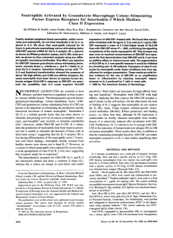
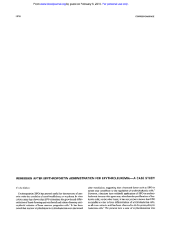
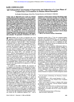
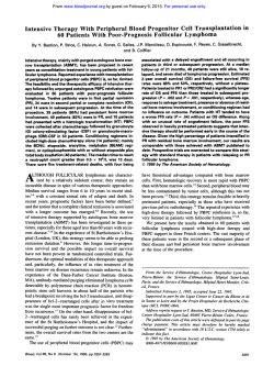
From www.bloodjournal.org by guest on February 6, 2015. For personal use only.
Genetic Basis of Hypo-Responsiveness of A/ J Mice to Interleukin-3
By Kevin B. Leslie, Sheila Jalbert, Paul Orban, Melanie Welham, Vincent Duronio, and John W. Schrader
Hematopoietic progenitor cells of the AIJ strain of mice
show a pronounced defect in the ability t o form colonies or
proliferate in response t o interleukin-3 (IL-3). Comparison of
immunoblots of AIJ mast cells and of mast cells from the
C57BL16 strain that respond normally t o IL-3 showed that,
in both strains, a 125-kD band of the expected size was recognized by an antibody against the p chain of the IL-3 receptor, the AlC2A molecule. However, in the C57BLl6 cells, there
was an additional 110-kD species not seen in cells of the
AIJ strain. Analyses using bone marrow-derived mast cells
from a panel of AIJ x C57BLl6 and A I J x C57BLl6 recombinant inbred (RI) mice showed that the hypo-responsiveness
t o IL-3 is governed by a single gene. However, the absence
of this 110-kD species in the A/J strain did not co-map with
IL-3 hypo-responsiveness but did indeed map t o the AlC2A
genetic locus. These data show that this trait in the AIJ
strain was due t o a polymorphism of the AIC2A gene unrelated t o IL-3 hypo-responsiveness. Typing of the RI strains
for the markers D14Mit98, D14Mit14, and D14Mit133
mapped the locus determining hypo-responsiveness t o IL-3
t o the subtelomeric region of chromosome 14, the region
that also bears the gene encoding the a chain of the IL-3
receptor (IL-3Ra). Immunofluorescence analyses indicated
that IL-3Ra protein was undetectable on fresh bone marrow
cells from A I J mice, although clearly detectable on cells from
the responder C57BLl6 strain. However, IL-3Ra was readily
detectable at normal levels on AIJ mast cells generated by
culture of AIJ bone marrow cells in a combination of IL-3
and steel factor. Moreover, IL-3Ra on these A/J mast cells
appears t o be functional in that IL-3 stimulation of these cells
results in tyrosine phosphorylation events characteristic of
IL-3 signaling, including tyrosine phosphorylation of the p
chain of the IL-3 receptor, Jak-2 kinase, and SHPTP2. Collectively, these data indicate that the hypo-responsiveness of
A/J mice t o IL-3 is due t o a defect in the gene encoding
IL-3Ra and that, although this defect gives rise t o reduced
expression of a chain on primary bone marrow cells, this
defect is not absolute and that, under certain circumstances,
AIJ cells can express functional receptors.
0 1996 b y The American Society of Hematology.
I
the highly homologous AIC2A protein.' AIC2A itself binds
IL-3 with low affinity and, unlike AIC2B, interacts only with
the a chain of the IL-3 receptor (IL-3Ra) and not that of
the GM-CSF or IL-5 receptors. Both IL-3Ra and one of the
two p chains must be expressed on the same cell to form a
functional, high-affinity IL-3 receptor, and the cytoplasmic
domains of both the cy and p chains are needed for generation
of the intracellular signals required for mitogenesis' (Orban
et al, unpublished observations).
Several inbred strains of mice have been reported to be
hyporesponsive to IL-3."' Hematopoietic progenitor cells of
the A/J, AKR, A.TH, and A.TL strains of mice are defective
in their ability to form colonies or proliferate in response to
IL-3.'."' FI hybrids between A/J and the responder C57BL/
6 strain exhibit normal responses to IL-3,4 indicating that
the defect is recessive. F1 hybrids between AKR and A/J
mice fail to respond to IL-3, indicating that these nonresponder strains share defects in a common gene. Although
A/J bone marrow cells respond poorly to 1L-3 alone, they
exhibit a synergistic response to a combination of IL-3 and
CSF-1:'
indicating that responses to IL-3 are not totally
absent.
Using recombinant inbred (RI) mice, we have shown that
the hypo-responsiveness of A/J cells to IL-3 fails to map to
the ,B chain locus and fails to correlate with a polymorphism
in the AlC2A chain of the IL-3 receptor, but instead maps
with 100% concordance to the subtelomeric region of chromosome 14 that bears the gene encoding IL-3Rn and correlates with the defective expression of a chain protein on the
surface of fresh bone marrow cells. These data are consistent
with the report of Ichihara et alHthat there is a 5-bp deletion
in intron 7 of the gene that encodes IL-3Rn in A/J mice and
in other IL-3 hypo-responsive strains' and that this results
in the expression of a defective a chain protein that does
not appear at the cell surface. However, we show that a
certain population of A/J-derived hematopoietic cells, bone
marrow-derived mast cells, can express normal levels of
functional IL-3Ra at the cell surface. Moreover, this IL-3Ra
NTERLEUKIN-3 (IL-3) is produced by immunologically
activated T lymphocytes and mast cells and appears to be
a major factor in the T-lymphocyte-dependent mastocytosis
associated with some parasitic infections. IL-3 stimulates
the survival and proliferation of pluripotential hematopoietic
stem cells and the progenitor cells of all hemopoietic lineages, with the possible exception of those of T lymphocytes.
IL-3 also regulates the survival and function of mature cells
of certain lineages, such as mast cells, basophils, eosinophils.
megakaryocytes, and macrophages.' The cell surface receptor for 1L-3 is composed of at least two chains, a specific a
chain and a common p (,&) chain.' In humans, the pc molecule is also a component of the receptors for IL-5 and granulocyte-macrophage colony-stimulating factor (GM-CSF) in
which it interacts with one of three specific a subunits, each
of which binds specifically to either IL-3, IL-5, or GMCSF.' In the mouse, the ,6 chain of the IL-3 receptor can be
provided by either the AIC2B molecule, which, like the ,Oc
molecule in the human system, is shared with the IL-5 and
GM-CSF receptors, or by a product of a duplicated gene,
~~~~
~~
~
From The Biomedical Research Centre, University of British Columbia, Vancouver, British Columbia, Canada; the Differentiation
Programme, EMBL, Heidelberg, Germany; the School of Pharmacy
und Pharmacology, University of Bath, Claverton Down Bath, UK;
and the Department of Medicine, UBC. Jack Bell Research Centre,
Vancouver, BC, Canada.
Submitted January 23, 1995; accepted November 29, 1995.
Supported by The Medical Research Council of Canada and the
National Cancer Institute of Canada.
Address reprint requests to Kevin B. Leslie, PhD, The Biomedical
Research Centre, 2505 Health Sciences Mall, Vancouver, British
Columbia, V6T IZ3 Canada.
The publication costs of this article were defrayed in part by page
charge payment. This article must therefore be hereby marked
"advertisement" in accordance with 18 U.S.C. section I734 solely to
indicate this fact.
0 1996 by The American Society of Hematology.
0006-4971/96/8708-0044$3.00/0
3186
Blood, Vol 87, No 8 (April 15).1996: pp 3186-3194
From www.bloodjournal.org by guest on February 6, 2015. For personal use only.
3187
HYPO-RESPONSIVENESS OF A/J MICE TO IL-3
appeared to be functional in that IL-3 stimulation of these
cells results in tyrosine phosphorylation events characteristic
of IL-3 signaling, including tyrosine phosphorylation of the
,B chain of the IL-3 receptor, Jak-2 kinase, and SHPTP2.
These results indicate that the hypo-responsiveness of A/J
mice to IL-3 results from a defect in the expression of IL3Ra but that this defect is not absolute and that, under certain
circumstances, A/J cells can express functional receptors.
MATERIALS AND METHODS
Animals and preparation of fresh cells. All mouse strains were
purchased from The Jackson Laboratory (Bar Harbor, ME). Mice
were killed by cervical dislocation or C 0 2 anesthesia, bone marrow
cells were flushed from the femurs using HEPES-buffered Hanks
solution, and red blood cells were removed by hypotonic lysis.
Cell culture. Bone marrow cells were cultured at 106/mL in
humidified incubators at 37"C, 5% CO, (vol/vol) in RPMI 1640
medium (GIBCO BRL, Grand Island, NY) supplemented with 10%
(voVvol) fetal bovine serum (GIBCO BRL), 20 pmol/L 2-mercaptoethanol, 100 U/mL penicillidstreptomycin, and 2 m m o m L-glutamine. Cytokines added to cultures included chemically synthesized
murine IL-3 (1 pg/mL; courtesy of Dr Clark-Lewis, The Biomedical
Research Centre (Vancouver, Canada), synthesized with cysteineto-alanine substitutions at positions 79 and 140 to facilitate folding
of the mature IL-3 protein) and, where indicated, recombinant murine Steel locus factor (SLF; 500 ng/mL). For the typing of RI
strains, bone marrow cells were cultured in JL-3 alone for 3 weeks
with cultures split weekly. Total cells were then counted and the
percentage of mast cells determined by staining with Toluidine Blue
and Astra Blue.
Bone marrow colony assays. Nucleated bone marrow cells were
placed in triplicate I-mL cultures at 2.5 to 5 X IO4 cells/mL in cell
culture medium supplemented with 0.3% agar, selected fetal calf
serum (20%), and growth factors as indicated (Fig lA, 1 pg/mL
synthetic murine IL-3,500 ng/mL recombinant murine SLF; Fig IB,
1 pglmL synthetic murine IL-3, 1 pg/mL factor "mix" consisting
of synthetic murine JL-3, 500 ng/mL recombinant murine SLF, 1.O
U/mL recombinant GM-CSF, 5% of a 10-fold concentrated medium
conditioned by L-cells as a source of CSF-I). Colonies were defined
as collections of 40 cells or greater and were counted on day 7.
Thymidine incorporation assays. Assays were performed according to the method described by Schrader and Crapper." Briefly,
nine aliquots of 3,000 A/J or C57BW6 mast cells (thrice washed
with RPMI 1640) were added to the wells of Terasaki trays (Lux
Corp, Newbury Park, CA) in 15 pL of culture medium containing
synthetic murine IL-3 (2 pg/mL) or recombinant murine SLF (500
ng/mL) or no growth factor. Trays were incubated at 37°C for 2
hours, pulsed for 18 hours with [3H]-thymidine, and harvested onto
glass fiber filters. Filters were washed then counted in a liquid scintillation counter.
Genetic mapping. Genetic mapping was performed using the RI
Manager program, version 2.3 (Kenneth F. Manly, Rosewell Park
Cancer Institute, Buffalo, NY), with linkage evaluation performed
as described by Silver." Mapping information on the AxB and BxA
sets of RI mice was largely from published data.'* However, Dr N.
Gough (The Walter and Eliza Hall Institute of Medical Research,
Melbourne, Australia) generously provided unpublished data on the
mapping of the gene encoding IL-3Ra obtained using a recombinant
inbred panel of C57BU6 x DBAl mice. A strain distribution map
ofthe loci D14Mit98, D14Mit14, and D14Mit133 (loci found in the
subtelomeric region of chromosome 14) for the RI strains in these
panels was constructed with data from polymerase chain reaction
(PCR) amplification of genomic DNA, based on information on
these loci provided on-line by The Whitehead InstituteMIT Genome
Center Genetic Map of The Mouse (Cambridge, MA). Oligonucleotide primers used in PCR amplifications were D14Mit98, 5'TCTTAAATACTCACTCTGTGGTGAGG3' and S'CTCCATGAAGCACCCACAC3'; D 14Mit14, S'GCACATTCCAAAACACATGC3'
and S'GGGATGGTGTCAATCAATCC3'; and D 14Mit133,5'1TGTCAAATAATTGCATGAGGC3' and S'AACTATGACTCAGATTCCAAGTTGG3'. PCR amplifications (30 cycles at 94°C for 45
seconds; 50°C for 1 minute; 72°C for 2 minutes; and 5 seconds of
extensiodcycle) were performed with Taq polymerase (Promega,
Madison, WI).
Cell labeling andflow cytometry. Cells were washed once with
FACS buffer (1 X phosphate-buffered saline [PBS], 2% [voVvol]
fetal bovine serum [FBS]), held for 20 minutes at 4°C with the
3 e ice-cold FACS buffer, held at
primary antibody, washed o ~ ~ with
4°C for 3 minuterwith fluorescein isothiocyanate (F1TC)-labeled
secondary antibody, washed once with ice-cold FACS buffer, and
then resuspended on ice in FACS buffer. The primary antibody
consisted of the F(ab'), fragments of an affinity-purified, rabbit antiserum against a synthetic peptide corresponding to residues Serl7
to Asp67 of the mature IL-3Ra. The peptide incorporated a Cterminal addition of GlyGlyCys to facilitate coupling and was kindly
synthesized by Dr Ian Clark-Lewis (The Biomedical Research Centre, Vancouver, British Columbia, Canada). For affinity purification
of antipeptide antibodies, this peptide was coupled to Sepharose."
F(ab'), fragments were produced by pepsin cleavage of the affinitypurified antiserum.I3 The secondary antibody was a FITC-labeled
goat antirabbit IgG (H&L) F(ab'), reagent (Sigma, St Louis, MO)
used at 1.1 pg/ml. Analysis of the specificity of primary antibody
binding was performed by determining the effect of addition of 500
pg/mL of the synthetic IL-3Ra peptide (Serl7 to Asp67) to diluted
antibodies at 4°C for 5 minutes before the addition of the antibody
mix to the washed target cells. Labeled cells were analyzed by flow
cytometry on a FACScan cell analyzer (Becton Dickinson, San Jose,
CA).
Cell stimulation and preparation of lysates. Stimulation of cells
with the synthetic IL-3 was performed as previously des~ribed.'~,''
Briefly, cells were deprived of growth factor (SLF and IL-3) by
culture for 18 hours in media in which the levels of SLF and IL-3
had been reduced 10-fold, washed three times, and then stimulated
with IL-3 at 10 pg/mL for 10 minutes, conditions previously determined to induce maximal levels of tyrosine pho~phorylation.'~~'~
Cells were washed and cell pellets were lysed in immunoprecipitation buffer (IP buffer: 50 mmol/L TrisHC1, pH 7.5; 10% (vol/vol)
glycerol; 1% (voVvol) NP-40, 150 mmol/L NaC1; 5 mmoVL EDTA;
1 mmoVL sodium orthovanadate; 1 mmol/L sodium molybdate; I O
mmoVL sodium fluoride; 40 pg/mL PMSF; 10 pg/mL aprotinin; 10
pg/mL soyabean trypsin inhibitor; 10 pg/mL leupeptin; 0.7 pg/mL
pepstatin) as previously de~cribed.'~
Nuclei and intact cells were
removed by centrifugation for 1 minute at 4°C in a microfuge at full
speed.
Sodium dodecyl sugaie-polyacrylamide gel electrophoresis (SDSPAGE) and immunoblotting. SDS-PAGE and immunoblotting
were performed as previously de~cribed.'~
The affinity-purified, rabbit anti-AIC2A antiserum was raised and purified using a synthetic
peptide corresponding to the N-terminal 74 residues of the mature
and was used at
murine AIC2A p chain, as described previou~ly,'~
0.5 pg/mL for immunoblotting. Goat antirabbit horse radish peroxidase-conjugated antibody (Dako, Dimension Laboratories, Mississauga, Ontario, Canada) was used at a concentration of 0.05 pg/mL.
Immunoblots were developed using the ECL system (Amersham,
Arlington Heights, IL). Kodak X-AR 5 film (Eastman Kodak,Rochester, NY) was used for detection of ECL signals.
RESULTS
Using colony assays, we confirmed the reports of oththat A/J bone marrow cells were hypo-responsive to
ers4.6.7
From www.bloodjournal.org by guest on February 6, 2015. For personal use only.
LESLIE ET AL
A
T
L
I
0
n
5
z
2,
C
0
0
0
C
Q
MA
11-3
SLF
IL3SLF
AN
BU6
AB1
AB4
B
Fig 1. Hypo-responsiveness of A/J cells to IL-3.
(A) Nucleated bone marrow cells from A/J or C57BL/
6 mice were cultured in semisolid medium in the
presence of 11-3, SLF, or both. Colonieswere counted
on day 7. IB) Nucleated bone marrow cells from A/
J or C57BL/6 or selected RI (AxB1, AxB4, and AxB13)
mice were cultured in semisolid medium in the presence of 11-3 or factor mix IIL-3, GM-CSF, 11-4, and
SLF). Colonies were counted on day 7. The bars indicate the mean colony count per 10' bone marrow
cells r SEM from triplicate cultures. Representative
experiments are shown.
z
100
2,
C
0
0
0
C
m
IL-3 but responded normally to other hemopoietic growth
factors such as CSF-1 or GM-CSF (data not shown). We
also confirmed that A/J cells responded synergistically to a
mixture of IL-3 and CSF-1 with the generation of macrophage colonies. Moreover, in experiments with certain
batches of fetal calf serum (FCS), we observed small numbers of clusters (<40 cells) growing in response to high
concentrations of IL-3 alone, perhaps reflecting synergistic
interactions of IL-3 and certain factors in FCS. We extended
the previous observations of synergistic responses of A/J
bone marrow cells to combinations of IL-3 and other factors
by showing a dramatic synergy of IL-3 and SLF, either factor
alone having no significant colony-stimulating activity (Fig
1A).
Similar results were obtained in liquid cultures. Whereas
liquid cultures of C57BU6 bone marrow cells stimulated
with IL-3 alone proliferated vigorously and, after 3 weeks,
generated homogeneous populations of mast cells, A/J bone
marrow cells generated significantly fewer cells and only a
small percentage of these were mast cells. However, experiments in which bone marrow cells were stimulated with a
mixture of IL-3 and SLF gave strikingly different results.
Thus, in the presence of IL-3 and SLF, A/J bone marrow
cells proliferated as vigorously as the C57BL/6 bone marrow
cells, with a 50- to 100-fold increase in total cell count over
an 8-day culture period. Cells from cultures of both A/J and
C57BU6 bone marrow cells had the histochemical characteristics of mast cells with prominent cytoplasmic granules
staining with toluidine blue or astra blue (data not shown).
50
n
AB13
Analysis of hypo-responsiveness to IL-3 using a panel of
recombinant inbred mice. The availability of a panel of RI
mice in which one parental strain was A/J (hypo-responsive
to IL-3) and the other C57BU6 (responsive to IL-3) offered
an opportunity to determine whether IL-3 hypo-responsiveness of A/J mice was controlled by a single gene and
the possibility of mapping this locus. Panels of strains of A/
J x C57BL/6 (AxB) and C57BU6 X A/J (BxA) RI mice'*
were typed by analysis of the ability of bone marrow progenitor cells to respond to IL-3 with colony formation in agarcolony assays (Fig 1B) and for their capacity to generate
mast cells in a 3-week liquid culture in the presence of IL3 alone. The results of these two assays were concordant.
The absence of an intermediate phenotype in the strain distribution pattern suggested that the trait was controlled by a
single IOCUS.'~
A polymorphism of the 0 chain of the IL-3 receptor in A/
J mice. Immunoblots of extracts from cells of C57BU6
origin that were analyzed with affinity-purified antibodies to
the N-terminus of AIC2A showed two major bands, one
corresponding to a molecular weight of about 125 kD and
a second of only 110 kD (Fig 2). The 125-kD protein corresponded to the expected electrophoretic mobility of the
AIC2A 0 chain of the IL-3 receptor, as identified in crosslinking studies with radiolabeled IL-3." Moreover, this
125-kD protein was rapidly tyrosine phosphorylated after
stimulation with IL-3, with a consequent retardation of electrophoretic m0bi1ity.I~In contrast, the lower 110-kD band
did not correspond to any species identified in cross-linking
From www.bloodjournal.org by guest on February 6, 2015. For personal use only.
HYPO-RESPONSIVENESS OF NJ MICE TO IL-3
125 kD110 kD-
3189
I
contain the AIC2A gene locus.” Collectively these data indicate that the presence of the I IO-kD form of AIC2A is
determined by a polymorphism of the gene encoding AIC2A.
However, the distinct strain distribution patterns of IL-3
hypo-responsiveness and the polymorphism in the IL-3 receptor 0 chain (Table 1 ) indicate that the AIC2A locus was
not linked to the trait of IL-3 hyporesponsiveness.
IL-3 h~poresponsivettessmaps to the locus containing the
IL-3Ra gene. The chromosomal localization of the gene
encoding IL-3Ra was mapped by Dr N. Gough to the subtelomeric region of chromosome 14 using a different panel of
RI mice (personal communication) and subsequently by Miyajima et d.’*There was a lack of information for the AxB
and BxA R1 panels on the strain distribution patterns of loci
in this region of chromosome 14. Thus, to test the hypothesis
that the hypo-responsiveness of A/J mice to IL-3 maps to
the gene for IL-3Ra, it was necessary to type the AxB and
BxA RI mice for the D14Mit98, D14Mitl4, and D14Mit133
loci, loci known to reside in the subtelomeric region of chromosome 14 and known to exhibit polymorphic differences
between A/J and CS7BW6 strains.
PCR amplification of genomic DNA from RI mice was
performed using primer pairs specific for each of the markers. These analyses showed tight concordance between the
most telomeric of these markers, D14Mit98, and the trait of
IL-3 hypo-responsiveness (P < .OOOS; Table 2). Thus, like
the gene encoding IL-3Ra. the hypo-responsiveness of A/J
mice to IL-3 mapped to the subtelomeric region of chromosome 14. These mapping experiments showed that the IL-3
hyporesponsiveness of the A/J strain was due to a defect in
a single gene and that the defective gene was either that
encoding IL-3Ra or a gene closely linked to the IL-3Ra
locus.
IL-3 receptor a chain cannot he detected onfresh A/J hone
marrow cells. Our genetic studies showing tight linkage of
the trait of hypo-responsiveness to IL-3 and the locus bearing
the IL-3Ra gene suggested that the hypo-responsiveness to
1L-3 was due to a defect in the IL-3Ra. To test this hypothesis, we investigated whether there were abnormalities in the
expression of the IL-3Ra protein in A/J mice. Fresh bone
marrow cells from CS7BL/6 or A/J mice were examined by
flow cytometry using F(ab’)2 fragments of an affinity-purified polyclonal antibody specific for the a chain of the IL3 receptor. In control experiments, these anti-IL-3Ra anti-
I
AB2
AJ
66
AB10
Fig 2. A polymorphism in the p chain of the IL-3 receptor from A/
J bone marrow cells. Nucleated bone marrow cells from A/J and
C57BL/6 mice and from the RI panels (representative RI strains shown
here: AxBZ and AxB10) were cultured for 14 days in SLF and IL-3 and
starved of IL-3 overnight, and cell lysates were prepared. After SDSPAGE of whole cell lysates through a 6.5% acrylamide gel, immunoblotting was performed with an anti-AICZA antibody. The relative
positions of AICZA-immunoreactive species are indicated.
experiments and was not phosphorylated on tyrosine in response to IL-3. Interestingly, unlike the 125-kD species, the
1 IO-kD species is not bound by a concanavalin-A Sepharose
column, suggesting that the two species are glycosylated
differently (Duronio et al, data not shown).
Mast cells were generated from hyporesponder A/J and
responder CS7BL/6 (BL/6) mice by culturing bone marrow
cells with IL-3 and SLF, and immunoblots were prepared of
whole-cell lysates from these cells using the anti-0 chain
antibody. The expected 125-kD AIC2A protein could be
detected in both CS7BL/6 and A/J samples. In the A/J samples, the larger 125-kD species migrated slightly faster than
the corresponding band in the CS7BL/6 samples and could
be resolved as a doublet when separated on a larger gel.
However, the additional I IO-kD band that was observed in
the CS7BL/6 responder mice was entirely absent in cells
from the A/J hyporesponder mice (Fig 2).
IL-3 h?po-responsiveness does not co-map with the 0
chain polvmorphism. To determine whether the absence of
the 110-kD form of AIC2A correlated with hypo-responsiveness to IL-3, mast cells were generated from fourteen
strains of the RI mice by culture of bone marrow cells in
IL-3 and SLF and analyzed by immunoblotting. As summarized in Table I , there was a clear discordance between
inheritance of the A/J-type absence of the 1 IO-kD isoform of
AIC2A and hypo-responsiveness to IL-3. Genetic mapping
analyses indicated that the absence of the 1 IO-kD band
mapped tightly to a region on chromosome IS close to c-sis
(Table 1 and Fig 2), the region subsequently reported to
Table 1. IL-3 HvDo-ResDonsivenessof the A/J Strain Does Not Mar, to ReceDtor 4 Chain Locus
Recombinant Inbred Strain
Locus
AB1
AB4
AB5
AB6
AB11
AB13
AB14
AB15
AB19
AB20
AB23
AB24
EA7
EA8
IL-3 resp.
A
B
B
A
A
A
B
B
B
B
B
B
B
B
X
X
X
X
X
X
X
A
X
X
AIC-PA
A
B
A
A
B
B
B
A
A
A
A
A
B
Sis
A
B
A
A
B
B
B
A
A
A
A
A
A
X
A
Bone marrow cells from recombinant inbred mice were typed for their responsiveness to IL-3 (IL-3 resp.) and for the AlC2A protein polymorphism (p110 protein) detected by immunoblotting. Published phenotypes are given for the locus C - S ~ S , ’ ~the gene tightly linked with the IL-3
receptor 0 chain locus.” Phenotypes are indicated as A, NJ phenotype of hypo-responsiveness; B, C57BU6 phenotype or responsiveness; U,
unknown. Crosses indicate mismatches between adjacent loci.
From www.bloodjournal.org by guest on February 6, 2015. For personal use only.
3190
LESLIE ET AL
Table 2. IL-3 Hypo-Responsiveness of the A I J Strain Maps to IL-3 Receptor a Chain Locus in the SuMelomeric Region of Chromosome 14
Recombinant Inbred Strain
Locus
IL-3 resp.
D14Mit98
AB1
AB4
AB5
AB6
AB11
AB13
AB14
AB15
AB19
AB20
AB23
AB24
BA7
BA8
cM
A
A
B
B
B
B
A
A
A
A
B
B
B
B
B
B
B
B
B
B
B
B
B
B
-
B
B
A
B
B
B
B
X
A
B
B
8.9
B
B
A
B
B
8.9
B
A
B
A
A
X
B
B
A
B
B
X
D14Mit14
A
B
X
0.0
X
D14Mit133
A
A
X
Np-2
Tcra
Xmv-19
B
B
B
X
A
A
A
B
B
B
A
A
A
B
B
B
B
B
B
B
B
B
U
B
B
B
B
B
B
B
B
A
X
B
B
X
X
X
A
A
A
B
B
B
A
A
A
A
U
A
16
18
18
Bone marrow cells from recombinant inbred mice were typed for their responsiveness to IL-3 (locus IL-3 resp.). Phenotypes (PCR polymorphisms) were determined for the loci D14Mit98, D14Mit14, and D14Mit133. Published phenotypes are given for the loci Np-2,12 Tcra,'2 and Xmv19.12 Phenotypes are indicated as A, A/J phenotype of hypo-responsiveness; B, C57BU6 phenotype of responsiveness; U, unknown. Crosses
indicate mismatches between adjacent loci. The column c M indicates the distance in centimorgans from the chromosomal telomere as estimated
by The Whitehead Institute/MIT Genome Center.
bodies were shown to bind to CTLL cells transfected with
the murine IL-3Ra, but not to nontransfected CTLL cells.
Moreover, this binding could be specifically inhibited using
the synthetic peptide corresponding to residues 17-67 of IL3Ra against which the antibody was raised, but not by a
control peptide (Orban et al, unpublished observations).
Flow cytometry showed that a small but significant proportion of the fresh C57BL/6 bone marrow cells were clearly
recognized by the anti-IL-3Ra antibody and this staining
could be specifically inhibited by competition with the IL3Ra peptide (Fig 3A). In marked contrast, there was only a
low amount of binding of the anti-IL-3Ra antibodies to the
A/J marrow cells, and this binding was not significantly
reduced in the presence of competing peptide ( P > .05; Fig
3B). This low or nonspecific binding of the anti-IL-3Ra
antibodies to A/J bone marrow cells was a consistent finding
in multiple, independent experiments. Together with the genetic studies that show a tight linkage of the IL-3Ra locus
and the trait of IL-3 hypo-responsiveness, these findings indicate that the hypo-responsiveness to IL-3 of A/J mice is due
to defective expression of the a chain of the IL-3 receptor.
IL-3 can induce tyrosine phosphorylation in A/J bone marrow-derived mast cells. Despite this apparent absence of
IL-3Ra protein on fresh bone marrow cells from A/J mice
(Fig 3B), we (Fig 1) and other^^,'.^ have shown that A/J bone
marrow cells are capable of responding to IL-3 when assayed
by colony formation in the presence of such synergistic
growth factors as CSF-1 or SLF. We therefore examined
whether mast cells generated by culture of A/J bone marrow
for 14 days with IL-3 and SLF were able to respond to IL3 with the same pattern of intracellular responses as do mast
cells from C57BL/6 mice. Stimulation of A/J mast cells
with IL-3 produced a pattern of tyrosine phosphorylation on
membrane and cytosolic proteins identical to that seen with
C57BL/6 mast cells (Fig 4A). In particular, stimulation with
IL-3 resulted in tyrosine phosphorylation of the 135- to 145kD p chain of the IL-3 receptor (Fig 4A) and of the 120kD protein tyrosine kinase Jak2 (Fig 4B). Also visible (Fig
4A) are other bands characteristically phosphorylated on ty-
rosine in response to IL-3, namely a 70-kD band that we have
previously shown is the tyrosine phosphatase SHPTP2,'4 a
55-kD band that we have shown is Shc" and an uncharacterized 90-kD band.I5 However, although the pattern of tyrosine
phosphorylation in A/J mast cells stimulated with IL-3 was
qualitatively identical to that seen in C57BL/6 mast cells,
the levels of tyrosine phosphorylation were significantly and
reproducibly lower. For example, the level of phosphorylation of Jak2 was several-fold lower in IL-3-simulated A/J
mast cells than in C57BL/6 mast cells (Fig 4B). Consistent
with these findings of qualitatively normal IL-3 receptormediated signal transduction and tyrosine phosphorylation,
PCR-cloning and sequencing of a cDNA fragment encoding
the trans-membrane region and cytoplasmic tail of A/J IL3Ra showed that the A/J-derived cDNA sequence was identical to the C57BL/6-derived cDNA sequence (data not
shown).
We then tested whether IL-3, in addition to inducing tyrosine phosphorylation, was also capable of delivering a mitogenic signal to A/J bone marrow-derived cells. A/J and
C57BL/6 bone marrow cells were cultured in SLF and IL3 for 7 days, after which they were washed extensively,
exposed to medium alone or IL-3 or SLF for 2 hours, and
then pulsed for 18 hours with [3H]-thymidinein the presence
of the respective cytokine. IL-3 stimulated both A/J and
C57BL/6 cells to incorporate comparable amounts of ['HIthymidine over the 18-hour incubation period (Fig 5). These
results suggest that, in these specific culture conditions, IL3 is capable of delivering a mitogenic signal to both A/J and
C57BL/6 cells. However, unlike the C57BL/6 cells, A/J cells
were incapable of sustained survival and proliferation in the
presence of IL-3 alone. Thus, parallel experiments in which
the ['HI-thymidine pulse was delayed showed that, after 3
days of culture in IL-3 alone, A/J cells were dead and incapable of incorporating [3H]-thymidine (data not shown).
IL-3 receptor a chain can be detected on A/J bone marrow-derived mast cells. Although we were unable to detect
IL-3Ra on fresh A/J bone marrow cells (Fig 3B), we hypothesized that the transient responsiveness of A/J cells derived
From www.bloodjournal.org by guest on February 6, 2015. For personal use only.
HYPO-RESPONSIVENESS OF AJJ MICE TO 11-3
A
B
C
D
E
3191
by culture in the presence of SLF and IL-3 might reflect a
higher level of the surface expression of IL-3Ra. To address
this hypothesis, nucleated bone marrow cells from C57BU
6 and A/J mice were cultured in the presence of SLF and
IL-3 for 7 days, starved of IL-3 by culture in SLF alone for
2 days, and then examined by flow cytometric analysis for
cell surface expression of IL-3Ra. IL-3Ra could readily be
detected on a significant proportion of cells from cultures
generated from both C57BU6 (Fig 3C) and A/J (Fig 3D)
bone marrow. However, if after culture for 7 days in SLF
and IL-3 the C57BU6 and A/J bone marrow-derived cells
were maintained in IL-3 alone for 2 days instead of in SLF
alone, although the level of IL-3Ra expressed on the surface
of C57BU6 cells was considerably reduced (Fig 3E), we
were unable to detect any IL-3Ra protein on the surface of
A/J cells (Fig 3F).
Cultures of C57BU6- or A/J-cell-derived bone marrow
cells supplemented with both SLF and IL-3 for 7 days generated similar numbers of IL-3Ra-positive cells ( 1 I % to 40%
in 4 independent experiments) and the levels of IL-3Ra
expressed on positive cells and the proportion of IL-3Rapositive cells were comparable. Similar findings were obtained when day 14 cultured bone marrow cells were analyzed, although the proportion of IL-3Ra-positive cells in
each culture had increased somewhat. Histochemical staining of these cultured cells from C57BL/6 and A/J bone marrow confirmed that the medium-sized cells that by flow cytometry stained with the IL-3Ra-specific antibodies
contained astra blue and toluidine blue-positive granules,
identifying them as immature bone marrow-derived mast
cells. Thus, whereas IL-3Ra cannot be detected on fresh A/
J bone marrow cells, it can readily be detected on immature
mast cells generated in vitro in the presence of IL-3 and
SLF.
L-&L
A.,L
L,L
L?,L
le
F
Fig 3. (I Chain of the 11-3 receptor can be detected on bone marrow-derived mast cells from A/J mice, but not on fresh nucleated
bone marrow cells. Bone marrow-derived cells were generated by
culture of bone marrow from C57BL/6 (C and E) or A/J (D and F)
mice for 7 days in IL-3 and SLF and then for 2 days in the presence
of SLF alone (C and D) or 11-3 alone (E and F). Cultured bone marrowderived cells or fresh bone marrow cells from C57BL/6 (A) or A/J
IB) mice were analyzed for surface expression of IL-3Ra. Cells were
stained with an anti-IL-3Ra antibody (unfilled regions), without (left
panels) or with (right panels) competing IL-3Ra synthetic peptide.
Solid regions represent fluorescence with secondary antibody alone
in the absence of the specific anti-IL-3Ra antibody.
DISCUSSION
A number of genetic defects result in impaired or altered
signaling from cell surface receptors in response to soluble
or membrane-bound cytokines. For example, achondroplasia
is due to a mutation in a receptor for fibroblast growth factor,2" some forms of human X-linked severe combined immunodeficiency result from defects in one chain of the receptor for IL-2,2' and mutations in c-kit, the receptor for steel
factor, result in anemia and defects in germ cells, skin, and
brain in mice2*and in the skin in humans." We confirmed
that the defect in IL-3 responsiveness in A/J mice is not
absolute, providing further evidence that IL-3 can synergize
with growth factors other than CSF-I by showing a synergy
between IL-3 and SLF on A/J cells. We have shown here
that the defective response to IL-3 of hematopoietic progenitor cells from NJ bone marrow cells fails to map to the B
,
chain of the IL-3 receptor or to correlate with a newly described polymorphism in AIC2A, but maps with 100% concordance to the subtelomeric region of chromosome 14 that
bears the gene for the a chain of the IL-3 receptor. Moreover,
cytofluorometric data showed that there was defective expression of the IL-3Ra protein on fresh NJ bone marrow
cells.
Since the submission of our manuscript, Ichihara et al'
From www.bloodjournal.org by guest on February 6, 2015. For personal use only.
LESLIE E T A L
3192
A
B
-AJ
IPAb :aJak-2
AJ B6
M c 3c3
B6
--
c3c3
2
o
o
r
Blot Ab :
116
97.4
1
1
qAic2A
qploo
200
aJak-2
116
Blot Ab : 97.4
aPY
66.2
45.
aAIC2A
Blot Ab :
116 1-bic2A
97.4
I 2 4 -.F
Blot Ab :
d a k - 2 116-
LJak-2
Fig 4. IL-3 induces tyrosine phosphorylation in A/J and C57BL/6 bone marrow-derived mast cells. Cells were generated by culturing
nucleated bone marrow cells from A/J and C57BL/6 mice for 14 days in SLF and IL-3 and starved of IL-3 overnight, and then cell lysates were
prepared before (C) or after (3) stimulation for 10 minutes with synthetic IL-3. (A) SDS-PAGE and immunoblotting of whole cell lysates with
an antiphosphotyrosine antibody (upper panel) or an anti-AICZA antibody (lower panel). (BI SDS-PAGE of anti-Jak2 immunoprecipitates
immunoblotted with an antiphosphotyrosine antibody (upper panel) or an antizlak2 antibody (lower panel). The relative positions of molecular
weight markers (left) and prominent bands (right) are indicated.
have described a 5-bp deletion in intron 7 of the gene encoding the a chain of the IL-3 receptor that is present in A/J
mice and other IL-3 hyporesponsive strains,' but not in the
responder CS7BL/6 strain. Ichihara et alx show that this intronic deletion results in aberrant splicing of the primary IL3Ra gene transcript resulting in a mature mRNA that lacks
the sequence corresponding to exon 8. They show that this
aberrant splicing results in the expression of a defective IL3Ra protein that does not appear at the cell surface, providing an explanation for the lack of responsiveness of A/J cells
to IL-3. However, using the AxB and BxA recombinant
inbred panels of mice, Ichihara et alx were unable to show
a significant linkage between the trait of IL-3 hyporesponsiveness and their most proximal chromosome 14 polymorphic marker, D 14Mit 14. They thus reasoned that recombination events must have occurred in the strains that show
different strain distribution pattems (SDP) between IL-3
hypo-responsiveness and D 14Mit14 genotype. By character3000
0 MA
izing the SDP of a more proximal polymorphic marker on
chromosome 14, D14Mit98. we showed that indeed the A/
J hypo-responsiveness to IL-3 maps with 100%concordance
2000
to the subtelomeric region of chromosome 14.
However, in contrast to the findings of Ichihara et al,' we
0
found that IL-3Ra protein could be detected in significant
levels on the surface of a subpopulation of primary A/J
lo00
hematopoietic cells identified as immature mast cells. Our
demonstration that A/J bone marrow-derived mast cells can
express normal levels of IL-3Ra at the cell surface suggests
that there may be tissue or cell-specific variation in the cell0
surface expression of IL-3Ra in A/J mice and therefore variAJ
B6
ation in the apparent phenotype of IL-3 hypo-responsiveness.
Fig 5. IL-3 induces uptake of ['HI-thymidine by A/J and C57BL/6
Short-term (4-hour or 24-hour) incubations of fresh A/J or
bone marrow-derived mast cells. Cells were generated from A/J and
CS7BL/6
bone marrow cells in SLF and IL-3 had no effect
C57BL/6 mice by culture of primary bone cells for 7 days in SLF and
11-3. Cells were washed extensively, exposed t o medium alone (0). on surface expression of IL-3Ra (unpublished observations),
IL-3 alone (W), or SLF alone (B)
for 2 hours, and then pulsed for 18
confirming the earlier findings of Sato et al." The expression
hours with ['HI-thymidine in the presence of that cytokine. Results
of IL-3Ra on a subpopulation of A/J cells generated in a 7are expressed as the mean of 9x replicates, less the mean counts
day culture of bone marrow cells with IL-3 and SLF most
for incorporation of ['HI-thymidine in the presence of medium alone
likely resulted from the generation of a cell population in
(A/J, 900 cpm; C57BL/6, 754 cpm). Bars indicate the standard error
of the mean of 9x replicate measurements.
which cell-surface IL-3Ra was expressed at levels compara-
E
From www.bloodjournal.org by guest on February 6, 2015. For personal use only.
HYPO-RESPONSIVENESS OF N J MICE TO IL-3
ble with those on a parallel population in cultures of C57BL/
6 cells. These cells are likely to be mast cells or their progenitors as the frequency of IL-3Ra-positive cells correlated
with the frequency of toluidine blue/astra blue-positive mast
cells. The enhanced expression of cell-surface IL-3Ra on
A/J bone marrow-derived mast cells generated in the presence of IL-3 and SLF was not shared by A/J serosal mast
cells in vivo. Thus, we were unable to detect surface expression of IL-3Ra protein on peritoneal mast cells (and macrophages) derived from A/J mice using flow cytometry (unpublished observations). We showed that IL-3Ra expressed on
these cultured A/J bone marrow-derived cells was functional
with respect to (1) the induction of phosphorylation on tyrosine on a characteristic set of membrane and cytosolic proteins in response to IL-3 (Fig 4A and B) and (2) IL-3stimulated DNA synthesis (Fig 5). The IL-3 -stimulated tyrosine phosphorylation events did not represent mere potentiation or prolongation of a response to any residual SLF not
removed by washing, because we observed tyrosine phosphorylation on proteins such as the p chain of the IL-3
receptor, the Jak2 kinase,25and the SHPTP2 phosphatase14
that are known to be phosphorylated in response to IL-3 but
not, in these cells, in response to SLF. Although the pattern
of phosphorylation on tyrosine on stimulation of A/J cells
with IL-3 was qualitatively indistinguishable from that seen
in C57BLJ6 cells, the level of phosphorylation of specific
substrates (such as Jak2) was reproducibly lower, consistent
with our flow cytometric data suggesting that the level of
IL-3Ra is in fact lower on these IL-3Ra-positive cultured
A/J cells than on similar C57BL/6 cells (Fig 3C and D).
Although exposure to IL-3 alone clearly stimulated DNA
synthesis at 24 hours in A/J bone marrow cells that have
been cultures in SLF and IL-3 for 7 days, this response was
transient, and at 3 days the cells were dead. It is possible
that the observed mitogenic response to IL-3 alone reflected
a synergistic effect on cells that had previously received a
signal from SLF. Alternatively, the failure of IL-3 to support
sustained mitogenesis of the A/J cells might result from the
inability of the A/J cells to maintain sufficient levels of
functional IL-3Ra on the cell surface when cultured in the
presence of IL-3 alone in the absence of SLF. This hypothesis is consistent with our observation that when cells generated from A/J bone marrow by culture in the presence of
SLF and IL-3 for 7 days were maintained for a further 2
days in the presence of IL-3 alone, the expression of IL3Ra on the cell surface of these A/J cells decreased to undetectable levels (Fig 3F). However, when the same cultured
cell population was maintained for 2 days in the presence
of SLF alone, the expression of IL-3Ra on the A/J cells was
maintained at normal levels (Fig 3E). These data would be
consistent with a direct or indirect effect of SLF on the cellsurface expression of IL-3Ra on A/J mast cells, perhaps
reflecting regulation of the frequency of the abnormal splicing events detected by Ichihara et a1.' Despite the inability
of IL-3 alone to sustain proliferation of A/J cells under these
culture conditions, when cell lines were derived from fetal
spleen of A/J and C57BL/6 mice using a retroviral vector
directing the expression of HOX11, one of two A/J-derived
lines exhibited sustained growth in the presence of IL-3
3193
alone, whereas all three of three C57BLJ6-derived lines proliferated in IL-3 alone (unpublished observations). The fact
that we were able to derive a cell-line from A/J mice that
grew continuously in IL-3 is consistent with the notion that
A/J mice can express IL-3Ra that participates in a fully
functional IL-3 receptor. Based on the findings of Ichihara
et a1,8 it is likely that the IL-3Ra we detected on N J bone
marrow-derived mast cells is the product of normally spliced
IL-3Ra mRNA and would be predicted to function normally.
However, it should be noted that, whereas Ichihara et a18
have shown that the product of the aberrantly spliced IL3Ra gene transcript is not expressed on the cell surface of
transfected COS cells or fresh bone marrow, the question
remains as to whether it can be expressed on cells such as
bone marrow-derived mast cells.
Our findings of decreased expression of IL-3Ra on fresh
bone marrow cells appear to conflict with those of an earlier
study that showed normal binding of Iz5I-IL-3 to bone marrow cells, as assessed by autoradiography.' We have shown
that the hyporesponsiveness to IL-3 of A/J mice is also evident when IL-3 is administered in vivo (unpublished observations), excluding the possibility that the defect in IL-3
responsiveness and in expression of the a chain of the receptor is restricted to the in vitro situation. However, it is possible that the autoradiographic study measured binding of IL3 to the AIC2A p chain that can bind IL-3, albeit with low
affinity.26
Our data clearly showed that the defect in A/J mice was
unrelated to AIC2A and did not map to the genetic locus
bearing the AIC2B gene. However, the difference we detected between the AIC2A molecule from A/J and C57BL/
6 strains is of some interest. Using specific anti-AIC2A antibodies, we showed that the 110-kD molecule was present in
C57BL/6 mast cells but was absent in AIJ cells. Our analyses
of recombinant inbred mice not only showed that the absence
of this 110-kD molecule was irrelevant to the defective response to IL-3 in A/J mice (Table l), but also established
that this trait mapped to a locus near sis, the region now
known to contain the AIC2A gene.I7These data indicate that
the 110-kD species is indeed an alternatively processed form
of the AIC2A p chain, resulting from a difference in coding
or noncoding sequences of the AIC2A gene in A/J and
C57BL/6 mice. The 110-kD species could result from an
alternatively spliced AIC2A message, an alternative glycoform of AIC2A, or a cleaved product of the mature AIC2A
protein. However, the failure of the 110-kD AIC2A species
to undergo tyrosine phosphorylation in response to IL-3 or
to be cross-linked to IL-3Ra with 1251-IL-3L5.27
(Duronio et
al, unpublished observations) indicates that the 110-kD species probably does not participate in IL-3 signal transduction.
Our data on fresh A/J bone marrow cells agree with the
findings of Ichihara et a18 and indicate that the genetic basis
of the hyporesponsiveness to IL-3 in A/J mice is a defect in
the gene encoding IL-3Ra. However, our finding that the
IL-3Ra protein can be expressed at normal levels on bone
marrow-derived mast cells and can function suggest that the
defect is ultimately a quantitative one. The existence of tissue-specific RNA splicing in cells of the hematopoietic system is well established and is perhaps best exemplified by
From www.bloodjournal.org by guest on February 6, 2015. For personal use only.
3194
LESLIE ET AL
the numerous splice-generated variants of the CD45 molecule found on the surface of certain lineages of hematopoietic
cells.** One possible explanation for the difference in the
levels of IL-3Ra seen between bone marrow-derived mast
cells and the bulk of other hematopoietic cells found in
primary bone marrow may relate to tissue-specific or cell
lineage-specific splicing of the primary IL-3Ra gene transcript, with a consequent tissue-specific or cell lineage-specific expression of the wild-type or defective forms of the
IL-3Ra protein.
ACKNOWLEDGMENT
We thank David Fong, Elizabeth Hajen, and Francis Lee for excellent technical assistance. We also thank Dr N. Gough for sharing
his unpublished data on the chromosomal mapping of the gene encoding IL-3Ra; Drs Joseph Nadeau, Emil Skamene, and Francine
Gervais of McGill University, Montreal, for helpful discussion on
genetic mapping with recombinant inbred mice; and Drs Ian ClarkLewis and Stewart Berger of The Biomedical Research Centre for
synthetic or recombinant cytokines.
REFERENCES
I . Kaushansky K, Karplus PA: Hematopoietic growth factors:
Understanding functional diversity in structural terms. Blood
82:3229, 1993
2. Miyajima A, Mui AL, Ogorochi T, Sakamaki K: Receptors for
granulocyte-macrophage colony-stimulating actor, interleukin-3 and
interleukin-5. Blood 82: 1960, 1993
3. Kitamura T, Miyajima A: Functional reconstitution of the human interleukin-3 receptor. Blood 80:84, 1992
4. Moms CF, Salisbury J, Kobayashi M, Townsend PV, Hapel
AJ: Interleukin 3 alone does not support the proliferation of bone
marrow cells from A/J mice: A novel system for studying the synergistic activities of IL-3. Br J Haematol 74:131, 1990
5. Kincade PW, Lee G, Fernandes G, Moore MA, Williams N,
Good RA: Abnormalities in clonable B lymphocytes and myeloid
progenitors in autoimmune NZB mice. Proc Natl Acad Sci USA
76:3464, 1979
6. Breen FN, Hume DA, Weidemann MJ: The effects of interleukin 3 (IL-3) on cells responsive to macrophage colony-stimulating
factor (CSF-I) in liquid murine bone marrow culture. Br J Haematol
74:138, 1990
7. Hapel AJ, Fung M-C, Mak N-K, Morris C, Metcalf D, Nicola
N: Bone marrow cells from A/J mice do not proliferate in interleukin3 but express normal numbers of interleukin-3 receptors. Br J
Haematol 82:488, 1992
8. Ichihara M, Hara T, Takagi M, Cho LC, Gorman DM, Miyajima A: Impaired interleukin-3 (IL-3) response of the A/J mouse is
caused by a branch point deletion in the IL-3 receptor CY subunit
gene. EMBO J 14:939, 1995
9. Hara T, Ichihara M, Takagi M, Miyajima A: Interleukin-3 (IL3) poor-responsive inbred mouse strains carry the identical deletion
of a branch point in the IL-3 receptor alpha subunit. Blood 85:2331,
I995
10. Schrader JW, Crapper RM: Autogenous production of hematopoietic growth factor “P cell stimulating factor” as a mechanism
for transformation of bone marrow-derived cells. Proc Natl Acad
Sci USA 80:6892, 1983
1 1. Silver J: Confidence limits for estimates of gene linkage based
on analyses using recombinant inbred strains and backcrosses. J
Heredity 76:436, 1985
12. Marshall JD, Mu J-L, Cheah Y-C, Nesbitt MN, Frankel WN,
Paigen B: The AxB and BxA set of recombinant inbred mouse
strains. Mammalian Genome 3:669, 1992
13. Goding JW: Monoclonal Antibodies: Principles and Practices.
London, UK, Academic, 1986
14. Welham MJ, Dechert U, Leslie KB, Jirik F, Schrader JW:
Interleukin (1L)-3 and granulocytehacrophage colony-stimulating
factor, but not IL-4, induce tyrosine phosphorylation, activation, and
association of SHPTP2 and Grb2 and phosphatidylinositol 3’-kinase.
J Biol Chem 269:23764, 1994
15. Duronio V, Clark-Lewis I, Federsppiel B, Wieler JS, Schrader
JW: Tyrosine phosphorylation of receptor p subunits and common
substrates in response to interleukin-3 and granulocyte-macrophage
colony-stimulating factor. J Biol Chem 267:2 1856, I992
16. Festing MFW: Notes on genetic analysis, in Festing MFW
(ed): Inbred Strains in Biomedical Research. London, UK, MacMillan, 1979, p 81
17. Gorman DM, Itoh N, Jenkins NA, Gilbert DJ, Copeland NG,
Miyajima A: Chromosomal localization and organization of the murine genes encoding the beta subunits (AIC2A and AIC2B) of the
interleukin 3, granulocytehacrophage colony-stimulating factor.
and interleukin 5 receptors. J Biol Chem 267:15842, 1992
18. Miyajima I, Levitt L, Hara T, Bedell MA, Copeland NG,
Jenkins NA, Miyajima A: The murine interleukin-3 receptor subunit
gene: Chromosomal localization, genomic structure, and promoter
function. Blood 85:1246, 1995
19. Welham MJ, Duronio V, Leslie KB, Bowtell D, Schrader JW:
Multiple hemopoietins, with the exception of interleukin-4, induce
modification of Shc and mSos1, but not their translocation. J Biol
Chem 269:21165, 1994
20. Chiang R, Thompson LM, Zhu YZ, Church DM, Fielder TJ.
Bocian M, Winokur ST, Wasmuth JJ: Mutations in the transmembrane domain of FGFR3 cause the most common genetic form of
dwarfism, achondroplasia. Cell 78:335, 1994
2 I. Noguchi M, Yi H, Rosenblatt HM, Filipovich AH, Adelstein
S, Modi WS, McBride OW, Leonard WJ: Interleukin-2 receptor
gamma chain mutation results in X-linked severe combined immunodeficiency in humans. Cell 73:147, 1993
22. Chabot B, Stephenson DA, Chapman VM, Besmer P, Bernstein A: The proto-oncogene c-kit encoding a transmembrane tyrosine kinase receptor maps to the mouse W locus. Nature 335:88,
1988
23. Giebel LB, Spritz RA: Mutation of the KIT (mastlstem cell
growth factor receptor) protooncogene in human piebaldism. Proc
Natl Acad Sci USA 8823696, 1988
24. Sato N, Caux C, Kitamura T, Watanabe Y, Arai K, Banchereau J, Miyajima A: Expression and factor-dependent modulation of
the interleukin-3 receptor subunits on human hemopoietic cells.
Blood 82:752, 1993
25. Silvennoinen 0, Witthuhn BA, Quelle FW, Cleveland JL, Yi
T, Ihle JN: Structure of the murine Jak2 protein-tyrosine kinase and
its role in interleukin 3 signal transduction. Proc Natl Acad Sci USA
90:8429, 1993
26. Hara T, Miyajima A: Two distinct functional high affinity
receptors for mouse interleukin-3 (IL-3). EMBO J 1 I :1875, 1992
27. Duronio V, Clark-Lewis I, Schrader JW: Two polypeptides
identified by interleukin 3 cross-linking represent distinct components of the interleukin 3 receptor. Exp Hematol 20:505, 1992
28. Thomas ML: The leukocyte common antigen family. Annu
Rev Immunol 7:339, 1989
29. Leclair KP, Rabin M, Nesbitt MN, Pravtcheva D, Ruddle FH,
Palfree RG, Bothwell A: Murine Ly-6 multigene family is located
on chromosome 15. Proc Natl Acad Sci USA 84:1638, 1987
From www.bloodjournal.org by guest on February 6, 2015. For personal use only.
1996 87: 3186-3194
Genetic basis of hypo-responsiveness of A/J mice to interleukin-3
KB Leslie, S Jalbert, P Orban, M Welham, V Duronio and JW Schrader
Updated information and services can be found at:
http://www.bloodjournal.org/content/87/8/3186.full.html
Articles on similar topics can be found in the following Blood collections
Information about reproducing this article in parts or in its entirety may be found online at:
http://www.bloodjournal.org/site/misc/rights.xhtml#repub_requests
Information about ordering reprints may be found online at:
http://www.bloodjournal.org/site/misc/rights.xhtml#reprints
Information about subscriptions and ASH membership may be found online at:
http://www.bloodjournal.org/site/subscriptions/index.xhtml
Blood (print ISSN 0006-4971, online ISSN 1528-0020), is published weekly by the American
Society of Hematology, 2021 L St, NW, Suite 900, Washington DC 20036.
Copyright 2011 by The American Society of Hematology; all rights reserved.

© Copyright 2026