
PDF (1 MB)
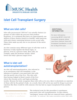
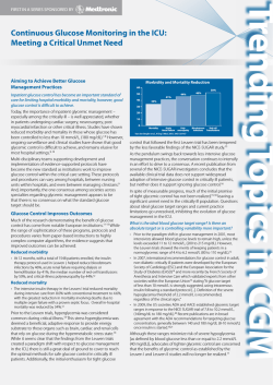
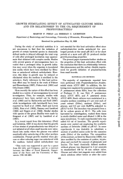
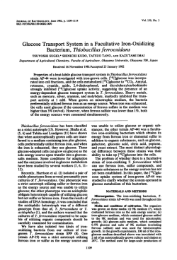
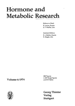
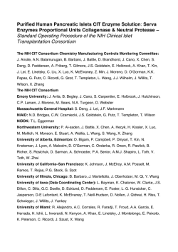
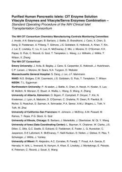
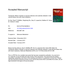
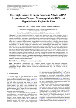
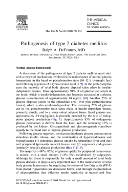
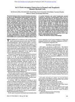
Article
Phospho-BAD BH3 Mimicry Protects b Cells and
Restores Functional b Cell Mass in Diabetes
Graphical Abstract
Authors
Sanda Ljubicic, Klaudia Polak, ...,
Adolfo Garcia-Ocan˜a, Nika N. Danial
Correspondence
[email protected]
In Brief
Ljubicic et al. show that BAD BH3 helix
phosphorylation simultaneously
enhances insulin secretion and protects b
cells from death induced by type-1diabetes-related stress. These cellautonomous benefits are mediated by
glucokinase, a direct target of the
phospho-BAD BH3 helix, and improve b
cell replacement in transplanted diabetic
mice.
Highlights
d
BAD BH3 domain phosphorylation imparts b cell
autonomous protective effects
d
The prosurvival effect of BAD phosphorylation is mediated by
glucokinase
d
Phospho-BAD BH3 mimicry improves islet engraftment in
transplanted diabetic mice
Ljubicic et al., 2015, Cell Reports 10, 497–504
February 3, 2015 ª2015 The Authors
http://dx.doi.org/10.1016/j.celrep.2014.12.056
Cell Reports
Article
Phospho-BAD BH3 Mimicry Protects b Cells
and Restores Functional b Cell Mass in Diabetes
Sanda Ljubicic,1,2 Klaudia Polak,1 Accalia Fu,1,2 Jessica Wiwczar,1 Benjamin Szlyk,1 Yigang Chang,3
Juan C. Alvarez-Perez,4 Gregory H. Bird,5 Loren D. Walensky,5 Adolfo Garcia-Ocan˜a,4 and Nika N. Danial1,2,*
1Department
of Cancer Biology, Dana-Farber Cancer Institute, Boston, MA 02115, USA
of Cell Biology, Harvard Medical School, Boston, MA 02115, USA
3Division of Endocrinology, University of Pittsburgh, Pittsburgh, PA 15260, USA
4Diabetes, Obesity and Metabolism Institute, The Mindich Child Health and Development Institute, Icahn School of Medicine at Mount Sinai,
New York, NY 10029, USA
5Department of Pediatric Oncology, Dana-Farber Cancer Institute and Boston Children’s Hospital, Boston, MA 02115, USA
*Correspondence: [email protected]
http://dx.doi.org/10.1016/j.celrep.2014.12.056
This is an open access article under the CC BY-NC-ND license (http://creativecommons.org/licenses/by-nc-nd/3.0/).
2Department
SUMMARY
Strategies that simultaneously enhance the survival
and glucose responsiveness of insulin-producing b
cells will greatly augment b cell replacement therapies in type 1 diabetes (T1D). We show that genetic
and pharmacologic mimetics of the phosphorylated
BCL-2 homology 3 (BH3) domain of BAD impart
b-cell-autonomous protective effects in the face of
stress stimuli relevant to b cell demise in T1D. Importantly, these benefits translate into improved engraftment of donor islets in transplanted diabetic mice,
increased b cell viability in islet grafts, restoration
of insulin release, and diabetes reversal. Survival of
b cells in this setting is not merely due to the inability
of phospho-BAD to suppress prosurvival BCL-2
proteins but requires its activation of the glucosemetabolizing enzyme glucokinase. Thus, BAD phospho-BH3 mimetics may prove useful in the restoration of functional b cell mass in diabetes.
INTRODUCTION
Autoimmune destruction of pancreatic b cell mass renders individuals with type 1 diabetes (T1D) insulin dependent. Strategies
that replace or regenerate b cells or preserve remaining b cell
mass are potential therapies in T1D (Claiborn and Stoffers,
2008; Halban et al., 2010; Hebrok, 2012; Pagliuca and Melton,
2013). However, the benefits of these approaches can be
thwarted by insufficient b cell proliferation, survival, and insulin
secretory response to glucose. As such, strategies that simultaneously enhance b cell mass and glucose signaling can be of
great therapeutic utility. Beyond stimulating insulin secretion,
increased b cell glucose metabolism stimulates b cell mass, at
least in part, through mitogenic effects (Levitt et al., 2011; Porat
et al., 2011; Terauchi et al., 2007). These observations suggest
shared molecular control of both b cell mass and function by
glucose.
A high-capacity glucose transport system and the high Km
glucose-phosphorylating enzyme glucokinase (GK, Hexokinase
IV)—the maturity onset diabetes of the young type 2 (MODY2)
gene product—enable b cells to sense blood glucose fluctuations and tightly couple insulin secretion to glucose metabolism
(Matschinsky, 2009). In addition to stimulating insulin secretion
and proliferation, increased glucose metabolism can enhance
b cell survival (Porat et al., 2011; Wei et al., 2009), which may
additionally contribute to glucose stimulation of b cell mass.
However, quantitative and mechanistic evidence for prosurvival
effects of glucose in b cells is limited. Moreover, the precise effect of glucose on b cell viability is dependent on the extent
and duration of increased glucose flux as well as the levels of
free fatty acids that, when abnormally and chronically elevated,
can cause b cell apoptosis through glucotoxicity and glucolipotoxicity (Bensellam et al., 2012).
The above observations indicate a complex link between
glucose metabolism and the core apoptotic machinery in b cells.
This complexity is further underscored by genetic manipulation
of several antisurvival and prosurvival BCL-2 family regulators
of apoptosis (Danial et al., 2008; Luciani et al., 2013; McKenzie
et al., 2010; Zhou et al., 2000). In particular, BCL-XL and BCL-2
prosurvival molecules are b cell protective but inhibit glucose
signaling and insulin secretion (Luciani et al., 2013; Zhou et al.,
2000). This may be explained, in part, by the capacity of BCL-2
and BCL-XL to modulate Ca2+ flux (Danial et al., 2010), which
is relevant for metabolic coupling of glucose and insulin secretion in b cells. Importantly, these findings highlight the challenge
of targeting BCL-2 proteins for promoting functional b cell mass
in a manner that ensures both resistance to apoptosis and
proper glucose modulation of insulin release.
We have previously shown that the BCL-2 family protein BAD
stimulates the b cell glucose response and insulin secretion
through phosphorylation of a defined residue within an amphipathic a helix known as the BCL-2 homology 3 (BH3) domain:
Ser155 in mouse BAD corresponding to Ser118 in the human
sequence (Danial et al., 2008). This modification neutralizes
BAD’s apoptotic function and simultaneously triggers its ability
to activate GK (Danial, 2008; Danial et al., 2008; Gime´nez-Cassina et al., 2014; Szlyk et al., 2014). At the molecular and
Cell Reports 10, 497–504, February 3, 2015 ª2015 The Authors 497
structural level, this is mediated by direct binding of the phospho-BAD BH3 helix near the active site of GK, resulting in the
increase of Vmax through a nonallosteric mechanism without
changes in the enzyme’s affinity for glucose (Szlyk et al., 2014).
Importantly, studies in islets derived from Bad / and Bad
S155A knockin mice and human donor islets indicate that the
phospho-BAD BH3 helix is required and sufficient for stimulation
of insulin secretion in response to glucose (Danial et al., 2008;
Szlyk et al., 2014). BAD phosphorylation is sensitive to fed/fasted
states and hormones known to regulate b cell survival (Danial
et al., 2008; Gime´nez-Cassina et al., 2014; Liu et al., 2009), suggesting that BAD’s function may be normally in tune with nutrient
and hormonal regulation of functional b cell mass. However,
whether beyond neutralizing BAD’s apoptotic activity, BAD
phosphorylation has active, cell-autonomous effects on b cell
survival has not been examined. Furthermore, the extent to
which BAD phosphorylation may be protective against stress
stimuli relevant to b cell demise in T1D is not known. This is
especially relevant given functional redundancies as well as
specialization among BCL-2 proteins in the regulation of cell
death/survival. In the present studies, we undertook genetic
and pharmacologic approaches to mimic BAD phosphorylation
within its BH3 helix and determine its acute contribution to b
cell survival in vitro, as well as its physiologic effects in replacing
and promoting functional b cell mass in an islet transplantation
model in vivo.
RESULTS
Survival-Promoting Effect of the BAD BH3
Phospho-Mimic Variant in b Cells
To examine the acute and cell autonomous outcome of BAD
phosphorylation in b cell survival, we assessed the effect of the
BAD S155D phospho-mimic variant on the viability of INS-1 cells
(Figures 1A–1D) and primary mouse pancreatic islets (Figures
1E–1H) subjected to stress stimuli relevant to b cell death in
T1D. These included inflammatory cytokines, oxidative, and
endoplasmic reticulum (ER) stress (Bedoya et al., 2012; Donath
et al., 2008; Eizirik et al., 2013) (Figures S1A–S1C; Table S1).
Compared with GFP-expressing controls, INS-1 cells and primary islets expressing BAD S155D were significantly protected
from apoptosis induced by a combination of tumor necrosis factor alpha (TNF-a), interleukin-1b (IL-1b), and interferon g (IFNg)
(Figures 1A and 1E), the nitric oxide (NO) donor GEA3162 (Figures 1B, 1F–1H, and S1C), hypoxia (Figure 1C), and the ER stress
inducer tunicamycin (Figure 1D).
At the molecular level, two scenarios may explain the protective effect of BAD S155D. First, because Ser155 phosphorylation blocks the BAD BH3 domain from binding and neutralizing
the prosurvival BCL-2, BCL-XL, BCL-w proteins and subsequent lowering of the threshold for apoptosis (Danial, 2008),
the protective effect of BAD S155D may be due to the inhibition
of BAD’s proapoptotic function. Second, the protective effect
of BAD S155D may result from its capacity to bind GK and
stimulate glucose metabolism. To distinguish between these
possibilities, we examined a second BAD BH3 phosphomutant,
BAD AAA (bearing the triple substitutions L151A, S155A, and
D156A). Similar to BAD S155D, the AAA variant is inert in its
498 Cell Reports 10, 497–504, February 3, 2015 ª2015 The Authors
apoptotic function as it cannot engage and inhibit the prosurvival BCL-2 proteins, but unlike BAD S155D, BAD AAA does
not bind or activate GK (Danial et al., 2008; Gime´nez-Cassina
et al., 2014). If blocking BAD from neutralizing prosurvival
BCL-2 proteins is sufficient to protect b cells, then the BAD
S155D and AAA variants should be equally protective. If, however, activation of GK is relevant for the protective effect of
phospho-BAD, then BAD AAA should lack b cell protective
properties. Indeed, unlike BAD S155D, the AAA variant failed
to protect b cells (Figures 1A–1H), indicating that blocking
BAD’s capacity to inhibit prosurvival BCL-2 proteins is not
sufficient to protect b cells. Collectively, these results indicate
that BAD phosphorylation has an acute and cell autonomous
effect on b cell survival that cosegregates with its ability to
engage GK.
Acute Modulation of the b Cell Response to Glucose by
BAD Phosphorylation
Processing of glucose-derived metabolites in b cell mitochondria generates metabolic signals such as ATP/ADP, NAD(P)H,
and mitochondrial GTP that couple glucose to insulin release
(Prentki et al., 2013). To examine the acute modulation of b cell
glucose responsiveness by BAD phospho mutants, we focused
on glucose stimulation of mitochondrial respiration and insulin
secretion. Real-time measurements of mitochondrial oxygen
consumption rate (OCR) indicated significantly higher respiration
in islets expressing BAD S155D compared with GFP- or BAD
AAA-expressing islets (Figure 1I). This increase in mitochondrial
glucose oxidation was associated with a 35% increase in insulin
release (Figure 1J, comparing GFP- and BAD S155D-expressing
islets at 20 mM glucose). Notably, BAD S155D expression did
not stimulate basal insulin release at 3 mM glucose (Figure 1J),
a concentration that does not activate GK (Matschinsky, 2009).
Insulin secretion in islets expressing the BAD AAA variant was
comparable to that of GFP controls (Figure 1J). Overall, these
data indicate an acute effect of BAD phosphorylation on mitochondrial handling of glucose and insulin release in primary
islets.
The BAD Phospho-BH3 Helix Is Sufficient to Protect
b Cells from Apoptosis
The survival-promoting and function-enhancing benefits of BAD
phosphorylation in b cells motivated the pursuit of phospho-BAD
BH3 mimicry for improving functional b cell mass. We have
previously shown that stabilized alpha-helices of BCL-2 domains (SAHBs) generated by hydrocarbon stapling of peptides
modeled after the BAD phospho-BH3 helix correct the insulin
secretory defect of Bad / islets in response to glucose, indicating that this domain is sufficient to emulate BAD’s effect on
b cell function (Danial et al., 2008). However, whether BAD
SAHBs influence b cell survival is not known.
The clear benefits of full-length BAD S155D over BAD AAA
in b cell survival and function prompted characterization of
their corresponding stapled peptides, BAD SAHBA (S155D)
and BAD SAHBA (AAA). Several quality-control assays were
performed to ensure the differential effect of the BAD BH3
domain on its metabolic target, GK, was preserved following
modification by hydrocarbon stapling. In vitro GK activity
A
B
C
D
E
F
G
H
I
J
Figure 1. Effect of BAD BH3 Phospho Variants on b Cell Viability and Glucose Handling
(A–H) Viability of INS-1 cells (A–D) and primary mouse islets (E–H) expressing GFP, BAD S155D, or BAD AAA treated with inflammatory cytokines (combination of
20 ng/ml TNF-a, 40 ng/ml IL-1b, and 10 ng/ml IFNg for 48 hr) (A and E), NO donor GEA3162 (43 mM for 72 hr) (B and F–H), hypoxia (2% O2 for 24 hr) (C), or
tunicamycin (5 ng/ml for 48 hr) (D). Cell viability/death was measured by annexin V and 7-AAD staining (A–F), caspase-3 (csp-3) cleavage (G), and loss of
mitochondrial membrane potential (DJm) (H).
(I and J) Effect of BAD BH3 phospho variants on glucose handling as examined by mitochondrial respiration (I) and insulin secretion (J) following stimulation
of primary islets with 20 mM glucose. Data are represented as means ± SEM. *p < 0.05; **p < 0.01; ***p < 0.001; n.s., nonsignificant; n = 3–8 independent
experiments per treatment.
See also Figure S1 and Table S1.
assays confirmed that BAD SAHBA (S155D) directly activates
recombinant GK, while BAD SAHBA (AAA) does not, as evidenced by changes in Vmax (Figure 2A; Table S2). This distinct
GK-activating capacity was consistent with the differential
effect of SAHBA (S155D) and SAHBA (AAA) on mitochondrial
glucose handling in primary islets (Figure 2B), effectively replicating the phenotype of the full-length BAD S155D and AAA
variants (Figure 1I).
Cell Reports 10, 497–504, February 3, 2015 ª2015 The Authors 499
A
B
Vehicle
BAD SAHB (S155D)
BAD SAHB (AAA)
1.5
150
n.s.
*
125
100
1.0
75
50
0.5
25
0
0
1
10
0
100
Glucose (mM)
C
*
pmole O2/min/islet
GK activity (% Control Vmax)
Vehicle
BAD SAHB (S155D)
BAD SAHB (AAA)
Vehicle + GEA3162
BAD SAHB (S155D) + GEA3162
BAD SAHB (AAA) + GEA3162
OCR
D
E
Vehicle
Vehicle + GEA3162
BAD SAHB (S155D) + GEA3162
ctrl shGk
Figure 2. GK-Dependent Protection of Islet
Survival by the Phospho-BAD BH3 Helix
(A) Activity of recombinant GK in the presence of
vehicle or 5 mM of the indicated BAD SAHBA. Data
show the means ± SD of a representative experiment. See Table S2 for the summary of enzyme
kinetic parameters derived from four independent
experiments performed similarly.
(B) Glucose stimulation of mitochondrial respiration in primary islets treated overnight with vehicle
or 3 mM of the indicated BAD SAHBA (n = 3).
(C) Viability of primary islets pretreated with 10 mM
of the indicated BAD SAHBA that were washed and
treated with 43 mM GEA3162 for 72 hr (n = 9).
(D and E) Viability of islets subjected to Gk knockdown (D) and treated with GEA3162 as in (C) (n = 7).
Data in (B)–(E) are represented as means ± SEM.
*p < 0.05; **p < 0.01; ***p < 0.001; n.s., nonsignificant.
See also Figure S2.
GK
n.s.
**
Improved Engraftment of Donor
Islets Treated with the Phospho100
ctrl
BAD BH3 Mimetic
120
80
80
shGk
To examine the physiological signif100
80
60
60
icance of phospho-BAD BH3 mimicry
80
60
in b cell function and survival, we under40
40
60
took transplantation studies using a mar40
40
ginal islet mass transplantation model.
20
20
20
In this model, 150 mouse islets com20
prise a suboptimal number of donor
0
0
0
0
islets that cannot reverse hyperglycemia
Adeno-ctrl
Adeno-shGk
when transplanted in mice rendered diabetic after streptozotocin (STZ)-mediated
To test the protective effects of SAHBA (S155D), we chose the destruction of endogenous b cells (Montan˜a et al., 1993). HowNO-induced islet death paradigm as a representative model of b ever, this limited number of donor islets can lead to diabetes
cell stress. NO production is a prime component of b cell oxida- reversal when used in conjunction with treatments that improve
tive stress and toxicity caused by inflammatory cytokines (Bed- islet function and viability (Plesner and Verchere, 2011).
We reasoned that the survival-promoting effect of BAD SAHBA
oya et al., 2012). Remarkably, pretreatment of islets with BAD
SAHBA (S155D) but not BAD SAHBA (AAA) was sufficient to pro- (S155D) may protect donor islets in early posttransplant periods
vide significant protection against death induced by the NO (days 2–4), when their viability is severely compromised due to
donor GEA3162 (Figure 2C). Of note, both SAHBA (S155D) and hypoxia prior to revascularization and inflammatory stress assoSAHBA (AAA) were taken up by islets with slightly higher uptake ciated with tissue trauma during transplantation (Biarne´s et al.,
of SAHBA (AAA) (Figure S2A), ruling out differences in islet uptake 2002; Plesner and Verchere, 2011). In addition, because only a
as an explanation for the observed differences in b cell survival. small percentage of transplanted islets that survive under these
Given the differential GK-activating capacity of BAD SAHBA conditions display physiologic insulin secretory characteristics
compounds and the attendant effects on mitochondrial glucose (Plesner and Verchere, 2011), we predicted that improved
handling (Figures 2A and 2B), we predicted that the survival-pro- glucose responsiveness and insulin secretion in donor islets premoting function of BAD SAHBA (S155D) would be dependent on treated with BAD SAHBA (S155D) would impart systemic beneglucose metabolism. To test this possibility, islets treated with fits in transplant recipients.
adenoviruses bearing Gk shRNA were analyzed in parallel (FigBlood glucose profiles were followed in a large cohort of
ure 2D). Molecular depletion of GK curtailed the protective effect STZ-treated diabetic mice that were transplanted with 150
of BAD SAHBA (S155D) in this setting (Figures 2E), indicating that WT islets pretreated overnight with BAD SAHBA compounds
GK is required for the survival-promoting effects of the phospho- or vehicle control. Pretreatment of donor islets with BAD
BAD BH3 mimetic. The protective effect of GK activation in SAHBA (S155D) prior to transplantation markedly improved
this setting is also consistent with the observation that the GK the glycemic profile in diabetic recipients, whereas mice transactivator (GKA) compound RO0281675 (Grimsby et al., 2003) planted with islets pretreated with BAD SAHBA (AAA) or
rescued NO-induced death in the absence or presence of vehicle remained hyperglycemic with blood glucose values
above 400 mg/dl (Figure 3A). Lowering of blood glucose levels
SAHBA (AAA) (Figure S2B).
100
% Survival
(Relative to untreated control)
HSP90
*** ***
Relative GK protein levels
(Integrated density)
% Survival
(Relative to untreated control)
120
500 Cell Reports 10, 497–504, February 3, 2015 ª2015 The Authors
100
A
B
Figure 3. Engraftment of Donor Islets Treated
with BAD SAHBA Compounds
C
(A) Glycemic profiles of diabetic mice transplanted
with donor islets pretreated overnight with vehicle or
10 mM of the indicated BAD SAHBA. Each line represents one transplanted animal (n = 6–11 mice per
group).
(B) Insulin levels in transplant recipients (n = 7–12
mice per group).
(C) Blood glucose levels in diabetic mice transplanted as in (A) and subjected to nephrectomy (red
arrow) on day 12 after transplantation. Each line
represents one transplanted animal (n = 3).
(D) b cell death in islets grafts 2 days after transplantation as quantified by cleaved caspase-3 and
insulin costaining (n = 3–5 per group). An average of
762 ± 252 cells were counted per condition.
Data in (B) and (D) are represented as means ± SEM.
*p < 0.05; **p < 0.01; ***p < 0.001; n.s., nonsignificant.
D
in the former cohort was associated with improved plasma
insulin levels (Figure 3B). Importantly, removal of islet grafts
once normoglycemia was achieved led to hyperglycemia (Figure 3C), indicating that reversal of diabetes in these mice was
specifically mediated by BAD SAHBA (S155D)-treated donor
islets.
To examine the contribution of islet graft survival to improved
glycemia in diabetic mice, we assessed b cell survival in the early
period posttransplantation, when donor islets are prone to undergo cell death prior to revascularization (Biarne´s et al., 2002;
Montan˜a et al., 1993; Plesner and Verchere, 2011). Assessment
of cells doubly positive for insulin and cleaved caspase-3
in grafts excised on day 2 revealed significantly lower b cell
death in grafts from BAD SAHBA (S155D)-treated donor
islets compared with grafts from vehicle-treated or BAD SAHBA
(AAA)-treated controls (Figure 3D). This paralleled the survivalpromoting effect of phospho-BAD BH3 mimicry in vitro (Figures
1A–1H and 2C).
To assess the overall outcomes of BAD SAHB pretreatment on
islet graft performance with respect to glucose homeostasis, mice
were subjected to intraperitoneal glucose tolerance test (i.p. GTT)
on day 49 after transplantation. Blood glucose levels during
GTT were significantly lower in recipients transplanted with BAD
SAHBA (S155D)-treated donor islets but not in control cohorts
transplanted with vehicle- or SAHBA (AAA)-treated islets (Fig-
ure 4A). Improved glucose tolerance was
associated with improved insulin release in
the first 30 min after glucose injection
when insulin normally peaks during i.p.
GTT, indicating glucose-responsive insulin
secretion by islet grafts in vivo (Figure 4B).
In contrast, mice transplanted with vehicleor SAHBA (AAA)-treated donor islets did not
elicit an insulin response to glucose (Figure 4B), which was consistent with their
GTT profile (Figure 4A). Overall, these
data show that the survival-promoting and
function-enhancing benefits of BAD phosphorylation in b cells extend to improved b cell replacement and
functional b cell mass in diabetic mice.
DISCUSSION
Our findings indicate that genetic and pharmacologic approaches to mimic BAD phosphorylation within its BH3 helix
impart b cell protective effects in an acute and cell autonomous
manner. The short-term benefits of phospho-BAD in b cells
include resistance to apoptosis in the face of inflammatory,
oxidative, and ER stress stimuli, as well as increased glucose
handling and insulin secretory capacities. In the long-term, these
benefits manifest in superior engraftment of donor islets in transplanted diabetic mice with an attendant increase in functional
b cell mass. Collectively, these observations highlight the utility
of BAD phospho-BH3 mimetic approaches in augmenting functional b cell mass in diabetes.
At the mechanistic level, parallel comparison of informative
phospho-BH3 BAD variants indicates that the effect of BAD
phosphorylation on b cell survival is linked to its activation of
GK and cannot be solely explained by the lack of its inhibitory
effects on prosurvival BCL-2 proteins. Consistent with these
observations, the protective effect of BAD SAHBA (S155D) is
abrogated following molecular depletion of GK. In addition,
GK gain of function is b cell protective in islets treated with
Cell Reports 10, 497–504, February 3, 2015 ª2015 The Authors 501
Vehicle
BAD SAHBA (S155D)
BAD SAHBA (AAA)
n.s.
A
600
60,000
GTT, glucose
400
40,000
300
* *
200
100
AUC
Glucose (mg/dl)
500
*
*
0
0
30
60
90
120
Time (min)
0.25
GTT, insulin
0.15
8
*
**
6
*
AUC
Insulin (ng/ml)
0.20
0.10
4
2
0.05
0
0
0
10
**
**
(A and B) Blood glucose (A) and insulin (B) levels
during i.p. GTT performed 49 days after transplantation (n = 11 per BAD SAHBA group). Asterisks in line graphs on the left compare the BAD
SAHBA (S155D) and SAHBA (AAA) groups. Area
under the curve (AUC) for glucose and insulin is
shown on the right.
Data are represented as means ± SEM. *p < 0.05;
**p < 0.01; ***p < 0.001; n.s., nonsignificant.
20,000
0
B
Figure 4. Glucose Homeostasis in Transplant Recipients
20
30
Time (min)
BAD SAHBA (AAA). The precise branch points of glucose metabolism downstream of the BAD-GK axis that may promote
b cell survival are currently under investigation. Glucosederived metabolites can feed a number of anabolic reactions
that may be relevant in this setting. In addition, changes in
ATP and Ca2+ flux following mitochondrial metabolism of
glucose-derived pyruvate may provide bioenergetic benefits
that can be b cell protective. Furthermore, whether and how
the metabolic outputs of glucose that promote b cell survival
overlap with those that mediate b cell proliferation such as
IRS-2, TORC1/S6K, CaMK, PKC, and sphingosine kinase
remain to be determined (Elghazi et al., 2010; Mastrandrea
et al., 2010; Sjo¨holm, 1997; Terauchi et al., 2007). Notably, under the experimental settings used in Figures 3 and 4, b cell
proliferation in islet grafts is not altered by pretreatment with
BAD SAHBA compounds (J.C.A.-P., A.G.-O., and N.N.D., unpublished data). Thus, improved b cell mass and function under
these specific conditions is predominantly associated with
increased survival and insulin secretory capacity of donor islets
treated with BAD SAHBA (S155D). However, thorough assessment of the potential connection between phospho-BAD and
b cell proliferation requires full examination of other in vivo
models of b cell proliferation.
Increased glucose flux through GK activation may have therapeutic utility in promoting b cell mass; however, these benefits
502 Cell Reports 10, 497–504, February 3, 2015 ª2015 The Authors
have to be carefully weighed against the
adverse effects of chronic elevation in
glucose metabolism, such as glucotoxicity-induced b cell dysfunction/loss. This
warrants characterization of the precise
conditions to derive the therapeutic benn.s.
efits of glucose metabolism for b cell
** ***
mass restoration. Within this context,
the mechanism of GK activation and the
duration of increased glucose flux are
important considerations. For example,
allosteric GKAs increase glucose flux
but drastically enhance the enzyme’s affinity for glucose, which may not be
beneficial in the long term (Matschinsky,
2009). A distinct class of GKAs with a
nonallosteric mode of action that do
not dramatically alter the affinity of GK
for glucose, such as phospho-BAD BH3
mimetics (Szlyk et al., 2014), may be
beneficial in this setting. In addition, defining the minimal metabolic outputs of glucose that promote b cell mass may provide
translational insights into the most effective strategies for harnessing the benefits of glucose signaling to pharmacologically
restore functional b cell mass.
EXPERIMENTAL PROCEDURES
Primary Islets, Cell Culture, and Insulin Secretion Assays
Primary mouse islets isolation and culture, as well as insulin release assays,
were performed as previously described (Danial et al., 2008). INS-1 cells
were a kind gift of Dr. Christopher Newgard (Duke University).
Treatment with Death Stimuli and Assessment of Cell Death/
Survival
INS-1 cells and primary mouse islets were seeded in six-well plates at 4 3 105
INS-1 cells per well and 100 islets per well, respectively, and treated with the
following combination of inflammatory cytokines (R&D Systems); 20 ng/ml
TNF-a, 40 ng/ml IL-1b, and 10 ng/ml IFNg for 48 hr. The NO donor GEA3162
(Enzo Life Sciences) was added at 43 mM in DMSO for 72 hr. Hypoxia was
induced by incubating cells at 2% O2 for 24 hr. Tunicamycin (Sigma) was
added at 5 ng/ml in DMSO for 48 hr. Control treatment for inflammatory cytokines was PBS, and that for GEA3162 and tunicamycin was 0.1% DMSO, as
described below and further detailed in Supplemental Experimental Procedures. Cell death/survival was quantified by several parameters, including annexin V and 7-amino-actinomycin D (7-AAD) staining, changes in mitochondrial membrane potential (DJm), and western blotting with an antibody to
cleaved (active) caspase-3.
For flow cytometric measurement of cell/death survival, cells were labeled
with annexin V (BD PharMingen) and 7-AAD (BD Biosciences) and subjected
to dual-color flow cytometry using a FACSCANTO II flow cytometer (Becton
Dickinson) and the BD FACSDIVA software (BD Biosciences). For each measurement, 104 cells were sorted, and gates were applied to generate four
quadrants based on the level of annexin V (PE) and 7-AAD (Cy5) staining intensities, and percentage survival was calculated based on annexin V and 7-AAD
double negativity. Figures S1A and S1B and Table S1 document the percentage of live cells or those undergoing apoptosis in control INS-1 cells and primary islets treated with the indicated death stimuli (see also Supplemental
Experimental Procedures).
For measurement of mitochondrial membrane potential, primary islets
were incubated in the presence or absence of GEA3162 as described above,
dispersed, and live stained with 10 nM tetramethylrhodamine, ethyl ester
(Molecular Probes) in RPMI for 30 min at 37 C. Treatment with 1 mM of
the protonophore carbonyl cyanide 4-(trifluoromethoxy) phenylhydrazone
(FCCP; Sigma) was used to dissipate the mitochondrial membrane potential
and to define the base line for the analysis of mitochondrial membrane potential (DJm). Cells were rinsed with RPMI (without phenol) containing 1%
fetal bovine serum and analyzed by flow cytometry using a FACSCANTO II
flow cytometer (Becton Dickinson) and the BD FACSDIVA software (BD Biosciences). Both mean intensities and percentage of cells that showed loss of
DJm were analyzed. Representative fluorescence-activated cell sorting
(FACS) plots of mitochondrial membrane potential changes in primary islets
expressing GFP, BAD S155D, or BAD AAA treated with DMSO or GEA3162
are shown in Figure S1C.
Adenovirus Production and Viral Infection
Recombinant adenoviruses carrying GFP, BAD S155D and BAD AAA variants,
as well as Gk and control shRNA have been previously described (Gime´nezCassina et al., 2014). INS-1 cells were infected using a multiplicity of infection
of 75 for 48 hr. Adenoviral transduction of primary mouse islets were carried
out at 1 3 105 pfu per islet for 48 hr.
Mitochondrial Respirometry
Real-time measurements of mitochondrial OCR were performed using the
XF24 extracellular flux analyzer instrument (Seahorse Bioscience) as
described in Supplemental Experimental Procedures.
SAHB Synthesis and Treatments
Peptide synthesis, olefin metathesis, fluorescein isothiocyanate (FITC)
derivatization, reverse-phase high-performance liquid chromatography purification, and amino acid analysis were performed as previously described
(Danial et al., 2008; Szlyk et al., 2014). For survival assays and donor islet
treatments, islets were treated overnight with 10 mM of the indicated FITC
BAD SAHBA in 1% DMSO in RPMI SAHB uptake medium containing
11 mM glucose, 10% serum, and 0.1% Tween 80 (pH 6.0) at 37 C. Control
islets were treated with vehicle composed of DMSO and SAHB uptake
medium. Islets were then washed and incubated in fresh media for 2 hr
before in vitro studies or transplantation. For OCR studies in Figure 1I,
islets were treated with 3 mM of the indicated SAHB compounds. For
GK activity assays in Figure 2A, BAD SAHBs were added at 5 mM final
concentration, and 5% DMSO was used as vehicle control. Recombinant
GK activity assays were performed as previously described (Szlyk et al.,
2014).
Islet Transplantation and Metabolic Studies
One hundred fifty islets derived from C57BL/6J mice were treated as indicated above and transplanted under the kidney capsule of male STZ-treated
diabetic C57BL/6J mice using established procedures (Montan˜a et al., 1993)
that are further detailed in Supplemental Experimental Procedures. Blood
glucose and insulin measurements and i.p. GTT were performed as previously described (Danial et al., 2008; Gime´nez-Cassina et al., 2014). Islet graft
viability was assessed by immunofluorescence as described in Supplemental
Experimental Procedures. All animal procedures were approved by the Institutional Animal Care and Use Committee of the Dana-Farber Cancer
Institute.
Statistical Analysis
Unless otherwise indicated, data are represented as means ± SEM. Statistical
significance was calculated using one-way ANOVA. Statistical significance
was defined as p < 0.05.
SUPPLEMENTAL INFORMATION
Supplemental Information includes Supplemental Experimental Procedures,
two figures, and two tables and can be found with this article online at
http://dx.doi.org/10.1016/j.celrep.2014.12.056.
AUTHOR CONTRIBUTIONS
S.L., A.F., and N.N.D. designed experiments. S.L., A.F., K.P., J.W., and N.N.D.
conducted in vitro studies with INS-1 cells and primary islets, performed islet
transplantation, and carried out in vivo studies. B.S. and N.N.D. purified recombinant GK and performed enzyme kinetic analyses. Y.C., J.C.A.-P., and
A.G.-O. performed immunofluorescence analyses. G.H.B. and L.D.W. designed and synthesized SAHB compounds. S.L., A.F., and N.N.D. wrote the
manuscript, which was reviewed by all authors.
ACKNOWLEDGMENTS
We thank Elaura Patton and Gabriella Casinelli for technical support and Jill
Fisher for help with islet transplantation surgeries. This work was supported
by the U.S. NIH grants R01DK078081 (N.N.D.), R01 DK067351 and R01
DK077096 (A.G.-O.), R01GM090299 (L.D.W.), Burroughs Wellcome Fund
Career Award in Biomedical Sciences (N.N.D.), Juvenile Diabetes Research
Foundation Grant 17-2011-595 (N.N.D.), Barry and Mimi Sternlicht Type 1 Diabetes Research Fund (N.N.D.), Claudia Adams Barr Award in Innovative Basic
Cancer Research (N.N.D.), and a Swiss National Science Foundation postdoctoral fellowship (S.L.). The authors also acknowledge generous support from
John H. Lippincott. L.D.W. is a consultant and scientific advisory board member for Aileron Therapeutics.
Received: May 26, 2014
Revised: November 21, 2014
Accepted: December 26, 2014
Published: January 29, 2015
REFERENCES
Bedoya, F.J., Salguero-Aranda, C., Cahuana, G.M., Tapia-Limonchi, R., Soria,
B., and Tejedo, J.R. (2012). Regulation of pancreatic b-cell survival by nitric oxide: clinical relevance. Islets 4, 108–118.
Bensellam, M., Laybutt, D.R., and Jonas, J.C. (2012). The molecular mechanisms of pancreatic b-cell glucotoxicity: recent findings and future research
directions. Mol. Cell. Endocrinol. 364, 1–27.
Biarne´s, M., Montolio, M., Nacher, V., Raurell, M., Soler, J., and Montanya, E.
(2002). Beta-cell death and mass in syngeneically transplanted islets exposed
to short- and long-term hyperglycemia. Diabetes 51, 66–72.
Claiborn, K.C., and Stoffers, D.A. (2008). Toward a cell-based cure for diabetes: advances in production and transplant of beta cells. Mt. Sinai J. Med.
75, 362–371.
Danial, N.N. (2008). BAD: undertaker by night, candyman by day. Oncogene 27
(Suppl 1), S53–S70.
Danial, N.N., Walensky, L.D., Zhang, C.Y., Choi, C.S., Fisher, J.K., Molina, A.J.,
Datta, S.R., Pitter, K.L., Bird, G.H., Wikstrom, J.D., et al. (2008). Dual role of
proapoptotic BAD in insulin secretion and beta cell survival. Nat. Med. 14,
144–153.
Danial, N.N., Gimenez-Cassina, A., and Tondera, D. (2010). Homeostatic functions of BCL-2 proteins beyond apoptosis. Adv. Exp. Med. Biol. 687, 1–32.
Donath, M.Y., Størling, J., Berchtold, L.A., Billestrup, N., and Mandrup-Poulsen, T. (2008). Cytokines and beta-cell biology: from concept to clinical translation. Endocr. Rev. 29, 334–350.
Cell Reports 10, 497–504, February 3, 2015 ª2015 The Authors 503
Eizirik, D.L., Miani, M., and Cardozo, A.K. (2013). Signalling danger: endoplasmic reticulum stress and the unfolded protein response in pancreatic islet
inflammation. Diabetologia 56, 234–241.
Elghazi, L., Balcazar, N., Blandino-Rosano, M., Cras-Me´neur, C., Fatrai, S.,
Gould, A.P., Chi, M.M., Moley, K.H., and Bernal-Mizrachi, E. (2010).
Decreased IRS signaling impairs beta-cell cycle progression and survival in
transgenic mice overexpressing S6K in beta-cells. Diabetes 59, 2390–2399.
Gime´nez-Cassina, A., Garcia-Haro, L., Choi, C.S., Osundiji, M.A., Lane, E.A.,
Huang, H., Yildirim, M.A., Szlyk, B., Fisher, J.K., Polak, K., et al. (2014). Regulation of hepatic energy metabolism and gluconeogenesis by BAD. Cell Metab.
19, 272–284.
Grimsby, J., Sarabu, R., Corbett, W.L., Haynes, N.E., Bizzarro, F.T., Coffey,
J.W., Guertin, K.R., Hilliard, D.W., Kester, R.F., Mahaney, P.E., et al. (2003).
Allosteric activators of glucokinase: potential role in diabetes therapy. Science
301, 370–373.
Halban, P.A., German, M.S., Kahn, S.E., and Weir, G.C. (2010). Current status
of islet cell replacement and regeneration therapy. J. Clin. Endocrinol. Metab.
95, 1034–1043.
Hebrok, M. (2012). Generating b cells from stem cells-the story so far. Cold
Spring Harb. Perspect. Med. 2, a007674.
Levitt, H.E., Cyphert, T.J., Pascoe, J.L., Hollern, D.A., Abraham, N., Lundell,
R.J., Rosa, T., Romano, L.C., Zou, B., O’Donnell, C.P., et al. (2011). Glucose
stimulates human beta cell replication in vivo in islets transplanted into
NOD-severe combined immunodeficiency (SCID) mice. Diabetologia 54,
572–582.
Liu, S., Okada, T., Assmann, A., Soto, J., Liew, C.W., Bugger, H., Shirihai, O.S.,
Abel, E.D., and Kulkarni, R.N. (2009). Insulin signaling regulates mitochondrial
function in pancreatic beta-cells. PLoS ONE 4, e7983.
Luciani, D.S., White, S.A., Widenmaier, S.B., Saran, V.V., Taghizadeh, F., Hu,
X., Allard, M.F., and Johnson, J.D. (2013). Bcl-2 and Bcl-xL suppress glucose
signaling in pancreatic b-cells. Diabetes 62, 170–182.
Mastrandrea, L.D., Sessanna, S.M., Del Toro, A., and Laychock, S.G. (2010).
ATP-independent glucose stimulation of sphingosine kinase in rat pancreatic
islets. J. Lipid Res. 51, 2171–2180.
Matschinsky, F.M. (2009). Assessing the potential of glucokinase activators in
diabetes therapy. Nat. Rev. Drug Discov. 8, 399–416.
504 Cell Reports 10, 497–504, February 3, 2015 ª2015 The Authors
McKenzie, M.D., Jamieson, E., Jansen, E.S., Scott, C.L., Huang, D.C., Bouillet,
P., Allison, J., Kay, T.W., Strasser, A., and Thomas, H.E. (2010). Glucose induces pancreatic islet cell apoptosis that requires the BH3-only proteins
Bim and Puma and multi-BH domain protein Bax. Diabetes 59, 644–652.
Montan˜a, E., Bonner-Weir, S., and Weir, G.C. (1993). Beta cell mass and
growth after syngeneic islet cell transplantation in normal and streptozocin diabetic C57BL/6 mice. J. Clin. Invest. 91, 780–787.
Pagliuca, F.W., and Melton, D.A. (2013). How to make a functional b-cell.
Development 140, 2472–2483.
Plesner, A., and Verchere, C.B. (2011). Advances and challenges in islet transplantation: islet procurement rates and lessons learned from suboptimal islet
transplantation. J. Transplant. 2011, 979527.
Porat, S., Weinberg-Corem, N., Tornovsky-Babaey, S., Schyr-Ben-Haroush,
R., Hija, A., Stolovich-Rain, M., Dadon, D., Granot, Z., Ben-Hur, V., White,
P., et al. (2011). Control of pancreatic b cell regeneration by glucose metabolism. Cell Metab. 13, 440–449.
Prentki, M., Matschinsky, F.M., and Madiraju, S.R. (2013). Metabolic signaling
in fuel-induced insulin secretion. Cell Metab. 18, 162–185.
Sjo¨holm, A. (1997). Glucose stimulates islet beta-cell mitogenesis through
GTP-binding proteins and by protein kinase C-dependent mechanisms. Diabetes 46, 1141–1147.
Szlyk, B., Braun, C.R., Ljubicic, S., Patton, E., Bird, G.H., Osundiji, M.A., Matschinsky, F.M., Walensky, L.D., and Danial, N.N. (2014). A phospho-BAD BH3
helix activates glucokinase by a mechanism distinct from that of allosteric activators. Nat. Struct. Mol. Biol. 21, 36–42.
Terauchi, Y., Takamoto, I., Kubota, N., Matsui, J., Suzuki, R., Komeda, K.,
Hara, A., Toyoda, Y., Miwa, I., Aizawa, S., et al. (2007). Glucokinase and
IRS-2 are required for compensatory beta cell hyperplasia in response to
high-fat diet-induced insulin resistance. J. Clin. Invest. 117, 246–257.
Wei, P., Shi, M., Barnum, S., Cho, H., Carlson, T., and Fraser, J.D. (2009). Effects of glucokinase activators GKA50 and LY2121260 on proliferation and
apoptosis in pancreatic INS-1 beta cells. Diabetologia 52, 2142–2150.
Zhou, Y.P., Pena, J.C., Roe, M.W., Mittal, A., Levisetti, M., Baldwin, A.C.,
Pugh, W., Ostrega, D., Ahmed, N., Bindokas, V.P., et al. (2000). Overexpression of Bcl-x(L) in beta-cells prevents cell death but impairs mitochondrial
signal for insulin secretion. Am. J. Physiol. Endocrinol. Metab. 278, E340–
E351.
© Copyright 2026