
Rheologic Properties of Senescent Erythrocytes: Loss of
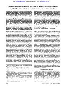
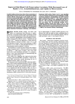
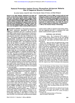
From www.bloodjournal.org by guest on February 6, 2015. For personal use only.
Rheologic Properties of Senescent Erythrocytes: Loss of Surface Area and
Volume With Red Blood Cell Age
By Richard E. Waugh, Mohandas Narla, Carl W. Jackson, Thomas J. Mueller, Takashige Suzuki, and George L. Dale
The rheologic properties of senescent erythrocytes have
been examined using two models of red blood cell (RBC)
aging. In the rabbit, aged erythrocytes were isolated after
biotinylation, in vivo aging, and subsequent recovery on an
avidin support. Aged RBCs from the mouse were obtained
using the Ganzoni hypertransfusion model that suppresses
erythropoiesis for prolonged periods of time allowing preexisting cells t o age in vivo. In both cases, the aged erythrocytes were found by ektacytometry t o have decreased deformability due t o diminished surface area and cellular
dehydration. The aged rabbit erythrocytes were further
characterized by micropipette methods that documented an
average surface area decrease of 10.5% and a volume decrease of 8.4% for the cells that were 50 days old. because
both the surface area and volume decreased with cell age,
there was little change in surface-to-volume ratio (sphericity)
during aging. The aged cells were found t o have normal
membrane elasticity. In addition, human RBCs were fractionated over Stractan density gradients and the most dense
cells were found t o have rheologic properties similar t o those
reported for the aged RBCs from rabbits and mice, although
the absolute magnitude of the bhanges in surface area and
volume were considerably greater for the human cells. Thus,
stringent density fractionation protocols that result in isolation of the most dense 1%of cells can produce a population
of human cells with rheologic properties similar t o senescent
cells obtained in other species. The data indicate that progressive loss of cell area and cell dehydration are characteristic
features of cell aging.
o 1992b y The American Society of Hematology.
T
their physical properties, specifically their in vitro deformability, membrane elasticity, and the ratio of surface area to
volume.
HE MAMMALIAN erythrocyte must survive a variety
of chemical and physical insults during its lifespan.
The potential chemical challenges include, among others,
oxidant stress, metabolic depletion, and loss of ion gradients,' all of which the cell resists via an impressive repertoire of enzymatic mechanisms. The physical challenges to
the erythrocyte are framed by the need for the cell to
deform from its resting diameter of approximately 8 pm to
diameters as small as 1.5 pm as it traverses capillary beds
and splenic sinusoids. The erythrocyte is remarkable in its
physico-elastic properties. The red blood cell (RBC) membrane maintains structural integrity while subjected to large
stresses and deformatiofis, yet remains highly deformable
with an elastic modulus softer than the softest latex rubber.'
However, there has been considerable d i s c ~ s s i o n as
~ . ~to
how well the cell's rheologic properties survive a lifespan
that involves approximately 1.7 X 10⁵ complete cycles
through the vascular circulation' over a period of 30 to 200
days, depending on the species.' Are there progressive
changes in cellular properties over the life of the cell that
might cause or contribute to the eventual removal of the
cell from the circulation?
Several precedents for this scenario of decreased deformability affecting the cell's lifespan are provided by a variety
of hemolytic anemias. For example, the RBCs from patients
with hereditary spherocytosis, hemoglobin CC disease,
sickle cell anemia, and autoimmune-mediated spherocytosis have all been documented to have decreased deformability in association with decreased RBC life~pan.~~'""
A major hindrance to the examination of erythrocyte
senescence has been the difficulty of isolating aged cells.'.'z
The vast majority of studies in the field of RBC aging have
used density fractionation to produce a population of dense
cells that has been assumed to be aged erythrocytes. Recent
work has shown that this conclusion is not acc~rate,'~.'~
and,
as a result, much of the work in the literature concerning
aged RBCs is of questionable value. Several techniques for
the unambiguous isolation of aged RBCs have been develped.'^,'^-^ In this report, we have used two of these
methods to isolate senescent RBCs from rabbits and mice.
These aged erythrocytes were then examined with regard to
Blood, Vol79, No 5 (March 1). 1992: pp 1351-1358
MATERIALS AND METHODS
Isolation of Aged Erythrocytes
The average lifespan of rabbit and mouse erythrocytes is 60
days? Aged erythrocytes from rabbits were isolated with a recently
reported method that involves the biotinylation of erythrocytes
with N-hydroxysuccinimido biotin?' Specifically, 2.5 kg rabbits
were injected on 3 consecutive days with 7.5 mglkg phenylhydrazine to produce a reticulocytosis; control experiments have
shown that 70% to 85% of all preexisting cells are destroyed by this
procedure (data not shown). Ten days later, the rabbit RBCs are
biotinylated in vitro by reaction with N-hydroxysuccinimido biotin
as previously described?' These biotinylated cells have been shown
to have a normal in vivo survival?1 After reinfusion, the biotinylated cells are allowed to age in vivo for various periods of time.
The animal is then bled again and the biotinylated RBCs are
recovered by binding to avidin-coated, plastic Petri dishes as
described." The recovered cells are removed from the Petri dishes
From the Department of Biophysics, University of Rochester, School
of Medicine and Dentistry, Rochester, W;the Division of Cell and
Molecular Biology, Lawrence Berkeley Laboratoy, University of
California, Berkelq, CA; the Departments of HematologylOncologv
and Biochemistry, St Jude Children's Research Hospital, Memphis,
TN; and the Department of Molecular and Experimental Medicine,
Research Institute of Scripps Clinic, La Jolla, CA.
Submitted December 6, 1990; accepted October 24, 1991.
Supported in part by Grants No. AG08545, HL18208, DK26263,
and HL30489from the National Institutes of Health, National Cancer
Institute (CORE)support Grant No. CA 21 765, and by the AmericanLebanese-SyrianAssociated Charities.
Address reprint requests to Richard E. Waugh, PhD, Department of
Biophysics, University of Rochester, School of Medicine and Dentisty,
601 Elmwood Ave, Rochester, NY14642.
The publication costs of this article were defiayed in part by page
charge payment. This article must therefore be hereby marked
"advertisement" in accordance with 18 U.S.C. section 1734 solely to
indicate this fact.
0 1992 by The American Society of Hematolog.
0006-4971I921 7905-0019$3.00/0
1351
From www.bloodjournal.org by guest on February 6, 2015. For personal use only.
WAUGH ET AL
1352
by collagenase digestion of the gelatin anchor’’ used to retain the
avidin on the plate surface. One advantage of this technique is that
when an age-synchronized cohort of cells is initially used for the
biotinylation, any desired age of erythrocyte can be obtained,
thereby allowing a temporal dissection of the aging process relative
to the parameter being examined?’.’’ As mentioned above, the
phenylhydrazine treatment does not quantitatively remove existing
cells so that the purity of the isolated RBC preparations is not as
great for an early isolation (ie, day 8) as it is for the more aged
preparations where any cells outside the initial age-window would
have been cleared by the natural senescence process. Isolated RBC
populations, as well as whole blood samples, were resuspended in
autologous plasma at 25% hematocrit and shipped on wet ice via
overnight carrier to the laboratories of N.M. and R.E.W. for
rheologic measurements.
Aged erythrocytes from the mouse were isolated with the
Ganzoni hypertransfusion p r o c e d ~ r e . ”With
~ ~ ~ this technique, a
large number of starting mice are split into two equal groups, and
one group is terminally bled to allow the other group of animals to
be hypertransfused. Two weeks later, one half of the surviving
animals are terminally bled to hypertransfuse the remaining mice;
this procedure is repeated every 2 weeks for approximately 60 days.
The model is based on the observation that hypertransfused
animals will not synthesize new eythrocytes, and, therefore, the
RBC population of the few surviving animals will have a continuously increasing mean age over the 60-day experiment.
There are important differences between the biotinylation and
hypertransfusion models. When the hypertransfusion procedure
has been proceeding for 30 days, the surviving RBCs represent a
range of ages from 30 to 60 days old. Only at the end of the
hypertransfusion experiment does the age-window of the remaining cells become relatively narrow. In contrast, the age-window in
the biotinylation system is determined by the starting population,
which is a phenylhydrazine-produced, young cohort of cells.
Therefore, the age-window for these cells is approximately 10 days
wide and will remain constant during the experiment. However, the
use of phenylhydrazine in the biotinylation model must be considered for the potential of introducing artifacts based on the use of
this oxidant challenge in the animal.
Density Separation of Human RBCs
Human RBCs were fractionated using discontinuous eight-step
stractan density gradients consisting of 1 mL fractions spanning a
density range of 1.084 to 1.120 g/mL in equal increments.= The
RBCs from the most dense fraction had a mean cellular hemoglobin concentration (MCHC) greater than 37 g/dL and constituted
0.8% of the total cell population.
Ektacytomehy Measurements
Deformability of intact RBCs was measured using osmotic
gradient ektacytometry, an assay in which whole-cell deformability
is measured as a continuous function of suspending medium
osmolality? For these measurements, we prepared gradients from
two solutions of 4% polyvinyl-pyrilidone (PVP; average molecular
weight 360 Kd and viscosity of 22 cp) in phosphate-buffered saline
(PBS), one adjusted to 50 mOsm/kg and the other to 900
mOsm/kg. The gradients were mixed in the first stage of the
three-stage mixing chamber of a Beckman gradient former (Beckman Instrument Co, Palo Alto, CA). Packed RBCs (70% to 80%
hematocrit) were pumped into the second stage of the chamber by
a Harvard infusion pump (Model No. 906; Harvard Apparatus,
South Natick, MA) and mixed with the gradient to a final
hematocrit of 0.2%. Thorough mixing was ensured by passage of
the cell suspension through the third stage of the mixing chamber.
The suspension was then pumped through a Wescan conductivity
meter (Wescan Instruments, Santa Clara, CA) to continuously
monitor its conductivity, and finally into the ektacytometer for
measurement of cellular deformability, at a constant shear stress of
170 dyne/cm2. The osmolality at which the deformability index
(DI) reaches a minimum in the hypotonic region of the gradient
has been shown to be the same as the osmolality at which 50% of
the cells will hemolyze in a standard osmotic fragility test? This
point is thus an index of the average surface area-to-volume ratio of
the population of cells studied. Cells attain their maximally
deformed state at or near the physiologically relevant osmolality of
290 mOsm. In the presence of normal membrane deformability,
this maximum value of DI has previously been shown to be related
to the membrane surface area.’ The hypertonic region of the curves
provides information on the state of cell hydration.
Variations in cell hemoglobin concentration distributions in
aging RBC populations was quantitated using the Technicon H-1
hematology analyzer (Technicon Instruments Corporation, Tarrytown, NY).*,
Micropipette Measurements
The surface areas and volumes of rabbit cells were measured on
six different occasions, and four or six samples were tested on each
occasion. The samples were grouped according to the age of the
labeled cohort: Day,, Day,, (days 22 to 30), and Day,, (days 49 to
51). For each age group there was a cohort sample and a whole
blood sample. Day, cells were tested on three occasions, and Day,
cells and Day,, cells were tested on four occasions each. In all, over
2,000 cells were measured. The whole blood samples do not
necessarily represent a “normal” cell population because the ages
of the cells in the blood depend on how soon after phenylhydrazine
treatment the blood is obtained. For example, for the Day, sample,
the control cells will represent a skewed age population because it
is only 18 days after the end of phenylhydrazine treatment of the
animal?’ By the time that a Day, sample is drawn for the isolation
of biotinylated cells, the animal will be 60 days removed from
phenylhydrazine treatment and will represent a more normal
distribution of cell ages. As a result, the whole blood samples from
Day,, are most representative of normal cells.
Details of the micropipette procedures are provided el~ewhere.~,
Cells were suspended in PBS (128 mmol/L NaC1,31.2 mmol/L Na
phosphate, pH 7.3; 285 to 295 mOsm) plus 3.0 mg/mL bovine
serum albumin at low hematocrit ( < 1.0%) and placed in a 1.0-mm
thick, U-shaped chamber on the microscope stage. A micropipette
(1.0 to 1.5 pm, inside diameter) was inserted into the chamber
through the open side of the “U”.
To measure the surface area and volume, cells were aspirated at
a pressure of 6,000 dyne/cm2(6.0 cm water), a pressure sufficient
to form the cell into a sphere plus a cylindrical projection into the
pipette. The outside diameter R, and projection length L were
measured for each cell, and the surface area (A), volume (V),
sphericity (S), and minimum cylindrical diameter (MCD) were
calculated according to the following relationships:
-
+ 2%L)
- Ri + 3R;L)/3
A = ~ ( 4 R -i R;
V
= p(4R:
S=
4.rrV2’)
(4~/3)A
~”
.rr(MCD)’ = 3 A . (MCD) - 12V
(1)
(2)
(3)
(4)
From www.bloodjournal.org by guest on February 6, 2015. For personal use only.
PROPERTIES OF SENESCENT RED CELLS
1353
where R, is the inside radius of the pipette. The MCD is the
diameter of the smallest tube through which the cell can pass. It
was found by numerical solution of equation 4 for given values of A
and V using Newton’s method. Some other measures of surface to
volume ratio can be calculated from the sphericity: “surface area
index” = S-’ and “swelling index” = S-”’. To avoid bias in cell
selection, every cell in an arbitrarily chosen field of view was
measured. Between 75 and 100 cells were measured for each
sample. Occasionally, cells were encountered that could not be
used for measurement. These included cells that creased or folded
when they were aspirated into the pipette, cells that were too rigid
to be deformed into the pipette, or cells that were so small that they
were sucked completely into the pipette. The sum of these rejected
cells amounted to less than 3% of any sample. Such cells are not
included in the statistics.
To measure the deformability of the membrane, cells were
aspirated near the dimple region at an initial pressure of 200
dyne/cm2. The aspiration pressure was increased in increments,
and the length of the projection into the pipette was measured as a
function of the pressure. The modulus (k) was calculated according to:
p=
(Ri/2.45) dP/dL
(5)
where dP/dL is the inverse of the slope of the length-pressure data
pairs.
RESULTS
Ektacytometry of Erythrocytes
Erythrocytes from all three species were examined by
ektacytometry to evaluate the deformability changes that
may occur during the aging process. The data presented in
Figs 1 through 4 address the changes that occur with
increasing erythrocyte age in the rabbit and mouse and with
increasing cell density in humans. Osmotic gradient deform0.6 0.5
DAY 22
CELL HEMOGLOBIN CONCENTRATION (g/dL)
Fig 2. Cell hemoglobin concentration for aged cell cohorts from
rabbit. The difference between whole blood samples (left curves) and
cohort samples increases progressively with the age of the cohort.
The data for the whole blood sample shown in each panel were
derived from blood samples of the animal from which the aged
cohorts of RBCs were isolated. The variation in these controls
represents biologic variation and differences in time since phenylhydrazine treatment. In addition t o the four in vivo aged samples shown
here, eight other samples were studied. The 12 samples were obtained from six different animals. The changes in cell hemoglobin
concentration documented here are representative of all samples
studied.
DAY 0
-
0.4-
z
$
8y
0.30.20.1 -
D
0.0 I
1
I
I
100
140
180
I
no
I
260
I
I
300
340
OSMOLALITY (mOsmoVkg)
I
I
380
420
Fig 1. Deformability of aged RBCs from the rabbit. Aged, biotinylated erythrocytes from the rabbit were isolated at the times
specified. The DI of these cells was measured at a constant shear
stress with variation of the osmolality of the suspending solution
(mOsm/kg). The oldest sample analyzed was 55 days; the lifespan of
the rabbit RBC is approximately 60 days. Data derived from four in
vivo aged blood samples is shown here. Similar patterns of deformability changes were seen in eight additional in vivo aged blood samples.
The 12 deformability measurements represent data obtained from six
separate animals.
I
I
I
100
140
180
I
1
I
I
220
260
300
340
OSMOLALITY (mOSmolkg)
I
I
380
420
Fig 3. Osmotic deformability profiles of aged RBCs from the
mouse. Aged RBCs were isolated from mice at various times after the
initiation of hypertransfusion. The measurements here are similar t o
those detailed in Fig 1. The lifespan of the mouse RBC is approximately 60 days. In addition t o the four in vivo aged samples shown
here, similar deformability changes were seen in four blood samples
of different ages obtained from four other animals.
From www.bloodjournal.org by guest on February 6, 2015. For personal use only.
WAUGH ET AL
1354
human RBCs are illustrated in Fig 4. With increasing cell
density there was a progressive decrease in maximum D1
and a leftward shift of the hypertonic arm of the deformability profile, characteristics similar to those seen for the aging
rabbit or mouse RBCs. The maximdm DI for the most
dense cell fraction (0.8% of total cells) is approximately
68% of control, a value quite similar to that observed for
the most aged rabbit RBCs. However, the absolute magnitude of the osmolality shift for the human cells is considerably greater than that observed for either the rabbit or the
mouse RBCs. This result is expected because in the case of
the human cells we have specifically selected for cells with
the highest concentration of hemoglobin, ie, the most
dehydrated cells.
OSMOiALIlY (mOSmobkg)
Fig 4. Osmotic deformability profiles of normal, denkity-fractionated human RBCs. Human erythrocytes were density separated over
Stractbn gradients and six different subpopulations of RBCs were
isolated. Fractions representing the most dense 0.8% of cells, the
least dense 52.3% of cells, and cells with densities intermediate
between these two extremes were analyzed. The deformability
profile of the whole blood sample from which these different density
fractions were isolated is represented by the dashed line. Densityfractionated RBCs from four other normal donors exhibited similar
deformabilityprofiles.
ability profiles of rabbit RBCs are shown in Fig 1. DI at a
constant shear stress was determined as a function of
suspending solution osmolality: These data indicate a loss
of cellular deformability for rabbit RBCs as a function of
cell age. Two distinct cellular factors account for the
decreased DI of these cells. First, there is a decline in the
maximum DI as the cells age, which is presumably due to a
loss of surface area (see below); this change occurs throughout the cell’s lifespan. Secondly, the hypertonic arm of the
deformability profile is shifted to the left (decreasing
osmolality values), indicating dehydration as the cell ages.
(This shift is due to the increased intracellular viscosity
resulting from the dehydration.) Increased cellular dehydration with increasing cell age was independently confirmed
by measuring cell hemoglobin concentration distribution
profiles of cohorts of cells of different ages (Fig 2). There
was a progressive increase in hemoglobin concentration
with increasing cell age. Cell density analysis using discontinuous stractan density gradients confirmed the finding of
a progressive increase in hemoglobin concentration with
increasing cell age (data not shown).
Deformability profiles of aging mouse RBCs are shown in
Fig 3. Here again, there was a progressive loss of surface
area and increased cellular dehydration as the cells aged in
vivo. It should be noted that the extent of dehydration
(leftward shift of the hypertonic arm of the deformability
profile) was not as large as that observed for the rabbit
RBCs. Cell density analysis confirmed the finding of a lesser
extent of dehydration of aging mouse RBCs (data not
shown). This result probably reflects different distributions
of cell age within the rabbit and mouse samples due to the
different methods of preparation.
Osmotic deformability profiles for density-fractionated
Micropipette Measurements
Aged cohorts of cells from rabbit. The distribution of
surface areas within a given RBC population was well-fit by
a log-normal distribution. Fitted distributions for the Day,,
whole blood sample and for aged cohorts for Day,, Day,,,
and Day,, are shown in Fig 5. The decrease in cellular
surface area with increasing age is clearly evident (Table 1).
If we treat each cell as a separate observation, all of the
mean values for area (except the Day, cohort and the
controls) are significantly different from each other at the
0.99 confidence level as assessed by the Student’s t-test.
However, if we recognize that there may be differences
between different samples of the same type and treat each
sample as a separate observation, only the areas and
volumes of the Day, (n = 3) and Day,, (n = 4) cohorts are
significantly different. The areas of the Day,, cohorts were
also significantly smaller than the areas of the Day,, whole
blood samples. Note that the distribution of areas within a
cohort was broad for all aged samples, that the decrease in
.... .....
.
Cell Area (pm2)
Fig 5. Analysis of cellular surface area by micropipette method.
The control sample (. * *) is a whole blood control drawn from the
rabbit 60 days after the end of phenylhydrazine treatment. The
remaining curves illustrate the diotributions of area for the aged
cohorts of biotinylated, rabbit RBCs drawn at Day, (-),
Day,
(---),and Day, (-). Data were obtained on cells from 11 different
rabbits, three sampled at Day., four at Day,, and four at Day,. Clearly,
the cells lose surface area as they age.
.
..
From www.bloodjournal.org by guest on February 6, 2015. For personal use only.
PROPERTIES OF SENESCENT RED CELLS
1355
Table 1. Micropipette Measurements of Different Populations of Erythrocytes
Minimum
No. of
RBC
Sample
Area
Cells
(samples)
Rabbit
Control*
Aged; Day,
Aged; DayZ4
Aged; Day,
Human
Controlt
Light fraction
(MCHC -31 g/dL)
Densest fraction
(MCHC >37 gldL)
Volume
(t")
421 (4)
270 (3)
372 (4)
427 (4)
(w")
Cylindrical
Diameter (pm)
Sphericity
60.9 k 12.0
64.9 f 14.3$
63.2 f 15.3
55.8 f 12.6
0.77 f 0.06
0.80 f 0.05
0.81 0.05
0.81 f 0.06
1.62 2 0.21
1.54 f 0.16
1.52 f 0.18
1.58 f 0.19
2.74
2.91
2.97
2.88
135 f 10
135 f 12
9 3 2 12
9 5 k 14
0.73 f 0.02
0.74 f 0.03
1.59 f 0.12
1.56 f 0.13
2.70 f 0.21
2.77 0.26
112 f 99
70 f 139
0.73 f 0.0611
1.77
2.46 f 0.38911
96.7
97.8
93.5
86.5
f 10.2*
f 13.6$
f 13.9
f 12.7
0.23911
t 0.40
f 0.37
f 0.38
f 0.48
*
*
All measurements are expressed as mean SD for all cells measured. Statistical comparisons for rabbit data are based on number and means of
individual samples. Statistical comparisons for human data based on number of cells.
*Rabbit control sample was taken 60 days after phenylhydrazinetreatment to ensure that a normal population of cells had been reestablished.
tHuman control sample was unfractionated blood.
*Mean significantly different from Day, cohort (P < .05, Student's t-test).
§Meansignificantly different from control (P < .01, Student's t-test).
IlVariance significantly different from control (P < .01, "F" test).
surface area appeared to occur for all cells in the distribution, and that the decrease in surface area appeared to be
continual and progressive over the life of the cell.
The same trends that were identified for changes in cell
area with aging could be documented for the cell volume.
The results are summarized in Fig 6 and Table 1. Like the
cell area, the distribution of cell volume for a given sample
was well-fit by a log-normal distribution. The mean volume
of the aged cells on Day,, was significantly smaller than the
mean for Day, cohorts and showed an 8.4% decrease in
volume as compared with the Day,, whole blood samples.
The distribution of sphericity within the different populations was approximately Gaussian. There was no significant
W
204
a3
?
3
Q
change in sphericity with increasing age (Table 1).In nearly
every experiment, the mean sphericity of the aged RBCs
was 1%to 2% higher than that of the whole blood sample.
This difference may have been due to a small loss of surface
during the process to recover the biotinylated cells from the
blood. Interestingly, although the sphericity (a dimensionless measure of surface-to-volume ratio) did not change
with age, there was a small increase in the ratio of AIV
(pm-I) between the Day,, aged cohort and the two younger
cohorts (Table 1). This result probably reflects the decrease
in cell size with aging. (For a given shape, the surface-tovolume ratio of a particle increases as the size of the
particle decreases.) The minimum cylindrical diameter
through which each cell could pass was also calculated. The
mean values and standard deviations for each cell group are
listed in Table 1. There was no significant difference in this
parameter among the different aged cohorts.
We could detect no change in the elastic deformability of
the membrane for any of the cell populations studied. The
results are tabulated in Table 2. In comparing measurements of membrane elasticity on different samples it is
important to recognize that the calculated value of the
modulus depends on the square of the pipette radius and
that the measurement of the pipette radius is susceptible to
errors because of the limits of optical diffraction. Thus,
reliable comparisons can be made only between samples
4
2
30
iio
Cell Volume (pm3)
50
70
90
Table 2. Membrane Elasticity of Different Aged Cohorts
of Rabbit Erythrocytes
130
Fig 6. Analysis of cellular volume by micropipette method. Fitted
distribution curve8 for the volume data were generated similarly t o
those in Fig 5. Distributions are shown for the various aged samples
and four at
from a total of 11 rabbits (three at Day,, four at Day,
Day),
and the Day, whole blood sample (results pooled from four
different rabbits). A progressive loss of cellularvolume is evident. See
Fig 5 legend for symbols.
Membrane Elasticity (p)
(mNlm)
Day,
~ay,,
Day,,
Control
Aged
0.00554 f 0.001 17 (n = 11)
0,00541 2 0.00123 (n = 11)
0.00548 2 0.00067 (n = i o )
0.00593 ? 0.00067 (n = 11)
0.00492 f 0.00053 (n = 9)
0.00555 2 0.00070 (n = 11)
Data are listed as mean f 1 SD. Controls are whole blood Samples
from each of the three rabbits tested.
From www.bloodjournal.org by guest on February 6, 2015. For personal use only.
WAUGH ET AL
1356
measured with the same pipette. The Day, and Day,
samples were measured on the same day with the same
pipette and so can be compared directly. The Day,, samples
were measured on a different day with a different pipette,
and so could not be compared directly with the other
samples. So that direct comparisons could be made among
the moduli of the different aged cohorts, we made the
assumption that the actual moduli for the control samples
were all the same. The value of the pipette radius for the
Day,, samples was adjusted so that the mean modulus for
the Day,, whole blood samples was equal to the mean of the
moduli for the Day,, and Day, whole blood samples. Thus,
direct comparisons can be made among the cohort samples.
Density-fractionated human cells. The surface areas and
volumes of human RBCs separated by density gradient
centrifugation were also measured. In this case, the distributions of surface area and volume as well as sphericity were
approximately Gaussian. When the densest 0.8% of the
cells were compared with either whole blood or with lighter
cell fractions, a statistically significant decrease in both the
cell area and the cell volume were observed (P < .OOl)
(Table 1). Consistent with the decrease in cell size, the ratio
A/V was larger for the densest cells, compared with either
of the other two samples (P < .001). However, the mean
value for the sphericity of the densest cells was not
significantly different from the other two samples. In
addition, the variance of the distribution of sphericities in
the densest fraction was significantly greater than the
variance of the whole blood or middle fractions (Fig 7).
This broadening was consistent with, and far more evident
than, the slight broadening in the distribution of sphericities of the oldest of the labeled cohorts of cells from the
rabbit. Finally, the minimum cylindrical diameter through
which each cell could pass was also calculated. The most
50
..
Sphericity
Fig 7. Distributions of sphericity for density-fractionated human
cells. The mean sphericities for the different populations were not
significantly different, but the variance of the oldest population was
significantly increased. Each curve representsmeasurements on 65 to
77 cells, all obtainedfrom a single individual. (-)
MCHC > 37 gldL;
(---) MCHC = 31 g/dL; (.
.)whole blood.
..
dense cells, on average, could pass through cylinders
significantly smaller than the less dense cells, an ability
attributable to the smaller size of the most dense cells.
DISCUSSION
The data presented here document that rabbit and
mouse erythrocytes become less deformable as they age.
This decrease in deformability can be attributed to two
factors, loss of surface area and dehydration. The surface
area loss in the aged rabbit cells was independently documented with micropipette measurements that showed a
surface area loss of approximately 10.5% for Day,, RBCs
from the rabbit when compared with a control population
of cells or a loss of 11.6% compared with cohorts recovered
on Day,. Cellular dehydration is evident in the decrease in
cell volume, the increase in MCHC, and the leftward shift
of the deformability curves from the ektacytometer.
Also analyzed by ektacytometry and the micropipette
method were density-fractionated human erythrocytes. The
most dense cells showed a loss of deformability, surface
area, and volume in a pattern similar to that seen for the
aged cells isolated from both rabbits and mice. These data
on the density fractionated cells agree closely with those of
Nash and Wyard6Xx and Linderkamp and Meiselman? The
first group reported losses of 8% in surface area and 11% in
volume when comparing the densest and lightest 10% of
the cells. The second group found losses of 12% in area and
20% in volume when comparing the densest 5% of cells
with unfractionated blood. These decreases are slightly
smaller than those found in the present study (177o in
’ area,
25% in volume) in which the most dense 1% of cells was
tested. These differences among the studies are most likely
due to differences in the stringency of the isolation protocols. In the two previous studies it was concluded that the
ratio of surface to volume did not change appreciably with
cell age, and our finding that the sphericity of the cells did
not change with age is in essential agreement with these
results. The increase in A/V observed in the present study
reflects the decreased size of the most dense cells. (Recall
that A/V is a dimensional quantity that increases as particle
size decreases.) This slight difference between present and
previous results may be due to the smaller size of the cells
obtained via the more stringent isolation protocols used in
the present study, or to small systematic differences in
measurement. Consistent with previous findings? we found
no change in the intrinsic elasticity of the membrane of
biologically aged cells from the rabbit. Linderkamp and
Meiselman7 found an increase in the apparent membrane
viscosity for the densest cells. The increase is due largely to
the increased cell hemoglobin concentration?’ Thus, it is
likely that similar increases would have been observed had
this parameter been measured in the present study.
The mechanism for the loss of surface area with RBC
aging is unknown. The loss may be due to pinching off of
membrane as the cells pass through the spleen, as is known
to happen with membrane containing Heinz bodies. A
second possibility is that membrane is lost as the result of
breaking adhesive contacts between the RBC and reticuloendothelial cells. Such contacts might arise as the result of
From www.bloodjournal.org by guest on February 6, 2015. For personal use only.
PROPERTIES OF SENESCENT RED CELLS
1357
leading many investigators to question the validity of
recognition of bound antibody or complement components
density fractionation as a method for obtaining aged cells.
on the cell s ~ r f a c e ~with
” ~ the actual membrane loss
In some instances, the failure to find characteristics of aged
occurring by a mechanical mechanism analogous to the
cells in dense populations may be due to less stringent
formation of membrane “tethers” in fluid shear fields.30The
density fractionation protocols. Several studies have comloss of cellular volume may be a consequence of the loss of
pared the most dense 10% of cells with the least dense lo%,
membrane area. For example, if the membrane area bewhereas we have restricted our measurement to the most
came too small to enclose the cell volume within the
dense 1%. However, the failure to find age-related changes
constraints of a small aperture or stenosis in the microcirculation, large membrane tensions and trans-membrane presin dense cells cannot always be attributed to the fractionsures could be generated?l sufficient to cause loss of cell
ation proto~ol.’~
In a recent study in one of our own
laboratorie~’~
it was shown that even in the most dense 1%
contents.
The physiologic significance of the loss of deformability
to 2% of cells from the rabbit, the enrichment of cells over
for the aged RBCs is difficult to quantitate. However,
50 days of age was less than twofold to threefold over the
attempts at evaluating the impact of this change are best
circulating fraction. In reconciling these observations it may
be important to distinguish between cells that are chronologapproached by examining other systems in which a loss of
ically old and cells that (whatever their age) exhibit properdeformability is due to similar changes in cellular factors,
ties or behaviors characteristic of old cells. Our data show
namely, loss of surface and cell dehydration. RBCs in
that as cells age, they become more dense. This is almost
hereditary spherocytosis come closest to meeting this critecertainly a stochastic process, and not all cells may increase
rion. Recent studies have shown that the severity of the
in density at the same rate. Thus, the densest cell fraction
hemolytic process in hereditary spherocytosis is related to
the extent of spectrin deficiency of the membrane, which in
will include cells that are different ages chronologically, but
exhibit rheologic properties characteristic of old cells.
turn is directly related to the extent of cell surface area
Although the present studies do not directly address the
~ O S S . ~ ’ Extrapolation
.~~
of these deformability data to our
immediate cause of senescent cell removal, we speculate
observations suggests that the observed decrease in surface
that the increased MCHC that occurs with aging may lead
area may be of sufficient magnitude to contribute to
indirectly to cell removal. Crosslinking of band-3 molecules
sequestration and removal of aged cells from circulation.
by hemoglobin and the formation of “senescent” antiHowever, the similarity in minimum cylindrical diameter
genzs.’9334
could be facilitated by increased MCHC. In the
for all of the aged cohorts from rabbit and the fact that the
most dense cells, formation of hemichromes and subsemean minimum diameter for the most dense human cells is
quent oxidative damage to membrane transport proteins
slightly but significantly smaller than control suggest that
could ultimately lead to increased membrane permeability
loss of deformability may not by itself account for the
and the loss of volume regulati0n.3~Such a mechanism is
immediate removal of cells from the circulation. The
supported by the broad range of sphericities we have
immediate events leading to cell removal remain unknown
observed in the most dense human cells, which are known
and need to be further explored.
The present results have implications with regard to the
to be within days or hours of removal from the circulation?6
current controversy over whether or not separation of cells
based on their density is an effective method for obtaining
CONCLUSION
aged cells. Clearly, the geometric changes that occur in cells
The
aging
process
for
rabbit and mouse erythrocytes
as they age are also evident in the most dense human cells
obtained by density fractionation. In addition, it has been
results in significant changes in the rheologic properties of
the cell. The present results show that cell aging involves a
found that membranes of these isolated human RBCs had
progressive loss of surface area and volume and an increase
significantly higher protein 4.la to 4.lb ratios (Mohandas,
in cell hemoglobin concentration. Although significant
unpublished observations), a biochemical feature of in vivo
changes in these parameters can be tolerated without the
aged RBCs.17This is in contrast to a variety of recent studies
immediate removal of the cell from the circulation, it is
that have compared the biochemical properties of dense
likely that these factors contribute to the ultimate demise of
cells with old cells obtained with the aging models used in
the present study. Few similarities in the biochemical
the cell. Identification of the immediate cause of cell
must await further investigation.
parameters of dense cells and old cells have been f ~ u n d , ’ ~ ~ ’ ~removal
~”
REFERENCES
1. Clark MR: Senescence of red blood cells: Progress and
problems. Physiol Rev 68503,1988
2. Evans E A Structure and deformationproperties of red blood
cells: Concepts and quantitative methods. Methods Enzymol173:3,
1989
3. Mohandas N, Groner W Cell membrane and volume changes
during red cell development and aging. Ann NY Acad Sci 554:217,
1989
4. Smith JE: Erythrocyte deformability, in Agar NS, Board PG
(eds): Red Blood Cells of Domestic Mammals. Amsterdam, The
Netherlands, Elsevier Science, 1983, p 55
5. Clark MR, Mohandas N, Shohet SB, Hoesch RM, Rossi ME:
Osmotic gradient ektacytometry: Comprehensive characterization
of red cell volume and surface maintenance. Blood 61:899, 1983
6. Nash GB, Wyard SJ: Changes in surface area and volume
measured by micropipette aspiration for erythrocytes ageing in
vivo. Biorheology 17:479, 1980
7. Linderkamp 0, Meiselman HJ: Geometric, osmotic and
From www.bloodjournal.org by guest on February 6, 2015. For personal use only.
1358
membrane mechanical properties of density-separated human red
blood cells. Blood 59:1121,1982
8. Allison A C Turnovers of erythrocytes and plasma proteins in
mammals. Nature 188:37,1960
9. Vacha J: Red cell life span, in Agar NS, Board PG (eds): Red
Blood Cells of Domestic Mammals. Amsterdam, The Netherlands,
Elsevier Science, 1983, p 67
10. Mohandas N, Clark MR, Jacobs MS, Shohet SB: Analysis of
factors regulating erythrocyte deformability. J Clin Invest 66:563,
1980
11. Cooper R A Loss of membrane components in the pathogenesis of antibody-induced spherocytosis. J Clin Invest 51:16,1972
12. Beutler E: Isolation of the aged. Blood Cells 141,1988
13. Morrison M, Jackson CW, Mueller TJ, Huang T, Docktor
ME, Walker WS, Singer JA, Edwards HH: Does red cell density
correlate with red cell age? Biomed Biochim Acta 42107,1983
14. Clark MR, Corash L, Jensen RH: Density distribution of
aging, transfused human red cells. Blood 74:217a, 1989 (abstr,
SUPP11)
15. Dale GL, Norenberg S L Density fractionation of rabbit
erythrocytes results in only a slight enrichment for aged cells.
Biochim Biophys Acta 1036:183,1990
16. Luthra MG, Friedman JM, Sears D A Studies of density
fractions of normal human erythrocytes labeled with iron-59 in
vivo. J Lab Clin Med 94:879,1979
17. Mueller TJ, Jackson CW, Docktor ME, Morrison M: Membrane skeletal alterations during in vivo mouse red cell aging.
Increase in the band 4.la:4.lb ratio. J Clin Invest 79:492,1987
18. Ganzoni AM, Oakes R, Hillman RS: Red cell aging in vivo. J
Clin Invest 50:1373,1971
19. Beutler E, Hartman G: Age-related red cell enzymes in
children with transient erythroblastopenia of childhood and with
hemolytic anemia. Pediatr Res 1944, 1985
20. Suzuki T, Dale G L Senescent erythrocytes: The isolation of
in vivo aged cells and their biochemical characteristics. Proc Natl
Acad Sci USA 85:1647,1988
21. Suzuki T, Dale G L Biotinylated erythrocytes: In vivo
survival and in vitro recovery. Blood 70:791,1987
22. Dale GL, Norenberg S L Time-dependent loss of adenosine
5'-monophosphate deaminase activity may explain elevated adenosine 5'-triphosphate levels in senescent erythrocytes. Blood 74:
2157,1989
23. Embury SH, Clark MR, Monroy G, Mohandas N Concurrent sickle cell anemia and a-thalassemia. 11. Effect of potential
properties of sickle red cells. J Clin Invest 73:116,1984
WAUGH ET AL
24. Mohandas N, Johnson A, Wyatt J, Croisille L, Reeves J,
Tycko D, Groner W: Automated quantitation of cell density
distribution and hyperdense cell fraction in RBC disorders. Blood
74442,1989
25. Waugh RE, Agre P: Reductions of erythrocyte membrane
viscoelastic coefficients reflect spectrin deficiencies in hereditary
spherocytosis. J Clin Invest 81:133,1988
26. Nash GB, Wyard SJ: Erythrocyte membrane elasticity during in vivo ageing. Biochim Biophys Acta 643:269,1981
27. Evans E, Mohandas N, Leung A Static and dynamic
rigidities of normal and sickle erythrocytes: Major influence of cell
hemoglobin concentration. J Clin Invest 73:477,1984
28. Kay MMB, Sorensen K, Wong P, Bolton P: Antigenicity,
storage, and aging: Physiologic autoantibodies to cell membrane
and serum proteins and the senescent cell antigen. Mol Cell
Biochem 4965,1982
29. Lutz HU, Fasler S, Stammler P, Bussolino F, Arese P
Naturally occurring anti-band 3 antibodies and complement in
phagocytosis of oxidatively-stressed and in the clearance of senescent red cells. Blood Cells 14:175,1988
30. Hochmuth RM, Mohandas N, Blackshear P L Measurement
of the elastic modulus for red cell membrane using a fluid
mechanical technique. Biophys J 13:747,1973
31. Fischer TM: Mechanical hemolysis of cross-bonded red cells
in the microcirculation. Int J Microcirc Clin Exp 8:159,1989
32. Agre PA, Asimos A, Casella JF, McMillan C: Inheritance
pattern and clinical response to splenectomy as a reflection of
erythrocyte spectrin deficiency in hereditary spherocytosis. N Engl
J Med 315:1579,1986
33. Chasis JA, Agre P, Mohandas N: Decreased membrane
mechanical stability and in vivo loss of surface area reflect spectrin
deficiencies in hereditary spherocytosis. J Clin Invest 82:617, 1988
34. Low PS, Waugh SM, Zinke K, Drenckhahan D: The role of
hemoglobin denaturation and band 3 clustering in red cell aging.
Science 227531,1985
35. Low PS: Interaction of native and denatured hemoglobins
with band 3, consequences for erythrocyte structure and function,
in Agre P, Parker JC (eds): Red Blood Cell Membranes: Structure,
Function, Clinical Implications. New York, NY, Dekker, 1989, p
237
36. TenBrinke M, DeRegt J: "Cr-half-life time of heavy and
light human erythrocytes. Scand J Haematol7:336,1970
From www.bloodjournal.org by guest on February 6, 2015. For personal use only.
1992 79: 1351-1358
Rheologic properties of senescent erythrocytes: loss of surface area
and volume with red blood cell age
RE Waugh, M Narla, CW Jackson, TJ Mueller, T Suzuki and GL Dale
Updated information and services can be found at:
http://www.bloodjournal.org/content/79/5/1351.full.html
Articles on similar topics can be found in the following Blood collections
Information about reproducing this article in parts or in its entirety may be found online at:
http://www.bloodjournal.org/site/misc/rights.xhtml#repub_requests
Information about ordering reprints may be found online at:
http://www.bloodjournal.org/site/misc/rights.xhtml#reprints
Information about subscriptions and ASH membership may be found online at:
http://www.bloodjournal.org/site/subscriptions/index.xhtml
Blood (print ISSN 0006-4971, online ISSN 1528-0020), is published weekly by the American
Society of Hematology, 2021 L St, NW, Suite 900, Washington DC 20036.
Copyright 2011 by The American Society of Hematology; all rights reserved.


© Copyright 2026