
Improved Red Blood Cell Preservation Correlates With Decreased
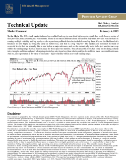
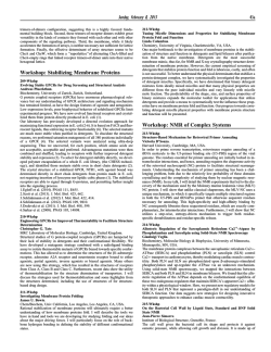
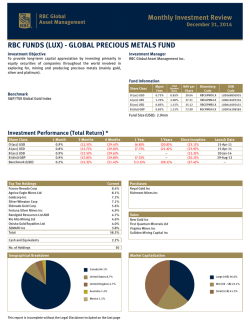
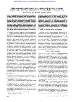
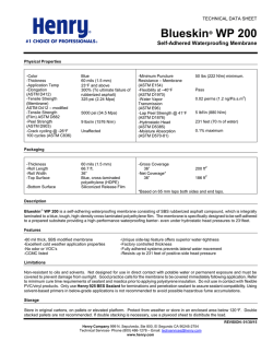
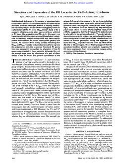
From www.bloodjournal.org by guest on February 6, 2015. For personal use only.
Improved Red Blood Cell Preservation Correlates With Decreased Loss of
Bands 3, 4.1, Acetylcholinestrase, and Lipids in Microvesicles
By U.J. Dumaswala, R.U. Durnaswala, D.S. Levin, and T.J. Greenwalt
In earlier studies we have shown that a final concentration of
0.69% glycerol in blood mixed with anexperimental additive
solution, EAS 25, improves the in vitro quality and in vivo
survival of red blood cells (RBCs).The objective of this study
was to determine if the better preservation of RBCs in EAS
25 is correlated with the improved maintenance of membrane lipids and proteins and decreased vesiculation. Split
units of RBCs were stored in Adsol or EAS 25(mmol/L: adenine 2/2, dextrose 122/110, mannitol 42/55, glycerol 0/150,
NaCl 154/50). After 12 weeks storage, RBC and microvesicle
membranes were analyzed for cholesterol, phospholipid, diphenyl hexatriene fluorescence anisotropy, and acetylcho-
linesterase (AchE) activity. Bands 3 and 4.1 were identified
in themicrovesicle membranes by immunoblotting. The RBC
membrane cholesterol, phospholipids, and AchE remained
higher in EAS 25 than inAdsol (P< .001). Vesicle membrane
lipids and AchE in EAS 25 were significantly less than in
Adsol ( P < .001). The fluidity of stored cells in both thesolutions was greater thanthe prestorage samples. Immunoblotting analyses showed that bands 3 and 4.1 were
greatly reduced in themicrovesicle membranes shed by the
RBCs stored in EAS 25 compared with those formed in Adsol.
0 1996 by The American Society of Hematology.
D
tion of approximately 7 5 mmoVL (0.69%) glycerol is less
affected, and AchE and lipids are better maintained than in
Adsol. Decreased total vesicle membrane proteins with less
AchE and bands 3 and 4.1 were found in EAS 25.
URING BLOOD BANK storage, red
blood
cells
(RBCs) are transformed from discocytes to spheroechinocytes. Associated changes in their membranes include
the loss of acetylcholinesterase (AchE), other membrane proteins, cholesterol, phospholipids, and the shedding of hemoglobin-containing 50 to 200 nm vesicles."' Kadlubowski and
Schrier et al"." reported the appearance of high molecular
weight complexes, increases in bands 1 plus 2, 4.2, 5 , and
6 and decreases in bands 7 and 8 in the membranes of RBCs
stored for 4 to 8 weeks in citrate-phosphate-dextrose or citrate-phosphate-dextrose-adenine-2 solutions.
The formation of vesicles has been related to the oxidative
damage of spectrin, band 4. l deficiency andthe cross-linking
of spectrin.','*." Only traces of bands l and 2 have been
found in the vesicle membranes and bands 3, 4.1, and glycophorin A have been demonstrated by immunoblotting and
periodic acid Schiff ~ t a i n i n g . ~ , ' ~ . ' ~
Our studies have suggested that improved preservation
correlates with decreased RBC vesiculation.15Goals for improvedRBC preservation include decreasing the loss of
membrane proteins, lipids, and vesicles.
We have previously reported that RBC adenosine triphosphate (ATP) is better maintained and there is less K' leakage
and hemolysis in a hypotonic experimental additive solution
containing 150 mmoVL glycerol (EAS 25) than in Adsol.l6
Itwas hypothesized that the beneficial effect was due to
better maintenance of membrane integrity and decreased vesiculation. We now report that the microviscosity of the
membranes of RBCs stored in EAS 25 with a final concentraFrom the Hoxworth Blood Center andthe Children's Hospital
Medical Center, University of Cincinnati Medical Center, Cincinnati,OH.
Submitted December 2, 1994; accepted October 2, 1995.
Supported in part by a National Institutes of Health Grant No.
HL 44987 and by a grant from the National Blood Foundation.
Address reprint requests to Umakant J. Dumaswala, PhD, Hoxworth Blood Center, University of Cincinnati Medical Center, PO
Box 670055, 3130 Highland Ave, Cincinnati, OH 45247-0055.
The publication costsof this article were defrayedin part by page
charge payment. This article must therefore be hereby marked
"advertisement" in accordance with 18 U.S.C. section 1734 solely to
indicate this fact.
0 1996 by The American Society of Hematology.
0006-4971/96/8704-0020$3.00/0
1612
MATERIALS AND METHODS
Blood donors acceptable by American Associationof Blood Banks and FoodandDrug Administration criteria
were used. The protocol was approved by the University Institutional
Review Board. Standard units of blood (450 +- 45 mL) were collected with 63 mL citrate-phosphate-dextrose solution in polyvinyl
chloride bags (PL 146, Baxter Healthcare Corp, Deerfield, IL) and
were centrifuged (RC-3C, Sorvall, Dupont, Wilmington, DE) at 8358
for 10 minutes. The platelet-rich plasma was expressed into a satellite
bag. The packed red blood cells (PRBCs) were split aseptically in
two aliquots by weight into 300 mL polyvinyl chloride transfer bags.
Using a sterile connecting device (SCD 312, Haemonetics Corp,
Braintree, MA), 100 mL of an experimental additive solution (EAS
2 5 ) was added to one aliquot, and 50 mL of the commercial additive
Adsol to the other. The aliquots were stored lying flat at 1 to 6°C
without mixing. Before sampling at 0 and 84 days, they were thoroughly mixed on a platelet agitator (Meddev Corp. Los Altos, CA).
Sterility was confirmed by inoculating 1.0 mL of the final samples
into tubes of thioglycolate and tryptic soy broth.
Preparation of EAS 25. Formulations of the preservation solutions are given in Table 1. The method of preparation was as described."
Preparation of RBCs, ghosts, and microvesicles. The RBCs
were purified by the method of Beutler et ai1' using a-cellulosemicrocrystalline cellulose columns, as described previously? to eliminate leukocyte and platelet contamination. The purified RBCs were
lysed in 30 volumes of 5 mmoUL Na,HPO, buffer, pH 8.0 according
to Dodge and Phillips.'x The ghosts were isolated by centrifugation
at 38,0008 for 20 minutes and washed five times in the same buffer
to obtain hemoglobin-free white ghosts.
Microvesicles were isolated as described previously." Briefly, the
supernatant was collected after centrifugation of blood at 2,000g for
10 minutes and passed through 0.8 p m nitrocellulose filters (Nuclepore Corp, Pleasanton, CA) in an Amicon filtration system (Amicon
Corp, Lexington, MA) at 5 to 10 lbs/sq in N, pressure. The filtrate
was centrifuged at 38,OOOg for 1 hour and the isolated vesicle pellet
was washed twice with phosphate buffered saline, pH 7.4 containing
2 mmoUL EDTA and 1 mmoUL phenylmethylsulfonylfluoride to
prevent proteolysis and stored at -70°C for further studies.
RBC and vesicle membrane lipid und juorescence unisotropy
analyses. After lipid extraction by the method of Broekhuyse,"
cholesterol was measured by a coupled enzymatic assay (Sigma
Chemical CO, St Louis, MO)" and phospholipid was measured as
Blood samples.
Blood, Vol 87, No 4 (February 15). 1996: pp 1612-1616
From www.bloodjournal.org by guest on February 6, 2015. For personal use only.
4.1,
LIPIDS
BANDS 3,
AND
PRESERVATION
IN RBC
1613
Table 1. ComDosition of Additive Solutions (Millimoles Per Liter)
Table 3. Effect of Storage on RBC and Vesicle Membrane Fluidity
~~~
Adenine
Dextrose
Mannitol
Glycerol
NaCl
pH at 25°C
Milliosmolality
Total
Nonpenetrating
EAS 25
Adsol
2.0
110.0
55.0
150.0
50.0
7.15
2.0
122.0
42.0
406.0
145.0
475.0
322.0
RBC Ghosts
Vesicles
Degree of Polarization
Pre
Adsol (12wk)
0.2121 2 0.0017
0.19722 0.0004*
<.001
0.20382 0.0022’
0.2136
<.001
P
154.0
5.86
=.073
Nonpenetrating milliosmolality represents components that do not
penetrate the cell membrane.
EAS 25(12 wk)
P
0.2058 2 0.0077
2
0.0008
n = 6;mean ? SD. The Pvalues forthe ghosts are basedon comparison to presamples.
* P value for Adsol v EAS 25 ghosts after 12 weeks of storage =
.04.
(IVF 12, courtesy Dr M.L. Jennings, The University of Texas Meditotal lipid phosphorus by the method of Bartlett.21Cholesterol and
cal Branch at Galveston, Galveston, TX) to human band 3. The
phospholipid concentrations were expressed as pmoVmL RBC, and
indicator antibody was a peroxidase-conjugated goat antimouse IgG
the molar fractions of cholesteroUphospholipidwere calculated.
(Organon Teknika, Durham, NC), and the color development was
Fluorescence anisotropy was determined according to the method
in the presence of 3,3’ diaminobenzidine and hydrogen peroxide.
of Williams and Hazel” using the lipid soluble probe, 1.6-diphenylThe amount of band 3 was determined by densitometric scanning
To mini1,3,5-hexatriene (Aldrich Chemical CO, Milwaukee, W).
(BioRad, Model 680 video densitometer, Hercules, CA).
mize probe-probe interaction, 300 pg of membrane/vesicle protein
Band 4.1 protein was localized by treating the blots with a polysamples were suspended in 5 mmoVL Tris-HCI buffer, and the samclonal rabbit antiserum to human band 4.1 (courtesy Dr P. Low,
ples were sonicated before fluorescence measurements. All measureIndiana University, Bloomington, IN). The secondary antibody was
ments were made at 37°C using a Perkin-Elmer fluorescence spectroperoxidase-conjugated goat antirabbit IgG (Organon Teknika, Durphotometer model 650-10s (Perkin Elmer, Norwalk, CT) equipped
ham, NC), and color development was as described above.
with a polarizing filter. Measurements were made at an excitation
wavelength of 366 nm and an emission wavelength of 430 nm.
Membrane proteins. Total proteins were determined by the
RESULTS
method of Bradford” with Coomassie Brilliant Blue G 250 phosTo simplify comparisons, data are presented in terms of
phoric acid reagent. The hemoglobin in the vesicle preparations was
units of PRBC volume represented as the denominator.
quantified by the micro-Drabkin method modified by the addition
of 0.5% Triton X-l00 to lyse the vesicles and clear the t ~ r b i d i t y . ~ ~ Effect of storage media on lipid composition andjuoresThe hemoglobin value was subtracted from the total protein value
cence anisotropy. RBC membranes lost more cholesterol
to obtain the amount of membrane protein.
than phospholipids during storage (Table 2). Though there
Acetylcholinesterase activity. The AchE was determined in the
was significantly less loss of both cholesterol and phosphomembranes of RBCs and vesicles bythe method of EllmanZ5as
lipids after storage for 12 weeks in EAS 25 than in Adsol,
modified by Chow and Islam.26
the RBC membrane C/P ratio in both media declined from
Localization and quanti$cation of band 3 and 4.1 proteins. Coma 0.83 prevalue to 0.73. Concurrent with changes in lipid
parable concentrations of vesicle proteins were analyzed by 10%
composition, there were significant changes in lipid fluidity.
polyacrylamide gel electrophoresis according to Tsang et al.27The
Steady state anisotropy values of RBC membranes decreased
fractionated proteins were electroblotted onto 0.2 p-nitrocellulose
with storage, the decrease being markedly greater in Adsol
membranes for 16 to 18 hours to assure quantitative transfer. The
than in EAS 25 (Table 3). The fluidity of the vesicle memblotted samples were reacted with a mouse IgG monoclonal antibody
Table 2. Comparison of the Effect of Storage Media on RBC Lipids
Cholesterol
Phospholipids
CIP
Iprnollrnl RBCs)
~~~~
branes in Adsol was greater than in EAS 25, but the difference was not significant (Table 3).
Cholesterol and phospholipid contents of the vesicles shed
in EAS 25 were significantly lower than those in Adsol, but
the C/P ratios were not significantly different (Table 4).
RBC vesiculation. RBCs stored in EAS 25 shed signifi-
~~
Pre
n = 6
,021
056
Adsol (12wk)
n = 11
P <.0001
EAS 25
(12
wk)
n = 11
P .3462
3.47 t 0.28
4.17
2
0.16
0.83
2.52 2 0.18’
3.48
<.0001
-c 0.38*
0.73
2.97 2 0.202*
4.08
,0007
2
2
0.06
t 0.11
Table 4. Effect of Storage Media on Lipids of Vesicles Collected
After 12 Weeks of Storage
Cholesterol
0.172’
0.73
PhosDholiDids
CIP
t 0.03
Mean t SD. All of the above Pvalues are based on comparing 12week data to presamples.
*P < ,001.comparison of Adsol v EAS 25 RBCs at 12 weeks of
storage.
Adsol
EAS 25
P
0.18 t 0.05
0.28
0.09 ? 0.04
0.13
<,0001
n = 12, mean 2 SD.
t 0.09
0.65
t 0.05
0.69
0.05
.l95
1.0001
t 0.09
From www.bloodjournal.org by guest on February 6, 2015. For personal use only.
DUMASWALA ET AL
1614
Table 5. Effect of Storage Mediaon Proteins of Vesicles Collected
After 12 Weeks of Storage
Adsol
€AS 25
P
0.27 2 0.14
<.0001
kDa
- 116
- 97
- 78
rnalrnL RBC
0.65 2 0.22
n = 13, mean 2 SD.
Band 4.1
>
- 66
cantly less vesicle membrane proteins ( P < .0001) than those
stored in Adsol (Table 5).
Immunohlot analyses of vesicle membrane hands 3 and
4. I . Band 3 and band 4.1 immunoblots are shown in Figs
1 and 2. Densitometric scanning showed that the amounts
of intact bands 3 and 4.1 were lower by 40% ? 7.2% and
51.6% ? 12.4% (n = 5), respectively, in vesicles formed in
EAS 25 than in Adsol. The Coomassie blue staining of the
- 45
- 14
kDa
Adsol EAS 25
at 12 wks at 12 wks
-
<Band 3
97
-45
-31
-14
Adsol EAS 25
at I2 wks
Fig l. Band 3 immunoblots of microvesicles after 12 weeks of
storage in Adsol andEAS 25. Blotted membranes were reacted with
monoclonal antibody llVF12)t o human band 3, followed by peroxidase-conjugated indicator antibody. The color development was in
the presence of 3.3' diaminobenzidine and hydrogenperoxide.
Fig 2. Band 4.1 immunoblots of microvesicles after 12 weeks of
storage in Adsol andEAS 25. Blotted membranes were reacted with
polyclonal rabbit antiserumt o human band 4.1,followed by peroxidase-conjugated indicator antibody. The color development was in
the presence of 3.3' diaminobenzidine and hydrogen peroxide.
vesicle membrane protein shows a similar decrease of band
3 protein only (Fig 3).
AchE activity. AchE activity (Table 6) remained significantly higher in RBCs stored in EAS 25 than
in Adsol
( P < .OOOI). At the end of12 weeks of storage, AchE
activity of RBCs stored in EAS 25 did notdiffer significantly
from the initial values. AchE in EAS 25 vesicles was 4 0 %
of that found in Adsol vesicles.
DISCUSSION
During blood bank storage or accelerated ATP depletion,
RBC membranes lose cholesterol, phospholipids, proteins,
and shed exocytic microvesicles.'.'.' The microvesicle membranes lack spectrin and ankyrin, but have demonstrable
amounts of most of the other peripheral and integral proteins.3.~n.~
I They are selectively enriched in A C ~ E . ' . ~ We
.~.'~
have previously shown that RBC vesiculation, hemolysis,
and potassium loss in vitro and in vivo recovery are improved in EAS 25.1".2xEAS 25 differs fromAdsolmainly
by containing 150 mmol/L (1.38%) glycerol, less NaCI, and
hypotonicity of the nonpenetrating ingredients (Table l ) .
The data in Table 2 indicate a greater loss of cholesterol
(27%) and phospholipids (16.5%) by RBCs stored in Adsol
than in EAS 25 (14% and 2%, respectively). The difference
in the loss of cholesterol maybe sufficient to account for
the greater increase in membrane fluidityfoundin RBCs
stored in Adsol (Table 3). Poznansky et al" reported that
From www.bloodjournal.org by guest on February 6, 2015. For personal use only.
4.1,
BANDS 3,
AND LIPIDS IN RBC PRESERVATION
1615
the cholesterol content of RBC membranes markedly affected K' transport. Relatively lower RBC K' leakage observed earlier16 in EAS 25 than in Adsol may be correlated
with reduced RBC cholesterol loss in EAS 25. The modest
change in the molar cholesterokphospholipid ratios was not
sufficient to cause the drastic changes in morphology reported by Cooper et al."
The markedly decreased loss of cholesterol, phospholipids, AchE, andproteins by RBC membranes in microvesicles
suggests better maintenance of membrane integrity in EAS
25 (Tables 4, 5, and 6). This is supported by the lower
amounts of bands 3 and 4.1 in the membranes of the microvesicles shed in EAS 25 thaninAdsol (Figs 1, 2, and
3). It is possible that significantly greater disruption of the
linkages between spectrin, bands 3 and 4.1 and between the
latter and the glycophorins occurs in Adsol than in EAS
97 -
116
66
-
4s
-
<Band3
- 97
-4s
-31
- 14
Adsal M25
at 12 wks
Fig 3. Coomassie blue staining of microvesicle membrane proteins after 12 weeks of storage in Adsol and EAS25. Polyacrylamide
gel electrophoresis performed as described in Materials and Methods. Lane 1, high molecular weight standards, lane 4, low molecular
weight standards.
Table 6. Effect of Storage Media on RBC and Vesicle AchE
RBCs
Vesicles
(mol. hydrolyzed per minute per mL RBCd
Pre
(1.872 0.31)X IO3
n
Adsol (12wk)
P
n
wk)
EAS 25 (12
P
n
7
(1.30 -C 0.19)X lo3"
0.0002
10
(1.60 2 0.25) X lo3*
.062
10
(2.042 0.51) X 10'
12
(1.002 0.39)X IO'
<.0001
12
Mean 2 SD. The P values for RBCs are based on comparisons to
presamples.
P < .0001,comparison of Adsol v EAS 25 RBCs at 12 weeks of
storage.
25. One possible reason for this could be the oxidation of
sulfhydryl groups in spectrin and protein 4.1.7*13Low levels
of protein band 4.1 found in vesicles released from RBCs
stored in EAS 25, suggest that there is a better maintenance
of this protein in RBCs, resulting in stronger spectrin-actin
interaction. This would be ameliorated if the glycerol in EAS
25 serves as a free oxygen radical scavenger."
Phosphoinositides have been shown to enhance the interaction of protein 4.1 with the g l y c ~ p h o r i n s . ~Possibly
* ~ ~ ~ this
relationship will prove to have a bearing on the release of
protein 4.1 in vesicle membranes. We have shown that phosphoinositide 4,5-P04, which has the greatest effect on the
protein 4.1 and glycophorin linkage, is selectively better
represented in RBC vesicles, suggesting perturbation of this
linkage.'
The effect of the hypoosmolarity of EAS 25 must also be
considered. The initial mean corpuscular volume (MCV) of
the RBCs in this preservative versus Adsol was 1 13 W90 fL,
and after 12 weeks, the values were 100/88
Meryman
et al" have suggested that increased membrane tension in
hypoosmolar solutions will reduce the shedding of vesicles
by RBCs during storage. This has not beenproven.Itis
difficult to conceive that this could be a factor in explaining
the differences in vesicle membrane composition thatwe
have described. Cell swelling and low pH activate the K-Cl
cotransport p a t h ~ a y . ~This
'
pathwayis active in reticulocytes and the young RBC population and plays a role in
regulatory volume decrease (RVD). Under blood bank storage conditions, RVD and the K-Cl cotransport pathway probably do not play a significant role.
The conclusions that may be drawn are that the formation
of vesicles by RBCs is a complex process that depends on
changes in the interactions occurring between the cytoskeleton, peripheral membrane proteins, and lipids. The quantity
and structure of the vesicles varies with the nature of the
storage medium in which the RBCs were kept. It is suggested
that less loss of critical membrane proteins is an indicator
of improved RBC preservation. Additional studies are
needed for understanding the results reported. They will be
the underpinning for the development of formulations for
the improved storage of RBCs.
From www.bloodjournal.org by guest on February 6, 2015. For personal use only.
DUMASWALA ET AL
1616
ACKNOWLEDGMENT
Appreciation is expressed to Margaret O'Leary for her assistance
in the preparation of this manuscript.
REFERENCES
I . Rimon S, Brok F, Rimon A: Changes in erythrocytes lipids
during storage. Clin Chim Acta 12:22, 1965
2. Trotter J, Rumsby MG: Lipids of the human erythrocyte membrane during storage for transfusion. Correlation of lipid changes
with the discocyte to spherocyte shape transformation. J Appl Biochem 3:19, 1981
3. Greenwalt TJ, Bryan DJ, Dumaswala UJ: Erythrocyte membrane vesiculation and changes in membrane composition during
storage in citrate-phosphate-dextrose-adenine-]. Vox Sang 47:261,
1984
4. Greenwalt TJ, Bryan BJ, Dumaswala UJ: Some changes in
RBC phosphoinositide metabolism during blood bank storage. Blood
66:277a, 1985
S . Dumaswala UJ, Greenwalt TJ: Effect of storage on red blood
cell membrane phosphoinositides (PIS) and acetylcholinesterase
(AchE). 28th International Conference on the Biochemistry of Lipids, Nottingham, UK, D28, 1987 (abstr)
6. Rumsby MG, Trotter J, Allan D, Michell RH: Recovery of
membrane microvesicles from human erythrocytes stored for transfusion. A mechanism for the erythrocyte discocyte-to spherocyte
shape transformation. Biochem Soc Trans 5:126, 1977
7. Wagner GM, Chiu J-H, Heath RH, Lubin BH: Spectrin oxidation correlates with membrane vesiculation in stored RBCs. Blood
69: 1777, 1987
8. Weitz M, Bjerrum OJ, Ott P, Brodbeck U: Quantitative composition and characterization of the proteins in membrane vesicles
released from erythrocytes by dimyristoylphosphatidylcholine.A
membrane system without cytoskeleton. J Cell Biochem 19:179,
1982
9. Hagelberg C, Allan D: Restricted diffusion of integral membrane proteins and polyphosphoinositides leads to their depletion in
microvesicles released from human erythrocytes. Biochem J
271:831, 1990
IO. Kadlubowski M: The effect of in-vivo ageing of the human
erythrocyte on the proteins of the plasma membrane. A comparison
with metabolic depletion and blood bank storage. Int J Biochem
9:79, 1978
1 1. Schrier SL, Sohmer PR, Moore GL, Ma L, Junga I: Red blood
cell membrane abnormalities during storage. Correlation with in vivo
survival. Transfusion 22:261, 1982
12. Wagner GM, Chiu DT-Y, Yee MC, Lubin BH: Red cell vesiculation-A common membrane physiologic event. J Lab Clin Med
108:315, 1986
13. Wolfe L, Byrne A, Becker P, John K, Lux S: Reversible
injury to the red blood cell membrane skeleton during storage. Clin
Res 31:486, 1983
14. Lutz HU, Liu S-C, Palek J: Release of spectrin-free vesicles
from human erythrocytes during ATP depletion. J Cell Biol 73:548,
1977
15. Greenwalt TJ, Zehner Sostok C, Dumaswala UJ: Studies in
redblood cell preservation. 2. Comparison of vesicle fOrInation,
morphology, and membrane lipids during storage in AS-l and
CPDA-1. Vox Sang 58:90, 1990
16. Dumaswala UJ, Bentley NL, Greenwalt TJ: Studies inred
blood cell preservation. 8. Liquid storage of red cells in a glycerolcontaining additive solution. Vox Sang 67:139, 1994
17. Beutler F, West C, Blume K-G: The removal of leukocytes
and platelets from whole blood. J Lab Clin Med 88:328, 1976
18. Dodge JT, Phillips GB: Comparison of phospholipids and of
phospholipid fatty acids and aldehydes in human red cells. J Lipid
Res 8:667, 1967
19. Broekhuyse RM: Improved lipid extraction of erythrocytes.
Clin Chim Acta 51:341, 1974
20. Allain CC, Poon LS, Chan CSG, Richmond W, Fu PC: Enzymatic determination of total serum cholesterol. Clin Chem 20:470,
1974
21. Bartlett GR: Phosphorus assay in column chromatography. J
Biol Chem 234:466, 1959
22. Williams EE, Hazel JR: Membrane fluidityand hemilayer
temperature sensitivity in trout hepatocytes during brief in vitro cold
exposure. Am J Physiol 266:R773, 1994
23. Bradford MM: A rapid and sensitive method for the quanititation of microgram quantities of protein utilizing the principles of
protein-dye binding. Anal Biochem 72:248, 1976
24. Greenwalt TJ, McGuinness CG, Dumaswala UJ: Studies in
red blood cell preservation: 4. Plasma vesicle hemoglobin exceeds
free hemoglobin. Vox Sang 61:14, 1991
25. Ellman GL, Courtney KD, Andres Jr V, Featherstone RM: A
newandrapid
colorimetric determination of acetylcholinesterase
activity. Biochem Pharmacol 7:88, 1961
26.Chow CM, Islam MF: Colorimetric determination of red
blood cell acetylcholinesterase activity. Clin Biochem 3:295, 1970
27. Tsang VCW, Peralta JM, Simons AR: Enzyme-linked immunoelectrotransfer blot techniques (EITS) for studying the specificities
of antigens and antibodies separated by gel electrophoresis, in Langdon JJ, Vanakie HV (eds): Methods in Enzymology, Immunological
Techniques, v01 92. New York, NY, Academic, 1983, p 377
28. Greenwalt TJ, Dumaswala UJ, Rugg N: A glycerol additive
solution improves red cell viability. Transfusion 34:66S, 1994 (abstr)
29. Poznansky M, Kirkwood D, Solomon AK: Modulation of red
cell K' transport by membrane lipids. BiochimBiophysActa
330:3S I , 1973
30. Cooper RA, Arner EC, Wiley JS, Shattil SJ: Modification of
red cell membrane structure by cholesterol-rich lipid dispersions. A
model for the primary spur cell defect. J Clin Invest 55:115, 1975
3 1. Oberley LW, Sierra E: Radiation sensitivity testing of cultured
eukaryotic cells, in Greenwald RA (ed): Handbook of Methods for
Oxygen Radical Research. Boca Raton, FL, CRC, 1989, p 417
32. Anderson RA: Regulation of protein 4.1-membrane associations by a phosphoinositide, in Agre P, Rarder JC (eds): Red Blood
Cell Membranes: Structure, Function and Clinical Implications. New
York, NY, Marcel Dekker, 1989, p 187
33. Pinder JC, Chung A, Reid ME, Gratzer WB: Membrane Attachment sites for the membrane cytoskeletal protein 4. I of the red
blood cell. Blood 82:3482, 1993
34. Meryman HT, Hornblower MLS, Syring RL: Prolonged storage of red cells at 4°C. Transfusion 26:500, 1986
35. Olivieri 0, de Franceschi L, de Gioncoli M, Cirelli D, Corrocher R: Potassium loss and cellular dehydration of stored erythrocytes following incubation in autologous plasma: Role of the KC1
cotransport system. Vox Sang 65:95, 1993
From www.bloodjournal.org by guest on February 6, 2015. For personal use only.
1996 87: 1612-1616
Improved red blood cell preservation correlates with decreased loss
of bands 3, 4.1, acetylcholinestrase, and lipids in microvesicles
UJ Dumaswala, RU Dumaswala, DS Levin and TJ Greenwalt
Updated information and services can be found at:
http://www.bloodjournal.org/content/87/4/1612.full.html
Articles on similar topics can be found in the following Blood collections
Information about reproducing this article in parts or in its entirety may be found online at:
http://www.bloodjournal.org/site/misc/rights.xhtml#repub_requests
Information about ordering reprints may be found online at:
http://www.bloodjournal.org/site/misc/rights.xhtml#reprints
Information about subscriptions and ASH membership may be found online at:
http://www.bloodjournal.org/site/subscriptions/index.xhtml
Blood (print ISSN 0006-4971, online ISSN 1528-0020), is published weekly by the American
Society of Hematology, 2021 L St, NW, Suite 900, Washington DC 20036.
Copyright 2011 by The American Society of Hematology; all rights reserved.


© Copyright 2026