
Erythrocytes by Dilauroylphosphatidylcholine-Induced
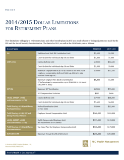
From www.bloodjournal.org by guest on February 6, 2015. For personal use only.
Generation of Phenotypically Aged Phosphatidylserine-Expressing
Erythrocytes by Dilauroylphosphatidylcholine-InducedVesiculation
By Cecilia Diaz, Jerzy Morkowski, and Alan J. Schroit
In vitro stored red blood cells (RBC) and RBC artificially induced to vesiculate by incubation with dilauroylphosphatidyl-choline were monitored for age- and vesiculation-dependent changes in celldensity, membrane lipid asymmetry,
and their ability to berecognized and cleared by reticuloendothelial cells. RBC demonstrated a progressive increase in
density on self-forming Percoll gradients upon vesiculation
and in vitro "aging." Uptake of vesiculated RBC by in vitro
cultivated macrophages was increased threefold over nonvesiculated control RBC. The clearance rate of dense vesiculated RBC was biphasic and contained a rapid component
and a slower second component consistent with the clear-
ance rates of normal control populations. Determination of
phosphatidylserine (PS) in the outer leaflet ofRBC by the
PS-dependent prothrombinase assay revealed that PS redistributed to thecell's outer leaflet upon in vitro storage and
vesiculation. Inhibition of PS movement by oxidation of
membrane sulfhydryls with pyridyldithioethylamine resulted in higher prothrombinase levels and enhanced.clearance of vesiculated RBC. These
experiments suggest that
vesiculation contributes to alterations in membrane lipid
asymmetry and cell density characteristic of the aged RBC
phenotype.
0 1996 by The American Society of Hematology.
T
Denselold RBC also exhibit significant reductions in volume/surface area that can be explained by decreased electrolyte content associated with osmotic water loss and loss of
membraneandcytoplasm by microvesiculation.' '.'i~z4 Because spontaneousformation of vesicles occurs in vivo'5 and
in i n vitro stored RBC,'"'' we hypothesized that vesiculation
could. at feast i n part, be responsible for the age-dependent
alterations in membrane lipid asymmetry associated with
macrophage recognition. To test this hypothesis, RBC were
artificially vcsiculated") with dilauroylphosphatidylcholine
(DLPC) and assessed for alterations in density, membrane
lipid asymmetry, and their propensity to be recognized by
macrophages in vitro and the RES in vivo.
HE MECHANISM(S) responsible for the recognition of
aged red blood cells (RBC) by the reticuloendothelial
system (RES) that results in their elimination from the circulation are not completely understood. Various moieties have
been shown to play a role in the determination of the aged
phenotype. These include several normal RBC components
that have undergone specific modifications such as loss of
carbohydrates that result in decreased surface charge'.' and
the appearance of asialoglycophorin,' irreversible oxidative
damage causing recognition of glycophorin A: proteolytic
3,"'' and
formation of "senescent cell antigen" from band
loss of plasma membrane phospholipid asymmetry resulting
in the progressive appearance of phosphatidylserine (PS) at
the cellsouter leaflet." Because theseprocesses are not
mutually exclusive, the mechanisms underlying phagocytic
recognition could be complex involving multiple receptorligand interactions. These could include the Fc receptor for
antibody-mediated recognition.'.' VCAM- I/vitronectin receptor for VLA-4 expressing apoptotic cells""" and differentiating erythroblasts,"
scavenger
receptors,"
oxidized
LDL receptors.""" and the putative PS receptor.' i.'"""
Because there seems to be a direct relationship between
RBC density and age,'"." most studies o n erythrocyte aging
have been bdsed on comparisons between density-separated
RBC populations. Using self-forming Percoll gradients,
we
have recently shown direct
a relationshipbetween
the
amounts of PS exposed on the surface of RBC, their ahility
to transport lipids between membrane leaflets, and their propensity to be bound by macrophages in vitro and cleared by
the RES in vivo.'
___
"
"
Fronl the Departrrrent of' Cell Biology, Tire Utlirc,r.\in; of Textrs
M.D.Anderson Cancer Cerfter. Houston.
Suhnritted June 7, 199.5: uccepted Novewher 7, 1995.
S~cpportedin part by Nutiond fnstitutes of Health Grunt No. D K
41714.
Address reprint requests to Alun J . Schroit, PhD, Department o f
CellBiology, B M 173, TheUniver.sity of Texas M.D. Anderson
C a m e r Center, 1515 Holcombe Blvd, Houston, TX 77030.
The publication costs of this article were defrayedin parr by puge
churgepuyment.Thisarticlemustthereforebeherebymarked
"advertisement" i n accordance with I8 U.S.C. section 1734 solely t o
indtcate this fact.
0 I996 by The American Socieiy of Hematology.
OUU~-4Y71/96/~707-0U47~3.00/0
2956
MATERIALS AND METHODS
Muterirr1.r t r l d routine
prowdure.\.
Pyridyldithioethylamine
as previously described.'" Factor V was
(PDA)wassynthesized
isolated from bovine plasma and activatedas previously described.''
Prothrombin and factor X were from Sigma (St Louis, MO) and the
throlnbin-sensitivcchronwgen. S223X. was purchased from Kabi
Laboratories (Franklin. OH), Percoll was obtained from Pharmacia
(Uppsala. Sweden). KBC were collectcd into heparinized tubes by
venipuncture frorn healthy volunteer5 or from the tail vein of mice.
l h e blood was diluted with HEPES-saline buffer (144 Inrnol/L NaCI,
I O mmol/L HEPES. I O mmol/L glucose, pH 7.3) and washed twice.
Percoll was prepared by mixing 213.5 g of Percoll with 25 niL of
1Ox concentrated phosphate-buffered saline (PBS) andwater to 250
nlL. The pH was adjusted to 7.4 and the osmolarity to 310 to 320
nlOsmdkg. Radiation was monitored by scintillation counting using
a Packard AutoGamma spectrometer (Packard, Downers Grove, IL).
&,,.sit!
w p m t ; [ m of KBC. RBC were separated by density on
self-forming Pcl-coll gradientsessentially B'\ described by Lutr et
al." Briefly. 0.5 tnL o f packed RBC were mixed with 9 mL of
Percoll (320 InOsnI, pH 7.4) at room temperature. The suspension
was then centrifuged at 38.700g for 20 minutes. RBC were obtained
from the gradients by puncturing the bottom o f the tube and washed
free of Percoll.
Vc,.sicu/nrionof' RBC. Smallunilamellarvesicles were prepared
by sonication of DLPC (1.5 mmol/L) in HEPES-saline buffer followed by centrifugation at 30,OOOg to remove metal fragments and
largevesicles.WashedRBCwereresuspended
in HEPES-saline
to a 10% hematocrit and incubated with DLPC (38 pmoIL final
concentration) at 4°C on an orbital shaker for 45 minutes. Remnant
RBC were collected by centrifugation at 85Og for I O minutes. The
renlnantcellswere
then washed with 1% bovineserumalbumin
(BSA) to remove residual DLPC from the cell membranes.
~ r o t l i r r ~ ~ ~ t hccc./ivit\'
i r ~ t r . c~s.sa\'.
~~~
Vesiculated andnonvesiculated
Blood, Vol 87, No 7 (April I), 1996: pp 2956-2961
From www.bloodjournal.org by guest on February 6, 2015. For personal use only.
VESICULATION-INDUCED PS EXPRESSION ANDAGING
RBC (2 X IO' cells) were incubated in 0.2 mLof prothrombinase
assay buffer (1 35 mmol/L NaCI, 3 mmol/L CaCl,, I O mmol/L
HEPES, pH 7.4) at 37°C for 3 minutes. A total of 0.05 mL of factor
Va (60 nmol/L) and 0.1 mL of factor Xa (0.25 U) was then added.
The suspension was incubated at 37°C for another 3 to 4 minutes
after which 0.1 mL of prothrombin (0.1 U) was added. After 5
minutes, a 0.1-mL aliquot of the suspension was transferred to a
cuvette containing I mL of TrisEDTA buffer ( I 75 mmol/L NaCI,
50 mmol/L Tris, 2 mmol/L EDTA, pH 7.9) to stop the production
of thrombin. The thrombin-dependent chromogen, S2238, was added
to the cuvettes (to 0.2 mmol/L), and the rate of chromophore formation was monitored at 405 nm with a Gilford Response Spectrophotometer using appropriate kinetic software. The initial rate of thrombin-dependent chromophore production, which is proportional to the
amount of PS present on the cell
was determined from the
slope of theabsorbance curve. To quantify the amount of endogenous
PS exposed on the surface of these cells, a standard curve of the
rates of thrombin productionlchromophore formation was generated
using PDA-treated RBC that contained known amounts of I-oleoyl-2-[N-(7-nitro-2,1,3-benzoxadiazol-4-yI)amino]caproyl]
(NBD)labeled PS exposed at the cells's outer
Thrombin-dependent chromophore production was directly proportional to the
amount of PS present in the cell's outer leaflet and linear within the
range of interest.
In vitro binding of 'ZsI-labeledRBC. Macrophages were obtained
from the peritoneal cavity of 8- to IO-week-old BalblC mice 4 days
after an intraperitoneal injection of 3 mL of thioglycolate medium.
The cells were collected and washed in serum-free RPMI-1640 medium. Cells (3X IO'lwell) were plated in 24-wellFalcon plastic
culture plates (Becton Dickinson, Lincoln Park, NJ). The nonadherent cells were removed from the plates after several hours. RBC
were labeled for I hour at 0°C with 0.25 mCi "'1 in the presence
of one iodobead (Pierce Chemical CO, Rockford, IL). The iodinelabeled RBC were then vesiculated and added to the macrophage
monolayers at a ratio of 10 RBChacrophage (-3 X 10" RBC) for
the indicated intervals. The cultures were then washed with HEPESsaline and lysed with 0.1% Triton X-100. The lysate was collected
and monitored for radiation.
In vivo clearanceof "Cr-labeled RBC. Two milliliters of mouse
RBC (50% hematocrit) were incubated with -0.5 mCi of "Cr for
45 minutes at 37°C and washed. The cells were vesiculated as described above and injected into the tail vein of syngeneic Balblc
mice (0.2 mL; 25% hematocrit). Mice were bled at the indicated
times and the fraction of "Cr-labeled RBC remaining in the circulationwas determined by scintillation counting. Livers and spleens
were collected at theend of the experiment to determine the presence
of cleared RBC. The same experiment was carried out with cells
pretreated with PDA and PDNdithiothreitol and with Percoll-separated in vitro-aged cells. The data points were fitted using Slidewrite
Plus (Advanced Graphics Software, Carlsbad, CA) curve-fitting routines. Initial rates were estimated from linear fits of data points
within the first hour after injection.
RESULTS
Clearance of in vitro stored RBC. To determine if in
vitro-stored RBCs exhibit density-dependent differences in
their ability to be cleared in vivo, mouse RBC were stored
at 4°C for 1 week, "Cr-labeled, separated into light and
heavy fractions on self-forming Percoll gradients (Fig l ) ,
and injected into syngeneic mice. Light cells separated from
freshly obtained blood were cleared with a Tln of -100
hours, whereas heavy cells from the same gradient were
cleared with half times of about 40 hours (Fig 2). Percoll
gradient centrifugation ofin vitro-stored cells showed a
2957
Fig 1. Percoll gradient separation of fresh and in vitro stored RBC.
Murine RBC were collected into heparin and stored for l week at
4°C. These cells and freshly obtained RBC were mixed with Percoll
and centrifuged at 38,7009 for 20 minutes at 20°C. (AI Fresh RBC; (B)
in vitro stored RBC. Arrow shows released vesicles.
prominent hemoglobin-containing band of vesicles at the top
of the gradient and RBC that localized at densities greater
than fresh RBC (Fig IB). In contrast to fresh cells, cells
recovered from these fractions exhibited TIQclearance rates
less than IO hours in a manner that was independent of
cell density (Fig 2). The rates of PS-dependent thrombin
production produced by the stored cells was also threefold
faster than the rates obtained with fresh cells (not shown),
suggesting that PS asymmetry is not rigourously maintained
after prolonged in vitro storage.
Effect of DLPC-induced vesiculation on RBC density and
PS asymmetry. The results presented above raise the possibility that a relationship between membrane vesiculation and
the appearance of the dense/aged phenotype with perturbed
membrane lipid distribution exists. To test this, freshly obtained RBC were induced to vesiculate with DLPC andseparated on Percoll (Fig 3). Similar to the pattern obtained after
in vitro aging, an overall shift in the distribution of the cells
to heavier densities occurred. The shift in density was also
dependent on the formation of microvesicles which accumulated near the top of the gradient (Fig 3B).
From www.bloodjournal.org by guest on February 6, 2015. For personal use only.
2958
DIM, MORKOWSKI, AND SCHROIT
n
same kinetics observed for control, nonvesiculated RBC.
Extrapolation of the calculated curves to the limit of percent
remaining cells approaching zero (Fig 6 inset) yielded a
lifespan of about 20 days for control cells andthe slow
component of vesiculated cells compared with approximately 5 days for the fast component of vesiculated cells.
Clearance of %"labeled RBC vesicles was also determined.
The data in Fig 6B shows that RBC vesicles were immedi-
A
S
A
0
W
0
A
m
0
lo
0
I
I
25
50
75
Time (hr)
Fig 2. Clearance of Percoll-separated fresh and stored RBC. Top
and bottom fractions of RBC separated by density on self-forming
Percoll gradients (Fig 1) were isolated, washed, and labeled with "Cr.
The labeled cell suspension was adjustedt o a 2546 hematocrit and0.2
mL was injected intravenously
into syngeneic mice. At the indicated
intervals minimal amounts of blood were collected from
tailthe
vein
and the fraction of
"Cr-labeled RBC remaining in the peripheralcirculation was determined by scintillation
counting. Fresh RBC, top fraction (A), bottom fraction (0);
in vitro-aged RBC, top fraction (A),
bottom fraction (0).
The effects of vesiculation on endogenous PS distribution
was determined by the PS-dependent prothrombinase assay.
Figure 4 shows that remnant DLPC-treated cells exhibited
significantly higher levels ofPS at the cell's outer leaflet.
PS content in the outer leaflet of the vesiculated RBC was
higher when cells were treated with PDA, a known inhibitor
of aminophospholipid transport. Upon the addition of dithiothreitol (DIT), the prothrombinase activity of the remnant
cells returned to values similar to vesiculated, non-PDAtreated cells suggesting that a fraction of the PS exposed at
the cell surface was transported back to the inner leaflet (Fig
4).
In vitro recognition and clearance of vesiculated RBC.
To determine whether RBC vesiculation results in their uptake by macrophages, '251-labeledcells were incubated with
macrophage monolayers. Figure 5 shows that the uptake of
the vesiculated cells was about threefold higher than the
uptake of nonvesiculated controls. Additional evidence suggesting that vesciculated RBC are recognized by cells of the
RES was obtained by determining the clearance rates of
vesiculated RBC in vivo. Figure 6 shows that control RBC
were cleared with a Tln of about 80 hours, whereas the
clearance kinetics of vesiculated cells suggested a two-component system. It can be seen that about half of the vesiculated cells were cleared within 12 hours (corresponding to
a T l n of <2 hours) with about 80% of the cleared radiation
recovered in the animals' liver and spleen (data not shown).
The remaining population seemed to be cleared withthe
Fig 3. Percoll gradient separation of DLPC-vesiculated RBC. Human RBC were collected into heparin and vesiculated as described
in the Materials andMethods. The cells were thenwashed and separated in Percoll. (A) ControlRBC; (B1 vesiculated RBC. Arrow denotes
vesicles that localized at the top of the
gradient.
From www.bloodjournal.org by guest on February 6, 2015. For personal use only.
2959
VESICULATION-INDUCED PS EXPRESSION AND AGING
600 r
control
500
0remnant
B r e m n a n t + DTT
400
h
CJ,
c
300
v)
a
200
100
changes lead to their recognition by macrophages and rapid
clearance from the peripheral circulation of syngeneic animals. a property characteristic of normal Percoll-separated
dense (aged) cells.' Thus. there seems to be a strong relationship between the presence of PS on cell surfaces and macrophage recognition/clearance mechanisms. Indeed, PS has
been shown to participate in various cell-cell interactions,"."
cell activation and hemostasis,24"" cell aging,"." membrane
fusion
and apoptosis."."
Similar to the redistribution ofPS obtained during Ca"
ionophore-mediated vesiculation of RBC,'" R B C vesiculation with DLPC is associated with a rapid redistribution of
cell-surface lipids. Analysis of cell-surface PS by prothrombinase activity showed that the cells immediately started to
correct abnormal lipid distributions. This can be seen from
the results showing that inhibition of PS transport byPDA
resulted in 1.7-fold higher prothrombinase levels which,
on treatment with DTT, returned to values of remnant cells
not treated with PDA. This conclusion is also supported by
the in vivo clearance studies which showed that removal of
vesiculated R B C from the circulation was a two-component
system: a fast component that was measurable immediately
after injection and a component with rates identical to control
values after -30 minutes.
R B C were treated with PDA at 0°C to monitor the clearance of vesiculated and transport inhibited cells. Although
PDA induced PS independent membrane alterations that re-
-
0
normal RBC
PDA-treated RBC
Fig 4. Prothrombinaseactivity of vesiculated RBC.Normal human
RBC and PDA-treated (2 mmol/L for 20 minutes at 0°C). PStransport
inhibited RBC were vesiculated as described in Fig 3. The cells were
then washed with 1% BSA at 8509 to remove vesicles and residual
DLPC. The PS concentration in the outer leaflet was then measured
by the prothrombinase activity assay. Some cells, before the prothrombinase assay, were washed with DTT (5 mmol/LI to reverse
the inhibitory effects of PDA. The resultsare expressed asnanograms
of PS equivalents/10' cells.
ately cleared with a TI/?of -20 min and completely eliminated from the peripheral circulation within 6 hours.
The data presented above are consistent with the concept
that vesiculation results in membrane scrambling that leads
to the exposure ofPSat the cell's outer leaflet. However,
the results shown in Fig 6 are consistent with the existence
ofat least two distinct R B C populations. Assuming that
vesiculation generates uniformly scrambled cells, then normal lipid transport mechanisms must be concurrently correcting vesiculation-dependent membrane disorganization.
To examine whether lipid asymmetry was reestablished
allowing the cells to escape recognition, the clearance of
vesiculated and transport-inhibited R B C was determined.
Figure 7 shows that vesiculated cells that were unable to
transport PS (PDA-treated RBC) were cleared faster (TI/?-3
hours) than vesiculated cells not treated with the transport
inhibitor (TI/?"14 hours). The addition of DTT, but not
glutathion, abrogated the inhibitory effects of PDA (Fig 7;
see Discussion).
DISCUSSION
It has previously been shown that dense R B C separated
on field-formed isodensity gradients correspond to distinct
cell populations of increasing age as determined by cell area
and cell volume" and relatively short lifespans in vivo.' In
this study we have shown that artificially induced vesiculation of R B C in vitro, a phenomenon that spontaneously occurs in vivo" and upon long-term storage of cells in vitro,'".2x
results in an increase in cell density associated with disruption of the normally asymmetric distribution of PS. These
0
1
2
3
Incubation time (hr)
Fig 5. Uptake of '*'I-labeled remnant RBC by macrophages. Murine RBC were labeled with '*'l and vesiculated with DLPCas described in the Materials and Methods section. A 10-fold excessof
RBC was incubatedwith themonolayersand washed at theindicated
times. The fraction of RBC remaining in the wells was determined
by scintillation counting. Control normal RBC ( 0 )and vesiculated
remnant RBC ( W .
From www.bloodjournal.org by guest on February 6, 2015. For personal use only.
2960
DIAZ, MORKOWSKI, AND SCHROIT
Time (hrs)
Fig 6. Clearance of S’Cr-labeled remnant RBC and RBC vesicles. Murine RBC were labeled with and
vesiculated with DLPC as described.
The cells and vesicles were separated by differential centrifugation, washed with 1% BSA t o remova residualDLPC, and injectedintravenously
into syngeneic mice. At the indicated intervals aliquots of blood were collected from the tail vein and the fraction of“Cr remaining in the
peripheral circulation was determined
by scintillation counting. Initial rates of
clearance were estimatedfrom theslopes obtained fromlinear
fits of the initial datapoints. (A) control normalRBC (0)and vesiculated remnant RBC (m); (B) vesicles. Inset shows the slopes of clearance
extrapolated t o 99% clearance (see text).
sulted in cell clearance, vesiculated and PDA-treated cells
were recognized and cleared more rapidly than the nonvesiculated PDA-treated controls. Consistent with the results obtained in the prothrombinase assay (Fig 4),DTT abolished
the effects of PDA causing the cells to be cleared at rates
identical to vesiculated cells (Fig 6). Reduced glutathion, on
A
the other hand, was without effect. Because glutathion cannot penetrate the membrane bilayer, this result indicates that,
similar to sulfhydryls critical to PS tran~port,~’
sites critical
to oxidation-dependent recognition of RBC also reside on
the luminal side of the membrane. These data are consistent
with results showing that in vivo clearance43and binding of
diamide-treated RBC to macrophages4 is inhibitable with
DTT and that oxidation of RBC with H,O, or malonyldialdehyde decreases PS-transport and induces partial scrambling
of phospholipids across the bilayer membrane.38
In summary, the results presented here suggest that vesiculation of RBC scrambles membrane lipids which results in
the exposure of PS at the cell surface. Although these data
suggest that PS can participate either directly or indirectly
in cell recognition, they do not rule out the participation of
other specific mechanisms in this process.
ACKNOWLEDGMENT
The assistance of Elizabeth Bruckheimer and Polina Khaskina is
gratefully acknowledged.
L
REFERENCES
1
0
25
50
Time (hr)
Fig 7. Effect of PDA on the clearance of normal and vesiculated
RBC. “Cr-labeled PDA-treated l2 mmol/L for 20 minutes at 0°C)
mouse RBC were vesiculated, washed with 1% BSA, and injected
of blood were
into syngeneic mice. At theindicated intervals aliquots
collected and the fractionof 61Cr remaining in the peripheralcirculation was determind by acintillation counting. (A),control normal
RBC; (W, normal vesiculated RBC; (W, PDA-treated RBC; W, vesiculated, PDA-treated RBC; (0).DlT-treated, veaiculatod PDA-treated
RBC.
1 . Bartosz G, Grzelinska E, Bartkowiak A: Aging of the erythroof bovine erythrocytes.
cyte. XIX. Decrease in surface charge density
Mech Ageing Dev 24:1, 1984
2 . Bartosz G: Erythrocyteaging:Physicalandchemicalmembrane changes. Gerontology 37:33, 1991
3. Dhermy D, Simeon J, WautierMP, Boivin P,Wautier JL:
Role of membrane sialic acid content in the adhesiveness of aged
erythrocytes to human cultured endothelial cells. Biochim Biophys
Acta 904:201, 1987
4. Beppu M, Takahashi T, Hyashai T, Kikugawa K: Mechanism
of macrophage recognition of SH-oxidized erythrocytes: Recognition of glycophorin A on erythrocytesby a macrophage receptorfor
sialosaccharides. Biochim Biophys Acta 1223:47, 1994
5. KayMMB: Role of physiologic autoantibodyintheremoval
of senescent human red cells. J Supramol Struct 9555, 1978
From www.bloodjournal.org by guest on February 6, 2015. For personal use only.
VESICULATION-INDUCED PS EXPRESSION AND AGING
6. Kay MMB, Goodman S , Whitfield C, Wong P, Zaki L, Rudloff
V: The senescent cell antigen is immunologically related to band 3.
Proc Natl Acad Sci USA 80:1631, 1984
7. Allen TM, Williamson P, Schlegel RA: Phosphatidylserine as
a determinant of reticuloendothelial recognition of liposome models
of the erythrocyte surface. Roc Natl Acad Sci USA 85:8067, 1988
8. Connor J, Pak CC, Schroit AJ: Exposure of phosphatidylserine
in the outer l e d e t of human red blood cells: Relationship to cell
density, cell age, and clearance by mononuclear cells. J Biol Chem
269:2399, 1994
9. Schroit A J , Madsen J, Tanaka Y: In vivo recognition and clearance of red blood cells containing phosphatidylserine in their plasma
membranes. J Biol Chem 2605131, 1985
10. Savill J, Dransfield I, Hogg N, Haslett C: Vitronectin receptormediated phagocytosis of cells undergoing apoptosis. Nature
343:170, 1990
11. Fadok VA, Savill JS, Haslett C, Bratton DL, Doherty DE,
Campbell PA, Henson PM: Different populations of macrophages
use either the vitronectin receptor or the PS receptor to recognize
and remove apoptotic cells. J Immunol 149:4029, 1992
12. Sadahira Y, Yoshino T, Monobe Y: Very late activation antigen 4-vascular cell adhesion molecule I interaction is involved in
the formation of erythroblastic islands. J Exp Med 18 1:411, 1995
13. Nishikawa K, Arai H, Inoue K: Scavenger receptor-mediated
uptake and metabolism of lipid vesicles containing acidic phospholipids by mouse peritoneal macrophages. J Biol Chem 265:5226,
1990
14. Stanton LW, White RT, Bryant CM, Protter AA, Endemann
G: A macrophage Fc receptor for IgG is also a receptor for oxidized
low density lipoprotein. J Biol Chem 267:22446, 1992
15. Sambrano GR, Steinberg D: Recognition of oxidatively damaged and apoptotic cells by an oxidized low density lipoprotein
receptor on mouse peritoneal macrophages: Role of membrane phosphatidylserine. Proc Natl Acad Sci USA 92:1396, 1995
16. Sambrano GR, Parthasarathy S , Steinberg D: Recognition of
oxidatively damaged erythrocytes by a macrophage receptor with
specificity for oxidized low density lipoprotein. Proc Natl Acad Sci
USA 91:3265, 1994
17. Savill J, Fadok V, Henson P, Haslett C: Phagocyte recognition
of cells undergoing apoptosis. Immunol Today 14:131, 1993
18. Fadok VA, Voelker DR, Campbell PA, Cohen JJ, Bratton DL,
Henson PM: Exposure of PS on the surface of apoptotic lymphocytes
triggers specific recognition and removal by macrophages. 148:2207,
1992
19. Pradhan D, Williamson P, Schlegel RA: Phosphatidylserine
vesicles inhibit phagocytosis of erythrocytes with a symmetric transbilayer distribution of phospholipids. Mol Memb Biol 11:181, 1994
20. Piomelli S, Seaman C, Reibman J, Tytun A, Graziano J,
Tabachnik N, Corash L: Separation of younger red cells with improved survival in vivo: An approach to chronic transfusion therapy.
Proc Natl Acad Sci USA 75:3474, 1978
21. Piomelli S , Seaman C: Mechanism of red blood cell aging:
Relationship of cell density and cell age. Am J Hematol42:46, 1993
22. Lutz HU, Stammler P, Fasler S , Ingold M, Fehr J: Density
separation of human red blood cells on self forming Percol1 gradients: Correlation with cell age. Biochim Biophys Acta 11 16:1,1992
23. Mohandas N, Groner W: Cell membrane and volume changes
during red cell development and aging. Ann NY Acad Sci 554:217,
1989
24. Waugh RE, Narla M, Jackson CW, Mueller TJ, Suzuki T,
Dale GL: Rheologic properties of senescent erythrocytes: Loss of
surface area and volume with red blood cell age. Blood 79: 1351,
1992
25. Dumaswala UJ, Greenwalt TJ: Human erythrocytes shed exocytic vesicles in vivo. Transfusion 25:490, 1984
2961
26. Rumsby MG, Trotter J, Allan D, Michell RH: Recovery of
membrane micro-vesicles from human erythrocytes stored for transfusion: A mechanism for the erythrocyte discocyte-to-spherocyte
shape transformation. Biochem Soc Trans 5:126, 1977
27. Lutz HU, Liu S-Ch, Palek J Release of spectrin-free vesicles
from human erythrocytes during ATP depletion. J Cell Biol 73548,
1977
28. Greenwalt TJ, Dumaswala UJ: Effect of red cell age on vesiculation in vitro. Br J Haematol 684.65, 1988
29. Ott P, Hope MJ, Verkleij AJ, Roelofsen B, Brodbeck U, van
Deenen LLM: Effect of dimyristoylphosphatidylcholine on intact
erythrocytes. Biochim Biophys Acta 641:79, 1981
30. Connor J, Schroit AJ: Transbilayer movement of PS in erythrocytes: Inhibition of transport and preferential labeling of a 3 1,OOO
Da protein by sulfydryl reactive reagents. Biochemistry 272348, 1988
31. Connor J, Bucana C, Fidler U, Schroit M: Differentiationdependent expression of phosphatidylserine in mammalian plasma
membranes: Quantitative assessment of outer leaflet lipid by prothrombinase complex formation. Proc Natl Acad Sci USA 86:3184,
1989
32. Utsugi T, Schroit AJ, Connor J, Bucana CD, Fidler U: Elevated expression of phosphatidylserine in the outer membrane leaflet
of human tumor cells and recognition by activated human blood
monocytes. Cancer Res 51:3062, 1991
33. Schlegel RA, Prendergast TW, Williamson P: Membrane
phospholipid asymmetry as a factor in erythrocyte-endothelial cell
interactions. J Cell Physiol 1123:215, 1985
34. Bevers EM, Comfurius P, Zwaal RFA: Changes in membrane
phospholipid distribution during platelet activation. Biochim Biophys Acta 73657, 1983
35. Rosing J, Bevers EM, Comfurius P, Hemker HC, van-Dieijen
G , Weiss HJ, Zwaal RFA: Impaired factor X and prothrombin activation associated with decreased phospholipid exposure in platelets
from a patient with a bleeding disorder. Blood 651.557, 1985
36. Sims PJ, Wiedmer T, Esmon CT, Weiss HJ, Shattil SJ: Assembly of the platelet prothrombinase complex is linked to vesiculation of the platelet plasma membrane. Studies in Scott syndrome:
An isolated defect in platelet procoagulant activity. J Biol Chem
264:17049, 1989
37. Shukla SD, Hanahan DJ: Membrane alteration in cellular
aging: Susceptibility of phospholipids in density (age)-related human
erythrocytes to phospholipase A*. Arch Biochem Biophys 214:335,
1982
38. Henmann A, Devaux PF: Alteration of the aminophospholipid translocase activity during in vivo and artificial aging in human
erythrocytes. Biochim Biophys Acta 1027:41, 1990
39. Farooqui SM, Wali RK, Baker RF, Kaka VK: Effect of cell
shape, membrane deformability and phospholipid organization on
phosphate-calcium-induced fusion of erythrocytes. Biochim Biophys
Acta 904:239, 1987
40. Song LY, Baldwin JM, O’Reilly R,LucyJA: Relationship
between surface exposure of acidic phospholipids and cell fusion in
erythrocytes subjected to electrical breakdown. Biochim Biophys
Acta 1104:1, 1992
41. Schewe M, Muller P, Korte T, Herrmann A: The role of
phospholipid asymmetry in calcium-phosphate-inducedfusion of human erythrocytes. J Biol Chem 2675910, 1992
42. Comfurius P, Senden JMG, Tilly RHJ, Schroit AJ, Bevers
EM, Zwaal RFA: 1990. Loss of membrane phospholipid asymmetry
in platelets and red cells may be associated with calcium-induced
shedding of plasma membrane and inhibition. Biochim Biophys Acta
1026:153, 1990
43. Johnson GJ, Allen DW, Flynn TP, Finkel B, White JG: Decrease survival in vivo of diamide-incubated dog erythrocytes. A
model of oxidant induced hemolysis. J Clin Invest 66:955, 1980
From www.bloodjournal.org by guest on February 6, 2015. For personal use only.
1996 87: 2956-2961
Generation of phenotypically aged phosphatidylserine-expressing
erythrocytes by dilauroylphosphatidylcholine-induced vesiculation
C Diaz, J Morkowski and AJ Schroit
Updated information and services can be found at:
http://www.bloodjournal.org/content/87/7/2956.full.html
Articles on similar topics can be found in the following Blood collections
Information about reproducing this article in parts or in its entirety may be found online at:
http://www.bloodjournal.org/site/misc/rights.xhtml#repub_requests
Information about ordering reprints may be found online at:
http://www.bloodjournal.org/site/misc/rights.xhtml#reprints
Information about subscriptions and ASH membership may be found online at:
http://www.bloodjournal.org/site/subscriptions/index.xhtml
Blood (print ISSN 0006-4971, online ISSN 1528-0020), is published weekly by the American
Society of Hematology, 2021 L St, NW, Suite 900, Washington DC 20036.
Copyright 2011 by The American Society of Hematology; all rights reserved.


© Copyright 2026