
Regulation of cy2 Integrin Gene Expression in Cells
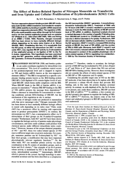
From www.bloodjournal.org by guest on February 6, 2015. For personal use only.
Regulation of cy2 Integrin Gene Expression in Cells With Megakaryocytic
Features: A Common Theme of Three Necessary Elements
By Mary M. Zutter, Audrey A. Painter, William D. Staatz, and Ying L. Tsung
The adl integrin mediates interactions between cells and
the extracellular matrix molecules, collagen and/or laminin.
The a& integrin is expressed in a variety of cell types, but
in cells of hematopoietic lineage, expression is restrictedto
megakaryocytes and platelets. Increased expression of the
a& integrin during megakaryocyticdifferentiationis a consequence of transcriptional activation of the az gene. We
have begunto characterizethe role of the 5’ flanking region
of the azintegrin gene in regulating expression during megakaryocyte differentiation. A 5-kb fragment of the 5’ region
directs both cell type and differentiation-dependentexpressionofareportergene
in the pluripotent hematopoietic
K562 cells upon megakaryocytic differentiation and in the
5’
megakaryocyticcellline, Dami. Analysisofaseriesof
deletion mutants indicatesthat expression ofthe azintegrin
gene in cells with megakaryocytic features requires acore
promoter region, a silencer region, and megakaryocyticenhancers in the distal 5‘ end. The organization of these
three
distinct regulatory regions the
of a2promoterlenhancer suggests a commontheme for megakaryocytic gene regulation
shared with other megakaryocyte-specific proteins, including allb integrin subunit and platelet factor 4.
0 1995 by The American Society of Hematology.
D
protin Iband the megakaryocyte/platelet-specific integrin
aIIbP3.’5
We now show that Damicells constitutively express
the azpI integrin protein and mRNA and that they use
the
receptor for adhesion to collagen.
We recently reported the characterization of the first 961
bpof the 5’ flanking region of the a2 integrin gene. This
region can direct cell-type-specific promoter and enhancer
activity in cells of epithelial origin, but not in K562 cells
induced to become megakaryocytic.’6 From these earlier
studies, three major questions remained unanswered: ( 1 ) Is
our phorbol-ester-induced model of megakaryocytic differentiation relevent to other models of megakaryocytopoiesis?
(2) Is there a silencer/repressor functioning in cells of hematopoietic lineage? (3) Where are the enhancers that are necessary for activity in megakaryocytes? To begin to answer
these questions, we have now compared the activity of an
expanded series of deletion mutants of the 961-bp a2 promoter construct in Dami cells to phorbol-ester-induced and
uninduced K562 cells. Within this region is a silencerhepressor active in both models of megakaryocytic differentiation.
In addition, we have extended our analysis of the 5’ flanking
region of the a2 promoter to include an additional approximate 4.0 kb of 5’ flank extending from -5,000 bp to + 109
bp. This genomic fragment contains enhancers necessary
for a2 integrin gene activity in cells with megakaryocytic
differentiation. Based on our results, we propose a model
for the megakaryocytic regulation of the a2 integrin gene
that requires a core promoter, followed by a strong silencer,
and megakaryocytic enhancers in the distal 5’ flank.
IFFERENTIATION and maturation of hematopoietic
precursors into megakaryocytes is associated with expression of megakaryocyte-specificproteins, development of
a granules, endomitosis, and, subsequently, the release of
platelets from the cell surface.’.’ The a Z p Iintegrin, a member
of the integrin family of heterodimeric adhesive protein receptors, is expressed on platelets and megakaryocytes during
this process of megakaryocytic maturation. The a2p,integrin
on platelets serves as a cell surface receptor for collagen,
required for normalhemostasis.”’Although
a$, is expressed on a variety of cell types, including epithelial and
mesenchymal cells, on cells of the hematopoietic lineage, it
is restricted to megakaryocytes and platelets.’,’”
Our understanding of megakaryocytopoiesis has been facilitated by the development of cell lines with megakaryocytic properties. The leukemic cell line, K562, derived from
a patient with chronic myelogenous leukemia, represents a
pluripotent hematopoietic cell that acquires megakaryocytic
properties including increased surface expression of the a2pI
integrin as well as glycoproteins IIb-IIIa and Ib in the presence of phorbol dibutryate.“”’ In earlier studies, we showed
that the increased expression of the azpIintegrin after phorbo1 ester induction of K562 cells is a consequence of increased steady-state levels of a2 mRNA due to transcriptional activation of the a2gene.I4 In contrast to K562 cells,
which acquire megakaryocytic properties after phorbol ester
induction, the Dami cell line, developed from the peripheral
blood of a patient with megakaryoblastic leukemia, constitutively expresses megakaryocytic proteins, including glycoFrom the Department of Pathology, Washington University School
of Medicine, St Louis, MO.
Submitted January 12, 1995; accepted May 9, 1995.
Supported bygrantsfrom
the National Institutes of Health
(HL51450) and the American Heart Association.
Address reprint requests to Mary M. Zutter, MD, Box 8118, Department of Pathology, Washington Universiry School of Medicine,
660 S Euclid Ave, St Louis, MO 63110.
The publication costs ofthis article were defrayed
in part by page
charge payment. This article must therefore be hereby marked
“advertisement” in accordance with 18 U.S.C. section 1734 solely to
indicate this fact.
0 1995 by The American Society of Hematology.
0006-4971/95/8608-0126$3.00/0
3006
MATERIALS AND METHODS
Cell culture and transfection assays. The K562 cell line was
obtained from the American Type Culture Collection (Rockville,
MD). The Dami cell line was a gift of Dr Robert I. Handin (Harvard
Medical School, Boston, MA). Both cell lines were propagated in
RPM1 1640 medium. Megakaryocytic differentiation of K562 cells
was induced by the addition of 40 nmoVL phorbol dibutyrate in
dimethyl sulfoxide, as previously described.’’
The K562 and Dami cell lines were transfected by electroporation
using a BTX Electro Cell Manipulator 600 (BTX Inc, San Diego,
CA).” Approximately 1.0 to 1.5 x 10’ cells were transfected in
RPM1 medium containing 100 mg/mL salmon sperm DNA, 30 pg
of plasmid DNA, and 3 pg of Rous sarcoma virus (pRSV)-luciferase
DNA by electroporation at 275 V and 600 pF. Cell extracts were
Blood, Vol 86,N O 8 (October 75). 1995:PP 3006-3014
From www.bloodjournal.org by guest on February 6, 2015. For personal use only.
(I*
INTEGRINEXPRESSION
3007
BY MEGAKARYOCYTES
harvested after 48 hours. Luciferase activity produced by 10 pL Of
the cell lysate in 190 pL of assay buffer (10 mmoVL Mg[OAc]z,
50 mmoVL Tris-MES, pH 7.8, and 2 mmoVL ATP) was analyzed
using a Monolight 2010 luminometer (Analytical Luminescence
Laboratory, San Diego, CA), as described," and was used to normalize for transfection efficiency. Cell extracts containing identical
amounts of luciferase activity were then assayed for chloramphenical
acetyltransferase (CAT) activity by the standard method of Gorman
et
The conversion of chloramphenical to acetylated chloramphenicol was determined by both thin-layer chromatography and
differential extraction.
Flow cytometry and antibodies. Flow cytometry was performed
on a Becton Dickinson FACScan Analyzer and analyzed with Becton
Dickinson Consort-30 software (Becton Dickinson, Mountain View,
CA). Adherent cells were detached in Versene solution (0.5 mmoV
L EDTA in phosphate-buffered saline: GIBCO BRL, Grand Island,
NY) at 4°C and washed in calcium- and magnesium-free Hanks'
balanced Salt Solution (CMF-HBSS) with 1% bovine serum albumin
(BSA). Viable cells were enumerated by trypan blue exclusion, and
cells were resuspended in CMF-HBSS to a final concentration of 5
X 10s viable celldml. Cells (5 X lo5 per tube) were labeled with
a primary antibody concentration of 5 pg/mL. Fluorescein isothiocyanate (FITC)-conjugated goat antimouse IgG (5 p g / m l ) was used
as the secondary antibody. Histograms are based on the analysis of
10,OOO cells. Controls included omission of both first and
second
antibodies, omission of the first antibody, and use of an irrelevant
first antibody.
The monoclonal antibody (MoAb) 7E3, which recognizes the
a&
(IIb-IIIa) and a,@, integrins in a complex-specific manner,
was generously provided by Dr Barry S . Coller (State University of
New York at Stony Brook, Stony Brook, NY). The P1E6 antibody
directed against the az integrin subunit was obtained from GIBCO
BRL (Gaithersburg, MD). The 4B4 MoAb reactive with the PI integrin subunit was obtained from Coulter Inc (Hialeah, FL). The FITCconjugated goat antimouse IgG was obtained from TAGO, Inc (San
Jose, CA).
mRNA analysis. Total cellular RNA was prepared by guanidine
thiocyanate extraction and preparative ultracentrifugation, as described." Northern blot analysis was performed by electrophoretic
separation of total cellular RNA (15 to 20 pg) in denaturing formaldehyde-agarose gels, transfer of the RNA to Nitroplus membrane
(Micron Separation, Inc, Westboro, MA), baking, and hybridizing
overnight to 32P-random-primedcDNA probes. The hybridized blots
were washed and exposed to film for 1 to 5 days.
A partial-length az cDNA clone corresponding to nucleotides 1
through 1289 of the published azcDNA sequence" was previously
described.14 The amplified cDNA was ligated into the plasmid
pBSSK (Bluescript; Stratagene Cloning Systems, La Jolla, CA) and
subjected to double-stranded DNA sequencing using the dideoxynucleotide chain termination method of Sanger et alZ2to establish
authenticity. A cDNA corresponding to nucleotides 130 through
2626 of the PI integrin subunitz3was generously provided by Dr
Laurence Fitzgerald (University of Utah,Salt Lake City).
a2 CATfusion constructs. The promoter and enhancer activity
of the 5' flanking region was analyzed by inserting the EcoRI-Ava
I1 genomic fragment (-961 to +l09 in relation to the transcription
start site at + 1) upstream to CAT structural sequences in the CAT
which
expression vector pCAT-Basic (Promega, Madison, W),
lacks natural CAT regulatory elements.16 Nested deletion mutants
of the 1.0-kb construct (pz961-CAT) were generated by restriction
enzyme digestion of ~~1,961-CAT the
at restriction enzyme sites Psr
I(-30), Xma 111 (-92), Sac I1 (-122). Nar I (-223), S m I (-351),
Bgl I1 (-549), and Acc I (-776). Cytomegalovirus (CMV)-CAT
containing the CMV promoter directing transcription of the CAT
gene served as a positive control. Cotransfection in the pRSV-luciferase containing the Rous sarcoma virus promoter served as a control
for transfection efficiency in all assays. All transfection experiments
were performed at least three times and the results are reported as
average values relative to the paQ2-CAT construct.
A 5-kb BamHYAva I1 genomic fragment representing approximately 5 kb of the 5' flanking region (-5,000 to +l09 in relation
to the transcription start site at + l ) was inserted upstream to CAT
structural sequences in pCAT-Basic (Promega). Deletion mutants of
the 5-kb construct (pcuZ5000-CAT) were generated by restriction
enzyme digestion of paz5000-CAT at the restriction enzyme sites
Hind111 (-3,739). Stu I (-2,592), and Sac I (-1,426).
RESULTS
Dami cells express the a2PIintegrin. In our earlier studies, we exploited a model of megakaryocytic differentiation
using K562 cells to examine regulation of the azPlintegrin
during megakaryocytopoiesi~.~~~~~
The Dami cell line, which
was derived from a patient with megakaryoblastic leukemia,
is known to constitutively express the platelet-specific glycoprotein IIb/IIIa (01&)
and glycoprotein I b . 1 5 We have now
analyzed the Dami cell line for expression and regulation of
the azPIintegrin. Surface expression of the a2and PIintegrin
subunits was determined using flow cytometric analysis and
compared with the expression of the a I I bintegrin
~3
in Fig
1A. Both the a2and PI integrin subunits were expressed at
comparable levels, but at significantly lower levels than the
a,&, integrin. The relative difference in the two megakaryocyte antigens is consistent with the greater number of aId3
receptors on circulating platelets and comparable to the differences we reported earlier for the expression of azPIand
at& using the K562 cell model of megakaryocytic differentiation."
We also compared the levels of az and PI mRNAs expressed by Dami cells, uninduced K562 cells, and K562 cells
induced with phorbol 12,13 dibutyrate for 8 days, as shown
in Fig 1B. In contrast to the high level of az and PI mRNA
expressed by induced K562 cells, Dami cells expressed low
but readily detectable levels of az and PI mRNA. As reported
earlier, uninduced K562 did not express detectable levels of
az mRNA by Northern blot hybridi~ation.'~
The a2PIintegrin is functional on Dami cells. The a d l
integrin serves as a Mg2+-dependent receptor for collagen
on platelets and fibroblasts. We showed not onlythat induced
K562 cells expressed the azPl integrin, but also that the
receptor was f~nctiona1.l~
On the other hand, when uninduced K562 cells were transfected with a full-length az
cDNA, the ad1integrin was expressed on the cell surface
but did not bind ligand.24Thus, megakaryocytic differentiation is accompanied not only by increased a d l expression
but also by changes in the activation state of the receptor. We
therefore evaluated the functional state of the ad1receptor
expressed on Dami cells. Dami cells adhered to collagen in
the presence of Mg2+, asshown in Fig 2A. The cells failed
to adhere to collagen when CaZ+was substituted for Mg2+
and did not adhere to BSA substrates. To verify that the
Mgz+dependent adhesion was mediated by the a2P,integrin,
adhesion assays were conducted in the presence of inhibitory
MoAb. The Mg2+-dependentadhesion to collagen was com-
From www.bloodjournal.org by guest on February 6, 2015. For personal use only.
3008
ZUTTER ET AL
Fig 1. Dami cells express a& integrin protein and mRNA. (A) Cell surface protein expression was determined by indirect flow cytomehic
complex and FllC-conjugated goat antimouse lgG as a
analysis using the MoAbs directed against either the a2or f3, subunits or the a&
secondary antibody. (B)Northern blot analysis was performed on total cellular RNA from uninduced K562 cells cultured in medium alone for
8 days, induced K562 cells cultured in medium containing 40 nmol/L phorbol dibutyrate for 8 days, or Dami cells. The 8.0-kb a2and 4.2-kb
mRNA were detected using '*P-labeled cDNA probes. The corresponding ethidium bromide-stained gels illustrate that equivalent amounts of
RNA were present in each lane.
pletely blocked by the inhibitory anti-a2 MoAb 6F1 (Fig
2B). In contrast, a control anti-a5 MoAb (PlD6) failed to
inhibit adhesion. Therefore, Dami cells adhered to collagen
via an adIintegrin-mediated mechanism.
Regulation of the a2subunit gene in Dami cells is similar
to that in phorbol-ester-induced K562 cells. In our initial
characterization of the 5' flanking region of the a2integrin
gene, we identified a 961-bp fragment of the gene extending
from bp + 1 to bp -961 that directed cell-type- and differentiation-specific expression of a reporter gene in epithelial
cells but not in induced K562 cells.I6 In this earlier study,
the pa292-CAT construct directed high-level enzymatic activity in induced K562 cells. However, the longer constructs,
ie, pa2549-CAT, pa2776-CAT, and pa2961-CAT, generated
low to undetectable activity in either uninduced or induced
K562 cells.
To compare the K562 model of megakaryocytic differentiation to Dami cells, we compared the ability of the constructs
pa230-CAT, pa292-CAT, pa2549-CAT, and pa2961-CAT,
outlined inFig 3, to direct reporter gene activity when
transfected into uninduced K562 cells, K562 cells induced
for 6 days with phorbol dibutyrate, andDami cells. The
pattern of CAT activity generated by the mutant constructs
was similar inDami cells andin induced K562 cells, as
shown as an average of six experiments (Fig 4A and B).
Enzymatic activity directed by the pa230-CATconstruct was
undetectable in either Dami cells or K562 cells induced for
6 days with phorbol esters. In contrast, the pa292-CAT construct directed high-level CAT activity inbothDamiand
induced K562 cells and only low-level enzymatic activity in
uninduced K562 cells. The high level of enzymatic activity
directed by pa292-CAT was partially silenced by the addition
of 457 bp of sequence between -92 and -549. In fact,
the CAT activity directed by the pa2961-CAT construct in
induced K562 cells or Dami cells was only 7% or 12%,
respectively, of that directed by the pa292-CAT construct.
Thus, the region between bp -30 and -92 contains elements that are necessary for high-level gene activity in cell
lines with megakaryocytic differentiation, but are not celltype specific. As shown earlier, the pa292-CAT construct
was active in the breast carcinoma cell line T47-D.I6 Elements between -92 and -549 almost completely silenced
activity in induced K562 cells and Dami cells. The silencer
was inactive in the breast epithelial cell line. Eventhe longest
construct, pa2961-CAT, did not restore the high-level promoter activity necessary to account for the observed 20-fold
increase in transcriptional activation of the a2gene in K562
cells upon phorbol ester induction or in Damicells. The
From www.bloodjournal.org by guest on February 6, 2015. For personal use only.
INTEGRIN EXPRESSION BY MEGAKARYOCYTES
(I?
30r
3009
L
T
COLL
BSA
30
C
.o
U)
20
a)
c
.U
a
S
10
n
"
BSA
No Ab
P 16
DF61
EDTA
Collagen
Fig 2. Dami cells adhere to type I collagen via an a& integrinmediated mechanism. (A) The ability of Dami cells to adhere to type
I collagen or BSA in the presence of Mg2+f@), CB2+ (01, or EDTA ( W
was determined. (B) MoAb 6F1 directed against the a& integrin
reduced the Mg2'-dependent adhesion of Dami cells to collagen to
the low level observed in EDTA. A control antibody, PlD6, directed
against the aspl integrin had no effect.
inability of pa2961-CATto mimic the increased transcription
of the a2gene in induced K562 cells or Dami cells suggested
that one or more enhancer elements important for the regulation of the a2gene in our models of megakaryocytic differentiation must be present in yet unidentified regions of the a2
promoter.
Silencer activity for cells of hematopoieric lineage is located between bp -92 and -351. To begin to dissect the
silencer activity located between bp -92 and -549, we constructed a series of deletion mutants pa2122-CAT, pa2223CAT, and pa2351-CAT tightly spaced between bp -92 and
-549 (Fig 3). The promoter activity of these CAT deletion
mutants was determined in K562 cells withandwithout
phorbol dibutyrate and in Dami cells. CAT enzymatic activity in cells with megakaryocytic differentiation decreased
with the addition of increasing sequence between -92 and
-351 (Fig 4A and B). In contrast to the pa292-CAT construct, which directed the highest enzymatic activity, the
pa2122-CAT, pa2223-CAT, and pa2351-CAT directed enzymatic activity at approximately 27% to 34%. 5 % to 12%,
and 2% to 8%, respectively, of the paQ2-CAT construct in
both Dami cells or in K562 cells induced for 6 days with
phorbol dibutyrate. Sequence analysis of the region between
-92 and -351 showed a single API site between -92 and
-122, one NFI-like site between -122 and -223, and a
second NFI-likesite between -223 and -351. CCAATbinding sites for NFI have been implicated in silencing a
number of genes, including the rat growth hormone gene, the
human retinal binding protein, the mouse Sparc (osteonectin)
gene, the chicken &globin gene, the mouse Ren-Id gene,
and the mouse a2 (I) collagen gene.2s In these genes, the
NFI-like consensus sequence serves as anegative regulatory
element that suppresses gene activity in a cell-type-specific
manner. Further studies are under wayto assess the role
of these NFI-like sites in the silencing of the a2 gene in
hematopoietic cells. Other potential elements in the region
between -92 and -351 that could modulate increasing silencer activity are the AP2 and Spl sites. In summary, we
have identified a strong silencer active in cells of hematopoietic lineage that requires multiple elements for full activity.
Megakaryocyte-specijc enhancers are located in the far
distal 5' yanking region. As discussed earlier, the lack of
essential regulatory elements to override the silencer activity
in cells with megakaryocytic differentiation suggested that
we were lacking important regions of the a2promotedenhancer. We therefore sought to identify additional elements
in the distal 5' flanking region. A restriction mapof the
original 15-kb phageclone'' indicated that a BamHI site was
present approximately 4 kb 5' to the EcoRI site at -961. To
determine if the additional 4-kb region contained elements
important for megakaryocytic gene activity, the BamH1-Ava
I1 genomic fragment spanning the region from -5,000 to
+l09 was placed upstream of CAT structural sequences in
the construct pa25000-CAT (Fig 3). The promoter activity
of pa25000-CAT construct was analyzed in uninduced and
induced K562 cells. This region conferred strong promoter
activity nearly comparable to that of the strong viral promoter CMV in induced K562 cells but exhibited little activity in uninduced K562 cells (Fig 5). Additional 5' deletion
mutants between -961 and -5,000 (Fig 3) localized strong
megakaryocytic enhancer activity to the region between bp
-1,426 and -2,592. Although the pa21426-CAT construct
increased activity slightly, the pa22592-CAT construct restored high-level promoter activity in the induced K562 cell
model. The pa22592-CATconstruct was 13.3-fold more active than the pu2961-CAT construct in induced K562 cells
and 9.3-fold more active in induced versus uninduced K562
cells (Fig 5). Little additional activity was gained by elements extending beyond -2,592. The strong promoter activity generated by elements between - 1,426 and -2,592 indi-
From www.bloodjournal.org by guest on February 6, 2015. For personal use only.
3010
ZUTTER ET AL
BamHI
-5000
Avall
I
+!!AT‘,
m(t,
a
-5000
+l
*B
-3739
-2592
-e
-1426
‘h
integrin gene
Pa, 5000-CAT
Pa,
3739-CAT
pa, 2592-CAT
Fig 3. Aschematic diagram
of the a2 promoter-CATconPa2961-CAT
structs. The construct p&OOO-776
CAT contained the entire 5’
Po, 776-CAT
flanking regionwith 109 bp of 5’-54?A&
Po, 549-CAT
untranslatedregion upstreamto
the CAT structural gene.Con-35po2351-CAT
structs pa3739-CAT, pa22592-223*
Po, 223-CAT
CAT, and pa21426-CAT were
derived from pa,5000-CAT. Conpa,
122-CAT
structs
pa2778-CAT,
pa2549-92
CAT, wJ51-CAT. Wz122-CAT.
pop92-CAT
and aZ92-CATwere derived from
-3%
Po, 30-CAT
the construct, pa2961-CAT.’E
-961
pap 1426-CAT
a
+l
a
-122a
%
cates that enhancers required for activity of the a2 integrin
gene in cells with megakaryocytic differentiation are located
in this region.
Sequence analysis of the region between -961 and
-3,739 confirmed the location of numerous potential binding
sites for ubiquitous as well as megakaryocyte-specific transcription factors (Fig 6). Three perfect consensus sequences
for binding the GATA family of transcription factors were
identified between -961 and -2,592.26 In each case, these
potential GATA boxes are located in close proximity to
concensus sequences for either AP1 or AP2. One or more
upstream GATA sequences are also required for promoter/
enhancer activity of the megakaryocyte-specific prot e i n ~ . * ~The
- ’ ~ numerous AP1 concensus binding sites, two
of which are separated by only 8 bp, suggest NF-E2 binding
NF-E2,a second erythroidmegakaryocytic transcription factor, is required for megakaryocytic differentiation and platelet p r o d ~ c t i o n Other
. ~ ~ potential sites include
concensus sequences for NFKb, CAAT binding protein, and
one estrogen receptor (ER) half-site. In addition to potential
binding sites for transcription factors, two clusters of binding
sites for either the enzyme, topoisomerase I1 (Top0 11) or
binding proteins for scaffoldmatrix associated regions
(SARsMARs), are located in this distal 5‘
The megakaryocytic features of Dami cells can be augmented by phorboE esters. Exposure of Dami cells to phorbo1 esters is known to increase expression of platelet membrane glycoproteins Ib, cyllb, andvon Willebrand factor,
suggesting that the adl integrin may be similarly effected.’
We confirmed using flow cytometric analysis that the addition of 12,13 phorbol dibutyrate to Dami cell cultures for 2
days markedly increased the expression of the a2 integrin
on the cell surface (Fig 7A). We compared the activity of
the a2promoter constructs in uninduced and induced Dami
cells. Exposure of Dami cells to phorbol dibutyrate for 4
days increased the activity of the pa292-CAT construct containing the core promoter/enhancer region by 7.0-fold (Fig
7B). The silencer domain greatly diminished CAT activity
in induced Dami cells in a similar stepwise manner as that
seen in uninduced Dami cells and induced K562 cells. However, the silencer failed to completely repress all promoter
activity in induced Dami cells. The megakaryocyte-specific
enhancer elements in the distal 5‘ flank located between
- 1,426 and -2,592 conferred high-level promoter activity in
induced Dami cells (comparable to the high level of activity
observed in induced K562 cells) and low-level activity in
uninduced Dami cells (Fig 7B). In a manner similar to that
observed in uninduced K562 cells, the region between
-2,592 and -3,739 augmented activity in uninduced Dami
cells, butonly
twofold. Therefore, the region between
- 1,426 and -2,592 contains strong megakaryocyte-specific
enhancers necessary to mimic the low level of transcriptional
activation of the a2gene observed in uninduced Dami cells
and the high level of activity observed in induced Dami
cells.
DISCUSSION
The a2PIintegrin, a collagen receptor on platelets and
megakaryocytes, is essential for normal hemostatic function
of platelets. To understand the regulation of a& integrin
expression during megakaryocytic differentiation, we identified the 5’ flanking region of the a2integrin gene and showed
that the elements within the first 961 bp could direct celltype- and differentiation-dependent expression in cells of
epithelial origin but could not recapitulate the expression of
a2integrin in K562 cells induced with phorbol dibutyrate to
become megakaryocytic.I6To focus on the molecular mechanisms controlling expression of the a& integrin gene during
megakaryocytic differentiation, we have now compared the
expression of the a2integrin gene in induced K562 cells with
that in Dami cells. Dami cells, a committed megakaryoblast,
constitutively express megakaryocytic features, including the
a& integrin protein and mRNA.’’ To determine the molecular regulation of adl integrin in cells with megakaryocytic
differentiation, we compared the activity of the original
promoter/enhancer constructs (pa230-CAT, pa*92-CAT,
From www.bloodjournal.org by guest on February 6, 2015. For personal use only.
cr2
301 1
INTEGRINEXPRESSIONBYMEGAKARYOCYTES
pa2549-CAT, and pa2961-CAT) as well as a series of nested
deletions between bp -92 and -549 in Dami cells with that
of uninduced and induced K562 cells. Similar regions of the
a2integrin gene are required for expression and silencing of
expression in both Dami cells and induced K562 cells. These
studies suggest that regulation of the a2integrin gene in cells
9.0
8.5
8.0
-
7.5 -
1
\
'
8.9
8.8
8.7
2,
-->
8.6
8.5
f
"
2
t9
L
1.0
0.9
0.8
F 0.7
';m 0.6
0.5
0.4
0.3
0.2
0.l
nn
Fig 5. The distal 5' flank of theai gene directed promoter activity
in cells with megakaryocytic features. Activity of the constructs
pmZl426-CAT,pa22592-CAT,pa23739-CAT,
and pa25000-CAT was
compared with that of prraO-CAT, pa292-CAT, and pa2961-CATin
uninduced K562 cells (R) and K562 cells induced for 6 days with 40
nmol/L phorbol dibutyrate ( W . CAT activity relative to the pa292CAT construct in induced K562 cells is plotted.
6.3
6.2
6.1
6.0
2,
.->
5.9
L
I
t; 1.0
24
0.9
E
0.6
0.8
g 0.7
=a: 0.5
0.4
0.3
0.2
0.1
nn
Fig 4. The a, integrin promoter activity in cells with megakaryocytic differentiation. pa2961-CAT, pa2549-CAT, pa.&l-CAT, pa2-223CAT, pazl22-CAT, pa292-CAT, and pa,BO-CAT were transfected in
parallel with the
CMV-CAT, which contains the strong promoter,
viral
and CAT-BASIC, which lacks promoter activity, into either uninduced
( W or induced (M) K562 cells IA) or Dami cells (B). Cotransfection
with RSV-luciferase was used t o control for transfection efficiency.
After 48 hours ofincubation, cell extracts were
assayed. After normalization for transfectionefficiency, CAT activity of the constructs was
determined using thin-layer chromatography and differential
extraction. CAT activity of an average of six experiments relative t o the
pu292-CATconstruct in induced K562 cells (A) or Darni cells (B) is
plotted.
of megakaryocytic lineage is mediated, at least in part, by
three functionally distinct domains within the 5' flanking
region of the a2 gene. These three domains consist of a
strong core promoter located between bp -30 and -92. The
core promoter is not cell-type specific. It is followed by a
silencer between -92 and -351 that is able to repress promoter activity in cells of hematopoietic lineage. Interestingly, this region is not active as a silencer in nonhematopoietic cells (data not shown)." Additional enhancers in the far
distal 5' region are required for high-level megakaryocytic
expression of the a2integrin subunit. The identified enhancer
and silencer elements are important for the regulation of the
a2pIintegrin in cells of megakaryocytic origin, ie,Dami
cells, and not simply a response unique to K562 cells or the
addition of phorbol esters.
This complex scheme of a2 integrin gene regulation,
which requires three distinct regions with both positive and
negative elements for megakaryocytic activity, resembles the
regulation of other megakaryocyte-specific proteins, including the (Yllh integrin subunit and platelet factor 4 (PF4).27-32
Both of these megakaryocyte-specificproteins contain a core
promoter domain, followed by a strong silencer, and enhancers required for expression by megakaryocytes in the
distal 5' flank. The core promoter region of PF4 and the (Yllh
subunit reside between the transcription start site andbp
-97 or -99, respectively. This location is almost identical
to where we located the core promoter of the a2 integrin
subunit gene.
Both the PF4 and (Yllh promoters, which are restricted to
megakaryocytic lineage, contain a necessary GATA-like site
within the core ~ e q u e n c e . ~The
~ . ~GATA
'
family of transcrip-
From www.bloodjournal.org by guest on February 6, 2015. For personal use only.
3012
ZUTTER ET AL
Silencer
domain
Megakarycyte-specific
enhancers
-2592
3739
L
-1426
-961
L
AR
CCAN
CCAAT
Core
promoter
A
-30
-351
-921+1 '129
*l93
I
CCAAT
Fig 6. A schematic diagram of
the distal 5' flank from bp -3,739 through exon 1 of
the o2integrin gene. The locations
of numerous potential
binding sites for ubiquitous
as well as megakaryocyte-specific transcription factors are shown.
In addition to numerous potential binding
sites
for AP1 and AP2, three perfect consensus sequences for binding the GATA family of transcription factors are located between -961 and
-2,592. Additional potential binding sit- indude consensus sequences for NFA, CAAT binding protein, and four estrogen receptor half-sites.
In addition, there are two clusters of binding sites for the enzyme Top0 II and the SARslMARs. The complete DNA sequence of the region
from -961 to -3,739 has been submitted to GenBank, Accession No. U31518.
tion factors mediates hematopoietic differentiation along
erythroid, megakaryocytic, and mast cell lineages, but is
not required for myeloid or lymphoid differentiation.26In
contrast to the core promoter of the megakaryocyte restricted
almand PF4, the core promoter element of the a2integrin,
which does not contain a GATA-like sequence, is active in
nonhematopoietic cells. Activity of the core region of the cy2
integrin subunit, therefore, may require ubiquitous transactivating factors. Our earlier sequence analysis of this region
identified two Spl sites and an AP2 site that potentially are
active in this core region.16
Although the cy2 integrin subunit is not megakaryocyte
specific, common strategies appear to have evolved to confer
megakaryocytic expression. The silencer domain of both PF4
and the (YIIb is necessary for lineage restriction to megakaryocytes. The silencers of both PF4 and a [ l b may repress expression of these genes in ail nonmegakaryocytic cell types.".
82.41 In contrast, the a2 subunit is expressed bymany
cell
types butnotby
cells of hematopoietic lineage, with the
exception of platelets and megakaryocytes. Therefore, the
silencer activity of a2 may be hematopoietic cell specific,
because it is inactive in epithelial cells but active in K562
cells and Dami cells. There is no similarity between the
silencer domain of the a2gene and the poly-T-rich silencer
region of the PF4 p r o m ~ t e r . ~ A
' . ~short
~
poorly conserved
heptad sequence of the a I I b silencer is present within the
silencer region of the a2integrin p r o m ~ t e r . ~The
" ~ importance of the short sequence in silencing gene activity has yet
to be characterized.
The lack of essential regulatory elements necessary to
restore high-level promoter/enhancer activity in the first 961
bp of the 5' flanking region necessitated analysis of a much
larger region of the a2 integrin gene. Elements between bp
- 1,426 and -2,592 restored high-level promoter activity in
both induced K562 cells and in Dami cells. These observations suggest that enhancer elements between -1,429 and
-2,597 are required for a2gene activity in cells undergoing
megakaryocyte differentiation. The requirement for such distal regulatory elements suggests that dramatic changes in
chromatin conformation contribute to the changes in gene
regulation. Regulation of hemoglobin gene expression and
switching is also controlled by distal regions, the locus control elements, at sites of DNA hypersensitivity that are
thought to interact with the nuclear matri~!~".'~.~~These
interactions are mediated by DNA binding proteins called
SAIUMAR binding proteins.3741These DNA binding proteins have been recently identified and the consensus binding
sequences were
The presence of clustered
binding sites for SARIMAR binding proteins, as wellas
topoisomerase 11, adjacent to -2,592 suggest that this region
may also interact with the nuclear matrix, bindto SAR/MAR
binding proteins, and be involved in DNA conformational
changes. Such conformational changes could explain how
distinctly different regions of the 5' flanking sequence, which
are widely separated in linear sequence, function in megakaryocytic versus nonhematopoietic gene regulation.
The marked similarities between the promoter/enhancer
regions that regulate the a z , aim, and PF4 genes during
megakaryocytic differentiation suggest a common regulatory
theme that requires a core promoter/enhancer (Fig 6). Just
5' to the core promoter/enhancer is a silencer domain that
limits gene activity in either hematopoietic, nonmegakaryocytic cells or nonhematopoietic cells that do not express the
gene. Finally, megakaryocytic enhancers are located in the
distal 5' flanking region. The common structural motif for
these regulatory elements of genes expressed during mega-
From www.bloodjournal.org by guest on February 6, 2015. For personal use only.
u2 INTEGRINEXPRESSION
3013
BY MEGAKARYOCYTES
REFERENCES
-. -...
...
.-
- Uninduced Dami
...e..
Induced Dami
Fig 7. The megakaryocytic features of Dami cellscan be augmented by phorbol esters. (A) Cell surface expression of the a2integrin subunit on uninduced Dami cells or Dami cells induced for 2
days with phorbol dibutyrate 140 nmol/L) was determined by indirect
flow cytometric analysis usingMoAbs directed against the a2integrin
subunit and FITC-conjugated goat antimouseIgG asa secondary antibody. (B) The a2integrin promoter activity in Dami cells, either uninduced or induced for4 days with phorbol dibutyrate, was compared.
The constructs pa25000-CAT, paZ3739-CAT,paz2592-CAT, pa21426CAT, pa2961-CAT,pa2351-CAT,pa2223-CAT,1~~122-CAT,
and pa2%!CAT were transfected into either uninduced (B)or induced) .1 Dami
cells. Cotransfection with pRSV-luciferase was used to control for
transfection efficiency. CAT activity relative to the pa292-CAT construct in uninduced Dami cells is plotted.
karyocytic differentiation suggests that not only are specific
transcription factors common to pathways of hematopoietic
cell differentiation but also that structural organization of
these genes is required.
ACKNOWLEDGMENT
We thank Samuel A. Santoro for encouragement and advice during the course of this work and for constructive criticism and review
of the manuscript. We thank Marian Bentz for excellent secretarial
assistance.
1. Hoffman R: Regulation of megakaryocytopoiesis. Blood
74: 1 196, I989
2. Long MW: Regulation of human megakaryocytopoiesis. Ann
NY Acad Sci 5 5 4 192, 1989
3. Santoro SA, Rajpara SM, Staatz WD, Woods VL Jr: Isolation
and characterization of a platelet surface collagen binding complex
related to VLA-2. Biochem Biophys Res Commun IS3:217, 1988
4. Staatz WD, Rajpara SM, Wayner EA, Carter WG, Santoro SA:
The membrane glycoprotein la-lIa (VLA-2) complex mediates the
Mg"-dependent
adhesion of platelets to collagen. Cell Biol
108:1917, 1989
S. Coller BS, Beer JH, Scudder LE, Steinberg MH: Collagenplatelet interactions: Evidence for a direct interaction of collagen
with platelet GPIdIIa and an indirect interaction with platelet GPllbl
llla mediated by adhesive proteins. Blood 74182, 1989
6. Elices MJ, Hemler M E The human integrin VLA-2 is a collagen receptor on some cells and a collagennaminin receptor on others.
Proc Natl Acad Sci USA 86:9906, 1989
7. Kirchhofer D, Languino LR, Ruoslahti E, Pierschbacher MD:
a2pIintegrins from different cell types show different binding specificities. J Biol Chem 265:615, 1990
8. Languino LR, Gehlsen KR, Wayner E, Carter WG, Engvall E,
Ruoslahti E: Endothelial cells use a2pIintegrin as a laminin receptor.
J Cell Biol109:245S, 1989
9. Zutter MM, Santoro SA: Widespread histologic distribution of
the a2pI integrin cell-surface collagen receptor. Am J Pathol
137:113, 1990
10. Hemler M E VLA proteins in the integrin family: Structures,
functions, and their role in leukocytes. AnnRev lmmunol 8:365,
1990
1 I . Lozzio CB, Lozzio B: Human chronic myelogenous leukemia
cell-line with positive Philadelphia chromosome. Blood 45:321,
I975
12. Leary JF, Ohlsson-Wilhelm BM, Giuliano R, LaBella S , Farley B, Rowley PT: Multipotent human hematopoietic cell line K562:
Lineage-specific constitutive and inducible antigens. LeukRes
11:807, 1987
13. Burger SR. Zutter MM, Sturgill-Koszycki S , Santoro SA:
Increased cell surface expression of functional azpIintegrin accompanies the megakaryocytic differentiation of K562 leukemia cells.
Exp Cell Res 202:28, 1992
14. Zutter MM, Fong AM, Krigman HR, Santoro SA: Differential
regulation of the a2pIand a&
integrin genes during megakaryocytic differentiation of pluripotent K562 leukemia cells. J Biol Chem
267:20233, 1992
15. Greenberg SM, Rosenthal DS, Greeley TA, Tantravahi R,
Handin RI: Characterization of a new megakaryocytic cell line: The
Dami cell. Blood 72:1968, 1988
16. Zutter MM, Santoro SA, Painter AS, Tsung YL, Gafford A:
Characterization of the a2integrin gene promoter: Identification of
positive and negative regulatory elements important in cell-type and
developmentally-restricted gene expression. J Biol Chem 269:463,
1994
17. Ulrich MJ, Ley TJ: Function of normal and mutated gammaglobin gene promoters in electroporated K562 erythroleukemia cells.
Blood 75:990, 1990
18. Nguyen VT, Morange M, Bensaude 0: Firefly luciferase luminescence assays using scintillation counters for quantitation in
transfected mammalian cells. Anal Biochem 171:404, 1988
19. Gorman CM, Moffat LF, Howard BH: Recombinant genomes
which express chloramphenicol acetyltransferase in mammalian
cells. Mol Cell Biol 2:1044, 1982
From www.bloodjournal.org by guest on February 6, 2015. For personal use only.
301 4
20. Davis LG, Dibner MD, Battey JF (eds): Basic Methods in
Molecular Biology. New York, NY, Elsevier, 1986
21. Takada Y, Hemler ME: The primary structure of the VLA2/collagen receptor a2subunit: Homology to other integrins and the
presence of a possible collagen binding domain. J Cell Biol 109:397,
1989
22. Sanger F, Nicklen S, Coulson AR:DNA sequencing with
chain-terminating inhibitors. Proc Natl Acad Sci USA 74:5463, 1977
23. Argraves WS, Suzuki S, Arai H, Thompson K, Pierschbacher
MD, Ruoslahti E: Amino acid sequence of the human fibronectin
receptor. J Cell Biol 105:1183, 1987
24. Kawaguchi S, Hemler ME: Role of the a subunit cytoplasmic
domain in regulation of adhesive activity mediated by the integrin
VLA-2. J Biol Chem 268:16279, 1993
25. Roy W ,Gosselin P, Anzivino MJ, Moore DD, Guerin SL:
Binding of a nuclear protein to the rat growth hormone silencer
element. Nucleic Acid Res 20:401, 1992
26. Orkin SH: Cell-specific transcription and cell differentiation
in the erythroid lineage. Opin Cell Biol 2:1003, 1990
27. Doi T, Greenberg SM, Rosenberg RD: Structure of the rat
platelet factor 4 gene: A marker for megakaryocyte differentiation.
Mol Cell Biol 73398, 1987
28. Ravid K, Doi T, Beeler DL, Kuter DJ, Rosenberg RD: Transcriptional regulation of the rat platelet factor 4 gene: Interaction
between an enhancedsilencer domain and the GATA site. Mol Cell
Biol 11:6116, 1991
29. Ravid K, Beeler DL, Rabin MS, Ruley HE, Rosenberg RD:
Selective targeting of gene products with the megakaryocyte platelet
factor 4 promoter. Proc Natl Acad Sci USA 88:1521, 1991
30. Prandini MH, Denarier E, Frachet P, Uzan G, Marguerie G:
Isolation of the human platelet glycoprotein IIb gene and characterization of the 5’ flanking region. Biochem Biophys Res Commun
1.56595, 1988
3 1. Uzan G, Prenant M, Prandini M-H, Martin F, Marguerie G:
Tissue-specific expression of the platelet GPIIb gene. J Biol Chem
26693932, 1991
32. Prandini M-H, Uzan G, Martin F, Thevenon D, Marguerie
G: Characterization of a specific erythromegakaryocytic enhancer
within the glycoprotein IIb promoter. J Biol Chem 267:10370, 1992
33. Hickey MJ, Roth GR: Characterization of the gene encoding
human platelet glycoprotein IX. J Biol Chem 68:3438, 1993
ZUTTER ET AL
34. Lavelle D, Ducksworth J, Eves E, Gomes G, Keller M, Heller
P, DeSimone J: A homeodomain protein binds to gamma-globin
gene regulatory sequences. Proc Natl Acad Sci USA 88:73 18, 1991
35. Mignotte V, Wall L, DeBoer E, Grosveld F, Romeo P-H:
Two tissue-specific factors bind the erythroid promoter of the human
porphobilinogen deaminase gene. Nuclic Acids Res 17:37, 1989
36. Shivdasani RA, Rosenblatt MF, Vignali K, Zucker-Franklin
D, Orkin SH: The transcription factor NF-E2 is required in vivo for
normal megakaryocyte maturation and platelet production. Blood
84: 148, 1994
37. Elgin SCR: Chromatin structure and gene activity. Curr Opin
Cell Biol 2:437, 1990
38. Eissenberg JC, Elgin SCR: Boundary functions in the control
of gene expression. Trends Genet 7:335, 1991
39. Laemmli UK, Kas E, Poljak L, Adachi Y: Scaffold-associated
regions: Cis-acting determinants of chromatin structural loops and
functional domains. Curr Opin Genet Dev 2:275, 1992
40. Cunningham JM, Purucker ME, Jane SM, Safer B, Vanin EF,
Ney PA, Lowrey CH, Nienhuis AW: The regulatory element 3’ to
the *gamma-globin gene binds to the nuclear matrix and interacts
with special A-T-rich binding protein a (SATBl), an SAR/MARassociating region DNA binding protein. Blood 84:1298, 1994
inte41. Fong AM, Santoro SA: Transcriptional regulation of uIIb
grin gene expression during megakaryocytic differentiation of K562
cells: Role of a silencer element. J Biol Chem 269:18441, 1994
42. Grosveld F, van Assendelft GB, Greaves DR, Kollias B: Position-independent, high level expression of the human p globin gene
in transgenic mice. Cell 51:975, 1987
43. Tuan D, Solomon W, Li Q, London I: The “P-like-globin”
gene domain inhuman erythroid cells. Proc NatlAcad Sci USA
82:6384, 1985
44. Grosveld F, Antoniou M, Berry M, de Boer E, Dillon N. Ellis
J, Fraser P, Hurst J, Imam A, Meijer D, Philipsen S, Pruzina S,
Strouboulis J, Whyatt D: Cold Spring Harb Symp Quant Biol 58:7,
1993
45. Dickinson LA, Joh T, Kohwi Y, Kohwi-Shigematsu T: A
tissue-specific MAWSAR DNA-binding protein with unusual binding site recognition. Cell 70:631, 1992
46. Nakagomi K,Kohwi Y, Dickinson LA, Kohwi-Shigematsu
T: A novel DNA-binding motifinthe nuclear matrix attachment
DNA-binding protein SATB 1. Mol Cell Biol 14:1852, 1994
From www.bloodjournal.org by guest on February 6, 2015. For personal use only.
1995 86: 3006-3014
Regulation of alpha 2 integrin gene expression in cells with
megakaryocytic features: a common theme of three necessary
elements
MM Zutter, AA Painter, WD Staatz and YL Tsung
Updated information and services can be found at:
http://www.bloodjournal.org/content/86/8/3006.full.html
Articles on similar topics can be found in the following Blood collections
Information about reproducing this article in parts or in its entirety may be found online at:
http://www.bloodjournal.org/site/misc/rights.xhtml#repub_requests
Information about ordering reprints may be found online at:
http://www.bloodjournal.org/site/misc/rights.xhtml#reprints
Information about subscriptions and ASH membership may be found online at:
http://www.bloodjournal.org/site/subscriptions/index.xhtml
Blood (print ISSN 0006-4971, online ISSN 1528-0020), is published weekly by the American
Society of Hematology, 2021 L St, NW, Suite 900, Washington DC 20036.
Copyright 2011 by The American Society of Hematology; all rights reserved.


© Copyright 2026