
Anti-CD40 Antibody Binding Modulates Human Multiple
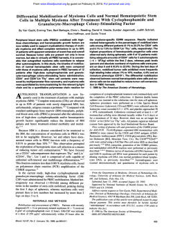
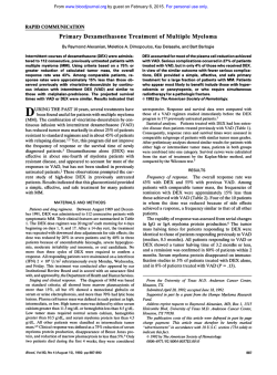
From www.bloodjournal.org by guest on February 6, 2015. For personal use only.
Anti-CD40 Antibody Binding Modulates Human Multiple Myeloma
Clonogenicity In Vitro
By Alex W. Tong, Bing-qing Zhang, Gabriele Mues, Max Solano, Terrie Hanson, and Marvin J. Stone
Ligand binding of theB-cell lineage antigen CD40 enhances
growth and interleukin-6 (IL-6) secretion in human B cells
(the CD40/11-6 loop). IL-6 has an autocrine and paracrine role
in human multiplemyeloma (MM)cell growth. With the
use
of the CD40 monoclonal antibody (MoAb)G28-5, we examined CD40 expression and theeffect ofCD40 binding onMM
clonogenic colony (MCC) formation t o characterize the IL-6/
CD40 loop activityin MM. CD40 was expressed on plasmacytoid cells in 21 of28 plasma cell dyscrasia (PCD) bone marrow (BM) biopsiestested ( I O of 14 MM, 2 of 2 Waldenstrom’s
macroglobulinemia [WM], 2 of 2 plasma cell leukemia [PCL],
6 of 8 monoclonal gammopathy of undetermined significance IMGUSI, and 1 of 2 primary amyloidosis [ALII. G28-5
binding increased MCCs by 35% t o 150% in 11 of 17 CD40+
PCD BM cultures, but did not affect MCC formation in CD40specimens or normal BM
colony forming units(CFU-GEMM,
CFU-GM, BFU-E). Responsive cultures originated from BM
of patients with MM (2 of 5 cases tested), WM (2 of 21,PCL
(2 of 21, and MGUS (5 of 6). CD40-responsivenesswas not
significantly inhibited by thepresence of an anti-IL-6 MoAb
(2 of 2 MGUS cultures tested), and did not correlate with
the capacity t o respond t o IL-6 stimulation (n = 17, P > .05)
or a detectable level of endogenous IL-6 fn = 15, P > .05).
Additional studies were performed with PCD cell lines t o
characterize the interrelationship of CD40 activation and IL6 production. Fifty percent t o greater than 95% of cells from
the RPM1 8226 and ARH77 lines expressed CD40, whereas
6% of U266 cells were CD40f. For RPM1 8226, ARH-77, and
U266 cells, the increased MCC formation after anti-CD40
stimulation was notaffected by the presence of an anti-IL6 neutralizing MoAb and was not accompanied by detectable IL-6 secretion. There was no apparent increase in IL-6
mRNA transcription following G28-5 treatment of U266 or
RPM1 8226 cells. Our observations indicate that CD40 is expressed in a subset of humanmyeloma cells present in various PCDs. Cell-line studies suggest that theCD40’ myeloma
cell mayregulate MM clonogenic colony formation without
activating theIL-6 pathway.
0 1994 by The American Society of Hematology.
C
human B cells and a murine lymphoma line transfected with
the human CD40 gene.15 Conversely, binding of 1L-6 to the
high affinity IL-6R (which is distinct from CD40) results in
increased phosphorylation of CD40, which in turnstimulates
IL-6 prod~ction.’~
This apparent interconnected signaling
between IL-6 and CD40 led to the hypothesis of a “CD40/
IL-6 loop”, by which CD40 functions to receive and regulate
IL-6-dependent signals in B cells.”
At present, it is unclear whether CD40 plays a similar
antiapoptotic role for malignant B cells or influences their
autocrine IL-6
This issue is clinically relevant in MM, in view of the observations that MM disease
progression may be affected by autocrine or paracrine action
of IL-6.I6An elevated IL-6 serum level was correlated with
MM disease ~everity,’”’~
and treatment with an anti-IL-6
MoAb produced a short-term remission in one plasma cell
leukemia (PCL) patient.z”
This study characterizes the distribution of CD40 in bone
marrow (BM) samples from various PCD patients to explore
the role of CD40 activation in IL-6-related growth modulation of MM. The relationship between CD40 activation and
IL-6 production also was examined with MM cell lines for
the purpose of characterizing activity of the CD40AL-6 loop.
D40 IS A 277-amino acid type I membrane glycoprotein that is expressed on normal B cells but not on
normal plasma cells.’,’ CD40 expression in human multiple
myeloma (MM) has been shown in a limited number of
cases,3” although the distribution of CD40 in other plasma
cell dyscrasias (PCDs) has not been defined. CD40 was detected in human B leukemias and lymphomas,8-” where it
was shown to be expressed on the clonogenic tumor subset.’
The natural ligand for CD40, termed gp39, is a type I1
transmembrane glycoprotein that is expressed transiently on
activated T-helper cells.’.’2 Triggering of CD40 by gp39 or
the CD40 monoclonal antibody (MoAb) G28-5 provides a
costimulatory signal that rescues germinal center B cells
from apoptosis and allows the resting B cell to proceed in
the maturation p a t h ~ a y . ’ ~
Expression
.’~
of both CD40 and
the interleukin-6 receptor (IL-6R) is increased in activated
B cells.15 (328-5 binding increased IL-6 secretion in normal
From the Cancer Immunology Research Laboratory, Charles A.
Summons CancerCenter,BaylorUniversityMedicalCenter,
and
the Mary C. Crowley Laboratory, Baylor ResearchInstitute, Dallas,
m.
Submitted January 18, 1994; accepted July 6, 1994.
Supported in part by the Tri Delta Cancer Research Fundand
the Edward and Ruth Wilkof Foundation.
Presented in part at the 34th Annual Meeting of the American
Society of Hematology, 1992, and the 85th Annual Meeting of the
American Association for CancerResearch, San Francisco, CA,
1994.
Address reprint requests to Alex W. Tong, PhD, Cancer Immunology Research Laboratory, Charles A.Summons Cancer Center,Buylor University Medical Center, 3500 Gaston Ave, Dallas, lX 75246.
The publication costsof this article were defrayedin part by page
chargepayment. This article must therefore be hereby marked
“advertisement” in accordance with 18 U.S.C. section 1734 solely to
indicate this fact.
0 1994 by The American Society of Hematology.
0006-4971/94/8409-0039$3.00/0
3026
MATERIALS AND METHODS
Cell lines and antibody. The human MM-derived lines U266B 1
(EA) and RPM1 8226 (A light chain), the PCL line ARH-77 (GK)
and the plasmacytoma-derived line HS-Sultan (GK)were obtained
fromAmericanTypeCultureCollection.
The MM-derived line
GM1312 (GK)was obtained from the National Institute of General
Medical Sciences Human Genetics Mutant Cell Repository,Institute
of Medical Research (Camden,NJ). The CD40-reactive MoAb G285 was a generous gift from Dr J. Ledbetter (Bristol Myers Squibb,
Seattle, WA). Recombinant human IL-6 was purchased from Promega (Madison, WI).
Patient group. Primary BM cultures used marrow aspirates from
patients with untreated MM (n = 22), MM in remission (n = 2).
refractory MM (n = l ) , and relapsed MM (n = 1 ). We also cultured
Blood, Vol 84, No 9 (November l ) , 1994: pp 3026-3033
From www.bloodjournal.org by guest on February 6, 2015. For personal use only.
STIMULATION OF MYELOMA CLONOGENICITYVIA CD40
marrow aspirates of patients with monoclonal gammopathy of undetermined significance (MGUS) (n = 12), Waldenstrom’s macroglobulinemia (WM) (n = 3), and primary amyloidosis (AL) (n = 5 ) .
Peripheral blood mononuclear cells with extensive myeloma
involvement from 2 PCL patients also were used for MM clonogenic
colony (MCC) assays. Diagnosis and classification of PCD were
based on the criteria of clinical presentation, M-protein level in
serum andor urine, presence or absence of lytic bone lesions, and
histopathologic distribution of BM plasma cells.”~’’ BM aspirates
were obtained during routine procedures for diagnosis or treatment.
All BM samples were procured according to a protocol approved
by the Institutional Review Board for Human Protection, Baylor
University Medical Center.
Immunoperoxidase staining protocol. (328-5 reactivity with
PCD patient BM biopsies was determined by the immunoperoxidase
technique as described previously.23Briefly, Zenker formalin-fixed,
paraffin-embedded BMbiopsy sections were deparaffinized overnight, rehydrated, and treated with Lugol’s iodine and 5% sodium
thiosulfate. The sections were washed in distilled water, then treated
with 1% HzOzin absolute methanol and normal horse serum. Immunoperoxidase staining involved 60-minute incubations at room
temperature withthe primary antibody, then secondary antibody
(biotinylated horse anti-mouse IgG), followed by the enzyme avidin
biotin-conjugated horseradish peroxidase (Vectastin ABC Elite kit;
Vector Laboratories, Burlingame, CA) and substrate (0.02% 3amino-9-ethylcarbazole: 0.03% HzOz: 5% N,N-dimethylformamide). The sections were counterstained with Meyer’s hematoxylin.
Reactivity was determined by light microscopy, in comparison with
a section treated with the negative control MOPC21 mouse Ig. Plasmacytoid cells in each biopsy section were identified based on histopathologic features byan independent pathologist. Reactivity of
(328-5 with plasmacytoid cells was confirmed by concomitant determinations with the myeloma cell-reactive MoAb MM4.23CD40 reactivity with each biopsy was graded as -: <5% reactive plasmacytoid
cells; +: 5% to 25% reactive plasmacytoid cells; ++: 26% to 50%
reactive plasmacytoid cells; + + +: >50% reactive plasmacytoid
cells.
MCC-forming assay. MCC-forming assay of MM cell lines was
performed as previously de~cribed.’~
The number of clonogenic units
in treated and untreated cultures was determined with a limiting
dilution assay, using Spearman’s estimate. For culture of patient
MCC, BM mononuclear cells were obtained by hypaque ficoll centrifugation (400g for 20 minutes) and resuspended in Iscove’s Modified Dulbecco Media (IMDM) at 4 X 10⁶/mL.MCC cultures for PCL
patients were established with hypaque-ficoll interphased peripheral
blood mononuclear cells. One hundred-microliter aliquots of cells
were dispensed to individual wells of a 24-well culture plate containing methylcellulose (40% vol/vol) in 20% fetal calf serum (FCS)
and IMDM. Other culture conditions were tested, including variations of the double-layered agarose technique or the use of various
culture s ~ p p l e m e n t s . ’These
~ ~ ~ ~conditions did not substantially improve the generation or yield of MCCs. Cells were incubated with
or without IL-6 (0.5 to 5 ng/mL) and/or (328-5 (0.375 to 3.75 pg/
mL). Optimal concentrations of these reagents were established in
cell line studies with U266 and RPMI 8226 cells. Tenfold increases
in (328-5 or IL-6 concentrations did not significantly vary the level
of stimulation for either cell lines (data not shown). The number of
MCCs (>50 cells per colony) was counted by inverted microscopy
at days 10 to 14. Determinations of the mean number of MCCs with
or without added IL-6 or G28-5 were based on triplicate determinations, with intrareplicate variations of 0% to 15%. An increase of
35% or greater in the meannumber of MCCs was arbitrarily considered as a significant response. The role of IL-6 in MCC stimulation
was determined by coincubation with a blocking concentration (7.5
3027
pg/mL) of an anti-human IL-6 neutralizing MoAb (R & D Systems,
Minneapolis, MN).
Flow cytometric analysis. Surface antigen expression of MM
cells was determined by single color or dual color flow cytometric
immunofluorescence as described p r e v i o ~ s l y .Briefly,
~~
myeloma
cells were harvested at logarithmic growth, washed twice, and resuspended at 2 X 10⁷/mL inphosphate buffered saline (PBS). Incubation
(4°C for 30 minutes) was carried out with (328-5, followed by a
fluorescein-conjugated goat anti-mouse Ig antibody (FITC-GaMIg;
from Tag0 Inc, Burlingame, CA) with appropriate washings. The
reactants were then incubated with phycoerythrin (PE)-conjugated
MoAbs to surface Ig (sIg), CD10, CD19, CD56, or CD38 with
appropriate washings. The CD10 MoAb (J5; Coulter Immunology,
Hialeah, F‘L) reacts with the common acute lymphocytic leukemia
antigen (CALLA) that is expressed in non-T acute lymphocytic leukemias (ALL), lymphomas, and some chronic myelogenous leukemia (CML) blast crisis patients. The CD19 MoAb (BWclone 89B,
Coulter) defines a 90- to 95-kD antigen that is present in early and
mature B cells and expressed on non-T ALL and some CML blast
crisis cells. The CD56 MoAb ( N W - l , Coulter) recognizes a human
natural killer (NK) cell antigen of 200 to 220 kD that is expressed
in subpopulations of large granular lymphocytes with NK activity.
The CD38 MoAb (Leu-l7/HB7, Becton Dickinson, Mountain View,
CA) reacts with most B cells, subsets of T cells and monocytes, and
most NK cells. Expression of these CD antigens on human myeloma
cells has been documented previ~usly!.~ Single-color immunophenotyping analysis was carried out concomitantly using G28-5 and
FITC-GaMIg, or individual PE-MoAbs alone. Unconjugated MOPC21 mouse Ig (Cappel Laboratories, Cochranville, PA) + FITC GaMIg and isotype matched PE-conjugate of irrelevant mouse Ig
(Coulter) were used to establish background fluorescence. Immunofluorescence was determined by the Ortho 50H Cytofluorograf (Ortho Diagnostics, Raritan, NJ). Green-emitting, red-emitting, and dual
fluorescent subsets were discriminated by software-driven computer
analysis (Ortho 2151) of 5,000 events.
IL-6R expression was determined byflow cytometric analysis
with the recombinant human [r(h)] IL-6 Fluorokine kit (R & D
Systems), based on cell binding with PE-conjugated r(h)-IL-6. The
frequency of expression of ILdR on the U266, ARH-77, GM1312,
and RPMI 8226 cell lines was 46%, 6%, 3%. and 2%, respectively,
after background correction.
QuantiJcarion 0flL-6. Quantification of IL-6 was performed by
an enzyme-linked immunosorbence assay with anti-IL-6 antibodycoated microtiter plates (R & D Systems) as described previo~sly.~
MGUS, and AL patients was deterIL-6 level in BM of MM, W ,
mined with BMsupernatant fluids after pelleting of patient BM cells
(200g for 10 minutes). The IL-6 level of PCL patients was determined with plasma after pelleting of peripheral blood cells (200s
for 10 minutes). The threshold of detection ( 2 3 pg/mL) is within
the same order of magnitude as proliferative assays withIL-6dependent cell lines (-2 ~g/mL).’~
All normal subjects tested had
less than the detectable level of IL-6 ( 2 3 pg/mL) in BM (n = 15).
MCC culture media and FCS were prescreened and did not contain
a detectable level of IL-6. The IL-6 level of MCC cultures following
(328-5 incubation could not be determined because of the requirement of methylcellulose in MCC culture media.
Reversetranscriptionpolymerasechainreaction(RT-PCR). Semiquantitative determination of cellular IL-6 mRNA was canied out
by the PCR reaction, using MM cell cDNA synthesized by RT. MM
cells were cultured with or without (328-5 for 48 hours. Our earlier
3H-thymidine uptake studies showed an increased proliferation by
(328-5 in the same culture p e r i ~ dCellular
.~
RNA was extracted from
equal numbers of cells by the guanidine isothiocyanate/acid phenolchloroform method?’ Total RNA was annealed to an IL-6 specific
3’ primer and reverse transcribed with M-MLV reverse transcriptase
From www.bloodjournal.org by guest on February 6, 2015. For personal use only.
3028
TONG ET AL
(GeneAmp RNA PCR kit; Perkin Elmer Cetus, Norwalk, CT). The
Table 1. CD40 Expression in PCD Patients
reverse-transcription products and PCR reagents were kept at the
Disease Status*/
Percent PC
5'
primer-annealing temperature before mixing and addition of the
(isotype)t Patient Name
(23mer) and 3' (22mer) oligodeoxynucleotide primers that flank
MM§
nucleotides 57-639 of the mature IL-6 message (Human IL-6 Ampli1. AS
35 (KLCD)
mer Set; Clontech Lab, Palo Alto, CA). Amplification was camed
2. RE
30 (GA)
out with Taq polymerase and a Perkin Elmer-Cetus Thermocycler
3.
BC
90 ( G K )
(denaturing: 94"C, 1 minute; annealing: 5.5" or 6 0 T , 45 seconds;
4. LE
35 (KLCD)
elongation: 7 2 T , 45 seconds; 35 cycles). Amplified DNA of the
5.
MV
35 ( G K )
expected size (628 bp) was identified following agarose (1 S % ) elec6. TW
10 (GM
trophoresis and ethidium bromide staining, Internal standards in7.
DS
95
(GK)
cluded concomitant RT and amplification of mRNA for the heat
8. MW
20 (GA)
shock cognate protein HSC70, a constitutively expressed molecular
9. BC
90 (DM
chaperone,'" and p-actin mRNA. Equivalent levels of amplified
10. WB
30 (GK)
HSC70 cDNA or p-actin cDNA in treated and untreated samples
90
JB
11.
(GK)
indicated that the source of amplified IL-6 cDNA from G28-5-treated
12. JS
80 ( G K )
and untreated samples was derived from equivalent amounts of total
45 RNA
13. TC
(GK)
cellular RNA. The use of equal amounts and quality of total
14. RC
21 (KLCD)
could be further shown by gel electrophoresis of the RNA samples.
WM
20 (MK)
15. KA
RESULTS
16. DP
2 (MA)
CD40 expression in PCD BM biopsies. Previous studies
have carried out limited determinations of CD40 expression
in MM and PCL BM.'" In this study, we examined CD40
expression in 28 patient BM biopsies from MM, PCL, as
well as other PCDs, using the MoAb G28-5 and the immunoperoxidase technique. Clinical diagnosis, the frequency distribution of plasma cells and their isotype of each case are
shown in Table 1. Twenty-one of the 28 PCD cases tested
contained 2 5 % of G28-S-reactive plasmacytoid cells (Table
1). CD40 was expressed in >25% (++) of plasmacytoid
cells in 6 of 14 MM cases, 2 of 2 PCL cases, 1 of 2 cases
of WM, and 1 of 7 cases of MGUS tested. One of 2 cases
of AL amyloid had low CD40 expression (Table 1). No G285 reactivity was evident with BM granulocytic
and erythroid
10
WJ
components. These observations indicate that CD40 was expressed on plasma cells from various PCDs.
Effect of anti-CD40 MoAb binding on MCC formation.
In vitro MCC forming cells maybe closely related to the
MC stem cells in
The pathophysiologic role of
CD40 activation inhuman MM cell growth was explored
by examining the effect of G28-5 incubation on MCC formation of PCD patient primary BM cultures. MCCs wereidentified by their colony morphology, which is distinct from normal BM-CFUs, and by identification of cells with
plasmacytoid features following Wright-Giemsa staining.?,"
The plasmacytic nature of MCCs was confirmed by their
expression of the relevant cytoplasmic monoclonal light
chain as determined by immunohistology and confirmation
of Ig-heavy chain utilization by PCR with culture-harvested
MCCs (n = 7).
Seventeen of 24 PCD BM specimens tested cocomitantly
for CD40 expression andMCC formation had a positive
MCC culture, including 15 of 22 cases of primary BM cultures and 2 of 2 PCL peripheral blood cultures (Table 2).
With intrareplicate variations that ranged from 0% to 15%
(triplicate determinations), a 235% increase in the mean
number of MCC was arbitrarily considered as a significant
response. Eleven of the 17 (65%) MCC+-cultures had a
235% increase in myeloma MCCs following treatment with
(328-5 (Table 2). Responders included BM specimens from
CD40
Expression*
++
i
~
+
+
~
+++
t
i
~
++
+
+t
~
+t
+
++
PCL
17. JM
18. DC
95 ( G K )
68 (ALCD)
++
t++
MGUS
19. CD
20. NS
2 (GK)
5 (GK)
21. MH
22. FD
4 (AA)
3 (GM
6 (GM
23.
24.
25.
26.
SO
CB
+
~
+
+
++
+
HL
6 (GA)
7 (ALCD)
IJ
9 (GK)
-
4 (ALCD)
-
(ALCD)
+
+
AL
27. HH
28.
*At the time of analysis, based on laboratory and clinical criteria.
t Percentage PC = YO plasma cell in BM as determined by histopathology except for patients 20 and 21, where the %PC represents the
% plasma cells in peripheral blood; M protein isotype was determined
by serum and urine protein immunofixation.
Frequency of CD40' plasmacytoid cells a s determined by the imrnunoperoxidase technique with Zenker-fixedBM biopsy sections and
G28-5 (15 pg/mL). -: <5% reactive plasmacytoid cells; +: 5% to 25%
reactive plasmacytoid cells; ++: 26% to 50% reactive plasmacytoid
celels; +++: >50% reactive plasmacytoid cells.
§ All MM pts were untreated at thetime of analysis, with the exception ofTC who had relapsed disease.
*
2 of 5 cases of MM, 5 of 6 MGUS, 2 of 2 WM, and peripheral
blood cultures from 2 of 2 PCL cases. All 11 G28-5-responsive cultures originated from BM with 2 5 % CD40' plasmacytoid cells. Six of the 11 responder cultures (compared
with 3 of the 7 nonresponder cultures) had greater than 25%
CD40' plasmacytoid cells. Treatment with the same concentration of an isotype-matched irrelevant monoclonal mouse
Ig (MOPC-21) did not affect MCC growth. Conversely,
G28-5 did not affect the level of normal BM colony-forming
cells (CFU-GM, BFU-E, CFU-GEMM; n = 3).
Regression analyses indicated that
the
frequency of
CD40' myeloma cells was correlated with the percentage of
plasmacytosis in BM specimens examined (Fig lA, P = .04,
From www.bloodjournal.org by guest on February 6, 2015. For personal use only.
STIMULATION OF MYELOMACLONOGENICITYVIA
3029
CD40
Table 2. Stimulation of MCCs of PCD Patient
Primery BM Cultures by 11-6 and 628-5
No. MM-CFUst
Disease Status'/
Patient Name
MM
MW
W0
JS
TC
RC
DS
JB
RW
WM
KA
DP
PCL
JM
DC
MGUS
NS
MH
FD
so
CB
HL
VJ
EW
IJ
AL
HH
WJ
nw
IL-6 Concentration*
No
G28.5
+G28.5
+L6
(pg/mL)
45
985
37
44
61§
54
738
35
49
62 §
0
0
0
0
0
0
0
0
11
<3
189
<3
NT
<3
NT
13
51
82
1135
1656
45
85
3
<3
41
280
1015
390§
775
3149
86
88
134
21
48
82
18
68
138
31 §
101§
1349
295 (28)l'
925 (98)
135
23
40
84
25§ (17)
66 (72)
NT
52
62
31
44
44
0
0
0
31
28
0
0
6
0
0
0
0
0
0
106
13
<3
6
<3
13
13
36
35
0
35
26
0
<3
<3
<3
Abbreviation: NT, not tested.
* A t the time of analysis, based on laboratory and clinical criteria.
t The number of myeloma clonogenic colonies (>50 cells per colony) was counted by inverted microscopy at days 10-14. Determinations of the mean number of MCCs with or without added G28-5 were
based on triplicate determinations, with intrareplicate variations of
0% to 15%.
Determined by ELSA with ananti-IL-6 MoAb (see Materials and
Methods).
5 Increase of 235% over untreated control sample.
11 ( ): No. of MCCs of parallel cultures coincubated with an anti-IL6 neutralizing MoAb added at day 0.
*
r = .4, n = 28). However, the magnitude of increase in
MCC following anti-CD40 stimulation ofMCC' cultures
was not proportional to the frequency of CD40+ myeloma
cells (Fig lB, P > .05, r = .2, n = 17) or disease status.
BM biopsies from 7 PCD patients that did not produce MCCs
were examined retrospectively for CD40 expression. Six of
these patients (DS, JB, RW, EW, IJ, HW) expressed CD40,
with 3 of the 6 specimens (DS, EW, HW) containing 225%
CD40+ myeloma cells (data not shown). These observations
suggest that CD40 expression may not be the limiting determinant of MM clonogenic potential in vitro, and that only
a subset ofCD40'MM
cells maybe involved in MCC
formation.
Role of IL-6in MCC stimulation. The effect of IL-6 on
MCC formation was compared with that of (328-5 to explore
the relationship between IL-6 and anti-CD40 MoAb-mediated MCC stimulation. MCC formation did not appear to
depend on the presence of endogenous IL-6 ( P > .05), which
was detected in 7 of 21 uncultured BM and peripheral blood
specimens (MM, 2 of 6 cases; WM, 0 of 2 cases; PCL, 2 of
2 cases; MGUS, 3 of 8 cases; Table 2).
Exogenous IL-6-enhanced MCC formation ( 2 3 5 % ) in 5
of 17 BM and peripheral blood primary cultures tested (2
of 5 MM, 2 of 2 PCL, and 1 of 6 MGUS; Table 2). All 5 IL6-responsive cultures also responded to (328-5 stimulation.
However, responsiveness to anti-CD40 MoAb stimulation
of MCC' cultures did not correlate with IL-6 responsiveness
( P = .07, Fisher's exact test, n = 17) or a detectable IL-6
level ( P > S , Fisher's exact test, n = 15).
In two MGUS primary BM cultures tested so far, cotreatment with a neutralizing anti-IL-6 MoAb abrogated the IL6-enhanced MCC response (patient CB) but did not affect
the stimulatory effect of (328-5 (CB and HL, Table 2). Our
observations suggest that anti-CD40 stimulation of MCC
formation may not require an active IL-6 pathway.
G28-5 and IL-6stimulation of human MM cell lines. Established PCD cell lines were used to better define the interrelationship of MCC activation by (328-5 and IL-6 binding.
Five of five PCD cell lines tested contained CD40' myeloma
cells (Table 3). The CD40+ subset coexpressed CD10, CD19,
CD38, CD56, and sIg (Table 3). The low number of CD40'
U266 cells also coexpressed the IL-6R. By comparison, 50%
or greater of RPMI 8226, ARH-77, and GM13 12 myeloma
cells expressed CD40, of which only a minority expressed
the IL-6R (Table 3).
We examined the effect of(328-5 and IL-6 binding on
MCC formation of RPMI 8226 and ARH-77 cell lines, with
U266 cells serving as reciprocal negative control (Table 4).
Both RPMI 8226 and ARH-77 cells had a doubling of MCCs
(102% and 128% increase, respectively; P < .05) following
G28-5 treatment, which affected U266 MCC formation only
moderately (22%; P < .05; Table 4). By comparison, IL-6
did not significantly increase MCC formation of RPMI 8226
or ARH-77 cells ( P > .OS), whereas the level of IL-6stimulated U266 MCCs was 2.4-fold that of unstimulated
cultures (139%, P < .001) (Table 4).
Coincubation with an anti-IL-6 MoAb did not affect the
baseline level of U266 MCC formation, but was effective
in eliminating 75% of the increased MCC response by IL-6
(Table 4). By comparison, anti-IL-6 MoAb treatment did
not significantly alter the G28-5-stimulated MCC level in
U266, RPMI 8226, and ARH-77 cultures (Table 4). These
observations suggest that MCC stimulation by G28-5 was
independent of secretable IL-6.
Characterization of IL-6secretion and IL-6mRNA. To
define the activity of the CD40/IL-6 loop, we examined cell
culture IL-6 levels with the high responders RPMI 8226,
ARH-77 and low responder U266 cells. Cells were cultured
for 48 hours, which we found previously to be appropriate
for generating an enhanced 3H-thymidine uptake response
by C128-5.~The IL-6 level in culture supernatants of all three
cell lines was below the threshold of detection ( 2 3 pg/mL)
before and after incubation with(328-5 (data not shown).
From www.bloodjournal.org by guest on February 6, 2015. For personal use only.
3030
TONG ET AL
.
.
A
100 l
I .
75
.G
-I
B
A
e
I
150
A
125
B
MGUS
0
0
m
100
Fig 1. Correlation freof
quency of CD40 myeloma cells
with percent plasmacytosis and
increase inMCCformation.(A)
Frequency distribution of CD40'
plasma cells versus percent of
plasmacytosis in BM of patients
with MM 101, W M M , PCL (AI,
MGUS (01. or AL amyloidosis
In).(B) Freauencv distribution of
CD40' plasma cells in each B M
specimen and its corresponding
MCC response after G28-5 stimdation.
75
8
0
50
0
A.
0
25
0
A
. o
5%-25%
DISCUSSION
Our study showed that CD40 was expressed on human
myeloma cells in BM of patients with various PCDs. PreviTable 3. CD40 Expression in Human MM Cell Lines
% Reactivity*
CD40
CD10
11
CD56
slg
95
44
252
33
21
14
2
CD19
CD38
<l
<l
IL-6R
20
3
24
3
33 39
3
2
34
2
46
6
16
17
62
53
14
14
<1
<l
NT
NT
6
5
88
<l
<1
NT
NT
NT
NT
NT
NT
NT
NT
3
3
>9588
84
82
NT
NT
NT
NT
NT
NT
I
Frequency of CD40+ cells
The effect of CD40 binding on IL-6 transcription was further
evaluated by RT-PCR with U266 and RPMI 8226 cells. For
RPMI 8226, IL-6 cDNA was not detected before (Fig 2,
lane 8) or after (328-5 incubation (lane S), indicating that
IL-6 mRNA was either absent or remained at below the level
of PCR detection following anti-CD40 stimulation. IL-6
mRNA expression in U266 cells was successfully established, based on identification of amplification products of
the expected size (628 bp) with IL-6 primers (Fig 2, lane
4). There was noapparent increase in the level ofIL-6 amplification product from RNA of U266 cells that were cultured
in presence of G28-5 (lane 5). These findings suggest that
anti-CD40 stimulation of MCC was not accompanied by an
increased IL-6 transcription.
Cell Line
T
T
W%
5%-25% 26%-50% >50%
Frequency of CD40+ cells
RPM1 8226
50
% reactive cells
6
50
% CD40'
U266
% reactive cells
6
% CD40'
6
ARH-77
% reactive cells >95
>95
% CD40'
HS-Sultan
% reactive cells 90 94
% CD40'
94
G M 1312
80
% reactive cells
80
% CD40'
-
A
I
* Determined by flow cytometric analysis and theindirect immunofluorescence technique with G28-5 and FITC-Gamlg and PE-conjugated MoAbs to CD10, CD19, CD38, CD56, and slg (see Materials and
Methods).
>"%
.
.
ous determinations of CD40 expression in PCD wererestricted to limited numbers of PCL and MM cases. Hamilton
et ai4 showed that CD40 was expressed on approximately
40% of myeloma cells in 1 of 2 PCL patients tested, whereas
2 of 2 myeloma cases did not contain CD40'myeloma
cells. Jackson et a15 described weak to moderate cytoplasmic
staining of CD40 in myeloma plasma cells of 4 of 7 patients.
Two of 16 patient samples showed weak surface membrane
expression.' By comparison, the recent studies by Westendorf et al' and Bakkus et al' showed CD40 expression on
myeloma cells from 7 of 7 and 10 of 10 MM specimens,
respectively. Our study showed that 75% of the 28 cases of
PCD BM tested contained CD40' myeloma cells, based on
immunohistochemical analysis. G28-5 reactivitywith patients having MGUS, as well as AL-amyloidosis, WM, MM,
and PCL indicates that the CD40' myeloma cell is present
in asymptomatic as well as overtly malignant PCDs. With
the exception of the 2 cases of AL-amyloidosis BM tested,
each of other PCD clinical subsets encompassed specimens
with a high frequency of CD40' myeloma cells.
Heterogeneity of CD40 expression was evident within individual myeloma cell populations. (328-5 reactivity ranged
from 5% to greater than 90% of total plasmacytoid cells in
BM specimens tested. This heterogeneity was also manifested in a difference in CD40 antigenic density on individ-
Table 4. MCC Formation After Treatment With IL-6 and 628-5
Treatment
G28-5
IL-6
IpgImL)
(ngImL)
0.375
0.375
None
None
None
None
5
5
% Increase in MCC'
Anti-IL-6
MoAb
(pgIrnL)
None
7.5
None
7.5
U266
22
22
139
35
2 8t
2 17t
t 27t
t 20t
RPM1 8226
102 2 30t
96 t 18t
11 4
6 5 11
ARH-77
128
91
5
14
t 46t
2 36t
2 5
t 14
* Based on comparison of MM clonogenic units in treated with
untreated control. M M clonogenic units were determined by Spearman's estimate and the limiting dilution clonogenic assay. Values
represent mean 2 SEM; n = 4 to 8.
t F' < .05 compared with untreated cultures.
From www.bloodjournal.org by guest on February 6, 2015. For personal use only.
STIMULATION OF MYELOMA CLONOGENICITYVIACD40
1 2 3 4 5 6 7 89101112
Fig2.RT
PCR of IL-6 mRNA. The PCR reaction was performed
with RT-generated cDNA from U266 (lanes 2 through 5) and RPM1
8226 cells (lanes8 through 111. Thirty-five cycles ofamplificationwas
performed with Taq polymerase inpresenceof 5' (23mer) and 3'
(22merl oligodeoxynucleotideprimers that flank nucleotide 57-639
of the mature IL-6-message and RT products from untreated (lanes
4 and 8 ) and G28-5-treated (lanes 5.9) cells. Amplified DNA products
were identified after agarose (1.5%) electrophoresis and ethidium
bromide staining.Parallel evaluations of the constitutive p-actin
mRNA showed that evaluations of paired untreated and G28-5treated samples originated from equivalent amounts of cellular RNA
(U266: lanes 2.3; RPM1 8226, lanes 10,111. Lanes 1,12: DNA size standards; lane 2: U266 cells, untreated, with p-actinprimers;lane 3: U266
cells 628-5, withp-actin primers; lane 4 U266 cells, untreated, with
IL-6 primers; lane 5: U266 cells + 628-5, with 11-6 primers; lane 6 IL6 primers without cellular cDNA; lane 7: IL-6 primers in presence of
commercially purchasedIL-6cDNA;
lane 8 RPM1 8226 cells, untreated, with IL-6 primers; lane 9: RPM1 8226 cells + G28-5, with IL6 primers; lane 1 0 RPM1 8226 cells, untreated, with p-actinprimers;
lane 11: RPM1 8226 cells 628-5, with p-actin primers. The position
correspondingto the expected amplified productsfor 11-6 mRNA (628
bp) is indicated at the right margin.
+
+
ualMM cells, as reflectedby variations in immunohistochemical staining intensity. The presence of low antigenic
density CD40' myeloma cells may account for the low number of MM positives detected by Hamilton et al' and Jackson
et als in their limited evaluation by the indirect immunorosette technique. By using themore sensitive subset flow
cytometric analysis and gold-silver immunohistochemistry
techniques, Westerndorf et alh and Bakkus et al' detected a
higher frequency of CD40+ MM cells. By the same reasoning, our routine avidin-biotin immunoperoxidase analysis
may underestimate the frequency of CD40' PCDs that are
comprised of small numbers of myeloma cells withlow
CD40 antigen density. Dual color flow cytometric analysis
with MM cell lines indicated that phenotypic heterogeneity
exists within the CD40' myeloma population, which coexpresses CDIO, CD19, CD38, and/or CDS6. Unlike normal
activated B cells, the IL-6RTD40' cell constituted only a
minor portion of each of the MM cell line populations.
Our ohservation of an enhanced MCC response following
G28-5 treatment indicates that the CD40 pathway is functionally active in patient myeloma cells. For normal resting
B cells, interaction of CD40 with its natural T-cell ligand
Gp39'.'* provides a second signal for normal resting B cells
that leads to polyclonal B-cell activation' and heavy chain
switching.''.'* It is presently unclear whether CD40 serves a
similar antiapoptotic role for cell activation in hematologic
3031
malignancies. Uckun et alx showed that CD40 is expressed
on the clonogenic subset of human B lymphocytic leukemias
and lymphomas. Various reports indicated that CD40 stimulation by its MoAb or a synthetic ligand may stimulate,"."
inhibit,"' or had no effect"' on human B-cell lymphoma cell
growth. The generation of an optimal growth-inhibitory signal onhuman lymphoma cell lines required the artificial
MoAb cross-linking before the binding of CD40.'" By contrast, soluble anti-CD40 MoAb and IL-4-stimulated the proliferation of tumor cells isolated from follicular lymphoma
patients? Similar conditions also promoted 'H-thymidine uptake of leukemia cell clones derived from S of 7 B-chronic
lymphocytic leukemia patients, but poorly stimulated their
differentiation into Ig-secreting ceIk3' An enhanced 'H-thymidine uptake following CD40 binding alone has been previously described by us and others with the RPMI 8226 line'
andthe IL-6-dependent myeloma line ANBL-6." In this
study, approximately 65% of CD40' PCD BM cultures had
increased MCC formation following (328-5 binding. This
assessment may represent an underestimation of CD40 responsiveness because variations in the MCC assay limit the
definition of a positive response to increases of 35% or
higher. The 1 I G28-5-responsive cases were comprised of
specimens from MM, PCL, as well as MGUS, WM, and AL
amyloidosis patients. These findings provide indirect evidence that the clonogenic subset of monoclonal plasma cells
expresses CD40, which may remain active in pathways that
regulate myeloma cell proliferation and MCC formation in
benign as well as malignant forms of PCD. The limited
number of cases analyzed precludes definitive comparisons
on the relative frequency of CD40-responsiveness in each
PCD clinical subset.
According to Clark and Shu," CD40 may function to
receive and regulate IL-6-dependent activation signals in B
cells (the CD40/IL-6 loop). The CD40/IL-6 loop does not
appear to be the predominant mechanism of CD40-mediated
MCC stimulation in patient primary BM cultures. Only 7 of
15 MCC' patients primary BM cultures had detectable IL6 before culture, indicating that MCC formation wasnot
dependent on the presence of IL-6. Furthermore, responsiveness to (328-5 stimulation was not correlated with IL-6
responsiveness nor with a preexisting detectable level of IL6. Coincubation with a neutralizing anti-IL-6 MoAb also
didnot affect the anti-CD40 stimulated MCC response in
two MGUS patient primary BM cultures tested. These observations suggest that enhancement ofMCC formation via
the IL-6R and CD40 may use distinct activation pathways.
Additional evaluations with MM cell lines support this premise. For RPM1 8226 andARH-77 cells, the doubling of
MCCs by G28-5 didnot produce detectable levels of the
IL-6 protein. There was no detectable IL-6 transcription in
unstimulated and(328-S-stimulatedRPMI
8226 ccll cultures. For U266 cells that actively transcribed IL-6, there
was no apparent increase in IL-6 mRNAlevel in G28-Sstimulated cultures. These observations argue against the
possibility that 1L-6 may be produced as a consequence of
anti-CD40 MoAb binding andused autocritically without
secretion." Finally, cotreatment withan anti-lL-6 MoAb,
which eliminated the majority of the MCC-enhancing effect
From www.bloodjournal.org by guest on February 6, 2015. For personal use only.
3032
of exogenous IL-6 on U266 cells, was ineffective in altering
the MCC stimulatory response of G28-5 for RPMI 8226,
ARH-77, or U266 cells.
Our findings differed from those with the IL-6-dependent
MM line ANBL-6, which responded to CD40activation with
an enhanced growth pattern and increased IL-6 mRNA production andcytokine secretion.h UnlikeARH-77,RPMI
8226, and U266 cells, whose growth
is independent of exogenous IL-6,the anti-CD40growth stimulatoryeffect
on
ANBL-6 was partially blocked by an anti-IL-6 MoAb. This
difference may reflect use of distinct CD40 signaling pathalso difways,as IL-6-growth-dependentmyelomacells
fered from their IL-6-nonresponding counterparts in terms
of receptor expression, cytokine secretion, and disease aggressiveness.’7,34 For IL-6-dependent hybridoma cells, IL-6
gene expression and signaling involves a common pathway
with tyrosine phosphorylation of a 160-!dl protein (p160)
and activation of a protein kinase that is distinct from phosphokinase C and phosphokinase A. Thispathway appears to
be independent of phosphotidyl inositol turnover and Ca++
ion in flu^.'^,'^ By comparison, signaling via CD40 in normal
B cells results in tyrosine phosphorylation that depends on
phosphotidyl inositol and C a + + t u r n o ~ e r . ”Further
~ ’ ~ evaluations of these activationpathways will be useful in characterizing the interplay of IL-6R and CD40 in IL-6-growth-dependent and independent MM.
Multivariate analysis of CD40 expression and MCC responsivenesssuggests that variables inaddition toCD40
expression may be involved in CD40-mediated MCC formation and/or its regulation: CD40+ myeloma cells were detected in both MCC+ and MCC- patient primary BM cultures, and the magnitude of MCC stimulation by G28-5 did
not correspond to the frequency of CD40’ myeloma cells
before culture. Our findings of heterogeneity of the CD40
myeloma cell with respect to phenotype and CD40antigenic
density suggest that MCC formation and its regulation may
engender a finite subset of these CD40’ myeloma cells. Additional factors may also contributetowards an optimalMCC
response, such as cytokines other than IL-6 that may serve
anautocritic or paracritic role in promotingmalignant Bcell or myeloma-cell g r ~ w t h . ’ ’ ~ Further
~ ’ ~ ~ ~ studies in these
areas may help define the pathophysiologic role of CD40 in
the growth maintenance and disease progression of patients
with various PCDs.
TONG ET AL
3. TongAW,HansonT,Zhang
B, StoneMJ:Stimulation
of
human multiple myeloma clonogenic colonies by a monoclonal antibody to the B lineage antigen CD40. Blood 80:124a, 1992 (suppl I)
4. Hamilton MS, Ball J, Bromidge E, Franklin IM: Specific antigen expression of human neoplastic plasma cells includes molecules
associated with lymphocyte recirculation and adhesion. BrJ Haemato1 78:60, 1992
S. Jackson N, Ling NR, Ball J, BromidgeE, Nathan PD, Franklin
1M: An analysis of myeloma plasma cell phenotypeusing antibodies
defined at the IIIrd international workshop on human leucocyte differentiation antigens. Clin Exp Immunol 72:3.51, 1988
6. Westendorf JJ, Ahmann GJ, Armitage RI, Spriggs MK, Lust
JA, Greipp PR, Katzman JA, Jeniek D F CD40 expression in malignant plasma cells. Role
in stimulation of autocrine secretion by a
human myeloma cell line. J Immunol 152:l 17, 1994
7. Bakkus MHC, Riet IV, De Greef C, de Boer M, Thielemans
K, Van Camp B: Expression of activation molecules (CD40. B7 and
IV International
CD28) in humanmultiplemyeloma.Proceedings
WorkshoponMultipleMyeloma,Rochester,MN,Mayo
Medical
Center,1993,127
8. Uckun FM, Gajil-Peczalska K, Myers DE, Jaszca W, Haissig
S, Ledbetter JA: Temporal association of CD40 antigen expression
with discrete stages of human B-cell ontogeny and the efficacy of
anti-CD40 immunotoxins against clonogenic B-lineage acute
lymphoblastic leukemia as well as B-lineage non-Hodgkin’s lymphoma
cells. Blood 76:2449, 1990
9. Johnson PWM, Watt SM, Betts DR, Davies D, Jordan S . Norton AJ. Lister TA: Isolated follicular lymphoma cells are
resistant
be grown in vitro intheCD40/stromalcell
toapoptosisandcan
system. Blood 82:1848, 1993
IO. Funakoshi S, Longo DL, Beckwith M, Conky DK, Tsarfaty
G , Tsarfaty I, Armitage M,Fanslow WC, Spriggs MK, Murphy WJ:
Inhibition of human B-cell lymphoma growth by CD40 stimulation.
Blood83:2787,1994
I I . Holder MJ, Wang H, Milner AE, Casamayor M, Armitage
R, Spriggs MK, Fanslow WC, MacLennan ICM, Gregory CD. Gordon J: Suppression of apoptosis in normal and neoplastic human B
lymphocytes by CD40 ligand is independent of Bcl-2 induction.Eur
J Immunol 23:2368, 1993
12. Hollenbaugh D, Grosmaire LS, Kullas CD, Jan Chalupny N.
Braesch-Andersen S, Noelle RJ, Stamenkovic I, Ledbetter JA and
Aruffo A: The human T cell antigen gp39, a member of the TNF
genefamily, is a ligandfortheCD40receptor:Expression
of a
soluble form of gp39 with B cell co-stimulatory activity. EMBO J
11:4313, 1992
13. Liu YJ, Joshua DE, Williams GT, Smith CA, Gordon J, MacLennanICM:Mechanismofantigen-drivenselectioningerminal
centres. Nature 342:929, 1989
14. Splawski JB, Fu SM, Lipsky PE: Immunoregulatory role of
CD40 in human B cell differentiation. J Immunol
150:1276, 1993
ACKNOWLEDGMENT
IS. Clark EA, Shu G: Association between IL-6 and CD40 signalThe authors express their appreciation to the staff of the DepartJ lmmunol
ing. IL-6 inducesphosphorylationofCD40receptors.
ment of Pathology, Baylor University Medical Center, for their assis- 145:1400,1990
tance in procurement and histopathologic evaluation of patient BM
16. Kawano M, Hirano T, Matsuda T, Taga T, Horii Y, lwato K,
samples, and to Drs Virginia Pascual and J. Donald Capra, UniversityAsaoku H, Tang B, Tanabe 0, Tanaka H, Kuramoto A, Kishimoto
of Texas Southwestern Medical Center, Dallas, for the characterizaT: Autocrine generation and requirement of BSF-2/IL-6 for human
tion of VHgene family utilization in patient MCC samples by PCR.
multiple myeloma. Nature 332:83, 1988
17.Bataille R, Klein B: Serumlevels of beta-2microglobulin
REFERENCES
and interleukin-6 to differentiate monoclonal gammopathy of undetermined significance. Blood 80:2433, 1992
1. Clark EA, Ledbetter JA: Structure, function, and genetics
of
18. Ludwig H, NachbaurDM,Fritz
E, KrainerM,Huber
H:
human B cell-associated surface molecules. Adv Cancer Res S2:81,
Interleukin-6 is aprognosticfactorinmultiplemyeloma.
Blood
1989
77:2794, 1991
2. Noelle RJ, Ledbetter JA, Aruffo A: CD40 and its ligand, an
S, Gandolfo G ,
essential ligand-receptor pair for thymus dependent B-cell activation. 19. GrecoC, Citelli G, CianciulliA,Alvino
Ameglio F: Are serum interleukin-6 levels always
useful in differImmunol Today 13:427, 1992
From www.bloodjournal.org by guest on February 6, 2015. For personal use only.
STIMULATION OF MYELOMA CLONOGENICITY VIA CD40
entiating monoclonal gammopathies in unselected pts? Blood
79:2173, 1992
20.Klein B, Wijdenes J, Zhang ZG, Jourdan M, Boiron JM,
Brochier J, Liautard J, Merlin M, Clement C, Morel-Foumier B,
Lu ZY, Mannoni P, Sany J, Bataille R: Murine anti-interleukin-6
monoclonal antibody therapy for a patient with plasma cell leukemia.
Blood 78:1198, 1991
21. Stone MJ: Monoclonal gammopathies: Clinical aspects, in
Ritzmann SE (ed): Pathology of Immunoglobulins: Diagnostic and
Clinical Aspects. Protein Abnormalities, v01 2. New York, NY, Liss,
1982, p 161
22. Stone MJ: Amyloidosis: A final common pathway for protein
deposition in tissues. Blood 75531, 1990
23. Tong AW, Lee JC, Stone MJ: Characterization of a monoclonal antibody having selective reactivity with normal and neoplastic plasma cells. Blood 69:238, 1987
24. Tong AW, Lee JC, Wang RM, Dalton WS, Tsuruo T, Fay
JW & Stone MJ: Elimination of chemoresistant multiple myeloma
clonogenic colony-forming cells by combined treatment with a
plasma cell-reactive monoclonal antibody and a P-glycoprotein-reactive monoclonal antibody. Cancer Res 49:4829, 1989
25. Karp JE, Burke PJ, Saylor PL, Humphrey RL: Correlation of
proliferative and clonogenic tumor cells in multiple myeloma. Cancer Res 44:4197, 1984
26. Dune BGM: The biology of multiple myeloma. Hematol Oncol 6:77, 1988
27. Tong AW, Lee JC, Wang RM, Ordonez G, Stone MJ: Augmentation of lymphokine-activated killer cell cytotoxicity by monoclonal antibodies against human small cell lung carcinoma. Cancer
Res 49:4103, 1989
28. Schwab G, Siegall CB, Aarden LA, Neckers LM, Nordan
R P Characterization of an interleukin-6-mediated autocrine growth
loop in the human multiple myeloma cell line, U266. Blood 77587,
1991
29. Chomczynski P, Sacchi N: Single-step method of RNA isola-
3033
tion by acid guanidinium thiocyanate-phenol-chloroform extraction.
Anal Biochem 162:156, 1987
30. Jaatela M, Wissing D: Emerging role of heat shock proteins
in biology and medicine. Ann Med 24:249, 1992
31. Tong AW, Hanson T, Stone MJ: Interleukin-6-modulation of
human multiple myeloma clonogenic colony formation in patient
primary bone marrow cultures. AACR Roc 33:299, 1992
32. Purkerson J, Isakson P A two-signal model for regulation of
immunoglobulin isotype switching. FASEB J 6:3245, 1992
33. Fluckiger AC, Rossi JF, Bussel A, Bryon P, Banchereau J,
Defrance T: Responsiveness of chronic lymphocytic leukemia B
cells activated via surface Igs or CD40 to B-cell tropic factors. Blood
80:3173, 1992
34. Hitzler J, Martinez-Lavdez H, Bergsagel D, Minden M, Messner H: Role of interleukin-6 in the proliferation of human multiple
myeloma cell lines OCI-MY1 to 7 established from patients with
advanced stage of the disease. Blood 78:1996, 1991
35. Kishimoto T: The biology of interleukin-6. Blood 74:1, 1989
36. Nakajima K, Wall R: Interleukin-6 signals activating junB
and TIsll gene transcription in a B-cell hybridoma. Mol Cell Biol
11:1409, 1991
37. Uckun F, Schieven G, Dibirdik I, Chandan-Langlie M, TuelAhlgren L, Ledbetter JA: Stimulation of protein tyrosine phosphorylation, phosphoinositide turnover, and multiple previously unidentified serinehhreonine-specific protein kinases by the pan-B-cell
receptor CD40/BP50 at discrete developmental stages of human Bcell ontogeny. J Biol Chem 266: 17478, 1991
38. Ren C, Geha R: CD40 engagement activates LYN kinase and
phosphorylates PLC-72. J Cell Biochem 53:169, 1993 (suppl 17B)
39. Fluckiger AC, Gamone P, Durrand I, Galizzi JP, Banchereau
J: Interleulun 10 (L-10) upregulates functional highaffinity IL-2
receptors in normaland
leukemia B lymphocytes. J Exp Med
178:1473, 1993
40. Anderson KC, Jones RM, Morimoto C, Leavitt P, Barut BA:
Response patterns of purified myeloma cells to hematopoietic growth
factors. Blood 73:1915, 1989
From www.bloodjournal.org by guest on February 6, 2015. For personal use only.
1994 84: 3026-3033
Anti-CD40 antibody binding modulates human multiple myeloma
clonogenicity in vitro
AW Tong, BQ Zhang, G Mues, M Solano, T Hanson and MJ Stone
Updated information and services can be found at:
http://www.bloodjournal.org/content/84/9/3026.full.html
Articles on similar topics can be found in the following Blood collections
Information about reproducing this article in parts or in its entirety may be found online at:
http://www.bloodjournal.org/site/misc/rights.xhtml#repub_requests
Information about ordering reprints may be found online at:
http://www.bloodjournal.org/site/misc/rights.xhtml#reprints
Information about subscriptions and ASH membership may be found online at:
http://www.bloodjournal.org/site/subscriptions/index.xhtml
Blood (print ISSN 0006-4971, online ISSN 1528-0020), is published weekly by the American
Society of Hematology, 2021 L St, NW, Suite 900, Washington DC 20036.
Copyright 2011 by The American Society of Hematology; all rights reserved.


© Copyright 2026