
Differential Mobilization of Myeloma Cells and Normal
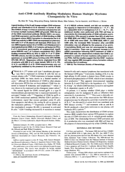
From www.bloodjournal.org by guest on February 6, 2015. For personal use only.
Differential Mobilization of Myeloma Cells and Normal Hematopoietic Stem
Cells in Multiple Myeloma After Treatment With Cyclophosphamide and
Granulocyte-Macrophage Colony-Stimulating Factor
By Yair Gazitt, Erming Tian, Bart Barlogie, Chris L. Reading, David H. Vesole, Sundar Jagannath, Judith Schnell,
Ron Hoffman, and Guido Tricot
Peripheral blood stem cells (PBsCs) mobilized with highdose chemotherapy and hematopoietic growth factors are
now widelyused t o support myeloablative therapyof multiple myeloma and effect complete remissions in up t o 50%
of patients with apparent extension of event-free and overall
survival. Because tumor cells are present not only in bone
marrow, but also in virtually allPBSC harvests, it is conceivable that autografted myeloma cells contribute t o relapse
after autotransplants. In this study, the kinetics of mobilization of normal hematopoietic stem cells were compared
with those of myeloma cells present in PBSC harvests of 12
patientsafter high-dose cyclophosphamide and granulocyte-macrophage colony-stimulating factor administration.
CD34+ and CD34+Lin-Thy+ stem cell contents were
measured by multiparameter flow cytometry, and myelomacells
were quantitated by
immunostaining for therelevant Ig light
chain and by a quantitative polymerase chain reaction for
the myeloma-specific CDRlllsequence. Results indicated
marked heterogeneity in the percentages of mobilized stem
cells among different patients(0.1% t o 22.2% for CD34' cells
and 0.1% t o 7.5% for CD34+Lin-Thy+cells, respectively). The
highest proportions ofhematopoietic progenitor cells were
observed early during apheresis, with 9 of 12 patients mobilizing adequate amounts of CD34' cells for 2 autotransplants
(>4 x 10⁶/kg) within the first 2 days, whereas peak levels
(percent and absolute numbers) of myeloma cells were present on days 5 and 6 (0.5% t o 22.0%). During the lastdays of
collection, mobilized tumor cells exhibited more frequently
high labeling index values (1% to 10%; median, 4.4%) and an
immature phenotype (CD19'). The differential mobilization
observed between normal hematopoietic stem
cells and myeloma cells can be exploited t o reduce tumor cell contamination in PBSC harvests.
0 1996 by The American Societyof Hematology.
A
completion of cyclophosphamide treatment and continued daily until
the completion of the PBSC collections. A Quinton double lumen
catheter was inserted in the subclavian vein for stem cell apheresis.
Apheresis procedures were performed on a Cobe Spectra Blood
Cell Processor (Lakewood, CO) and PBSCs were collected once the
leukocytes count exceeded 0.5 X 1Oy/Land platelets approached 50
X 10yL(untransfused). Collections continued until at least 6 X lo*
mononuclear cellsikg were obtained (usually within 4 to 6 days) or
for a maximum of 14 days. However, there was no set target for
CD34' or for CD34+Lin-Thy+ cells. All patients signed an informed
consent form approved by the local Institutional Review Board.
Flow sorting of plasma cellsand generation of the CDRIIIprimer
for ASO-PCR. Ficoll-Hypaque-separated BM mononuclear cells
(BMMCs) were stained for the CD38 and CD45 antigens (CD45fluorescein isothiocyanate [F'ITC]; CD3S-phycoerythrin [PE]; Becton Dickinson [BD], Mountain View, CA). The CD38b"gh'CD451""
cells were sorted using a FACStar Plus cell sorter (BD) as described
previously.'3.18DNA extraction, generation of the CDRIII primers,
and radiolabeled ASO-PCR reaction were performed as previously
de~cribed.'~."-~'
Dilution curves of myeloma cell DNA between 10%
and 0.001% myeloma cell DNA were generated for each patient by
diluting myeloma cell DNA into normal peripheral blood lymphocytes DNA, as previously described.l3.Iy Autoradiograms were
scanned densitometrically and quantitation of myeloma cells in
UTOLOGOUS TRANSPLANTATION is now frequently used in the treatment of patients with multiple
myeloma (MM)."4 Complete remissions (CRs) are observed
in up to 50% of patients with newly diagnosed MM, but,
unfortunately, relapses remain a pr~blem.',~"
Compared with
standard autologous bone marrow (BM) transplants, peripheral blood stem cells (PBSCs) procured after the administration of high-dose cyclophosphamide and/or hematopoietic
growth factors significantly reduce the duration ofBM
aplasia and hence treatment-related morbidity and mortality.'"'
Because MM is a disease considered to be restricted to
the BM, the concentration of myeloma cells in PBSCs was
felt to be negligible. However, we and others have documented tumor cells in PBSC harvests with a frequency of
0.01% to greater than
This observation prompted
the exploration of hemopoietic stem cell selection as a means
of reducing tumor cell ~ontamination.'~
We have focused
on a CD34+ subcompartment that expresses Thy+ and Lin(CD34', Thy+, Lin-) and is composed of cells capable of
unlimited self-renewal and multilineage differentiation.14"'
This fraction contains less than 0.001%myeloma cells, based
on polymerase chain reaction (PCR) amplification of the
CDRIII sequence.13
In the current study, high-dose cyclophosphamide and
granulocyte-macrophage colony-stimulating factor (GMCSF) were administered for PBSC mobilization. Marked heterogeneity was observed (>50-fold) among different patients in the number of stem cells mobilized, peaking during
the first 3 days of apheresis, whereas myeloma cells were
present then in low numbers but increased by more than 2
logs on days 5 to 6.
MATERIALS AND METHODS
Mobilization and procurement of PBSCs. Patients with recently
diagnosed (N = 5) or previously treated myeloma (N = 7) received
6 g/m' of cyclophosphamide over 12 hours. GM-CSF was initiated
at a dose of 250 pglm2 subcutaneously within 24 hours after the
Blood, Vol 87, No 2 (January 15), 1996: pp 805-811
From the Department of Medicine, Division of Hematology-Oncology, University of Arkansas for Medical Sciences, Little Rock,
AR; and SyStemix, Palo Alto, CA.
Submitted June 15, 1995; accepted August 30, 1995.
Supported by National Institutes of Health Grants No. POI-CA
55819 and CA 539340.
Address reprint requests to Yair Gazitt, PhD, Department of Medicine, Division of Hematology-Oncology, University of Arkansas for
Medical Sciences, 4301 WMarkham Slot 508,Little Rock, AR 72205.
The publication costs of this article were defrayed in part by page
charge payment. This article must therefore be hereby marked
"advertisement" in accordance with 18 U.S.C. section 1734 solely to
indicate this fact.
0 19% by The American Society of Hematology.
0006-4971/96/8702-0025$3.00/0
805
From www.bloodjournal.org by guest on February 6, 2015. For personal use only.
806
PBSCcollections was performed by log linearregressionas previously described."."
Quantitation of CD34'Lin-Thy' stem cells. Stem cells were de(1 0' cells) for CD34
tected by staining samples from apheresed blood
using anti CD34-PE(HPCA-2;BD)andforThy(CD,90)
using
biotin-labeled GM 201 (SyStemix, Palo Alto, CA), followed
by RED
FITC670-streptavidin(Systemix). Lin' cellswerestainedwith
conjugated antibodies to CD14,CDIS, CD2, CD16, CD19, and antiglycophorin (all from BD). Isotypic controls corresponding to the
antibody isotypes and the proper fluorochrome were used to deterIgG l-biotin and avidin-RED670 were
mine the background staining.
used as a control for the Thy antibody. Propidium iodide was added
to all tubes to exclude dead cells. Tubes were run on a FACSCAN
BOW cytometer (BD). Six parameters (FS, SS, PI, FITC, PE, and
Red 670) were examined in listmode using the ListView software
(Phoenix Flow Systems, San Diego,
CA). The cells were sequentially
gated from viable nucleated cells to the lymphoblastoid region and
then to each of the two color combinations,
ie. CD34 versus Lin
and CD34 versus Thy. Each fluorochrome
was colorgated. Fifty
thousand cells were analyzed.
Sruiningfor myeloma cells. Staining of myeloma cells was per(50,000 cellsfromapheresissamples)after
formedoncytospins
ethanol fixation (30 minutes at 4°C). Slides were stained for both K
and X light chains of human Ig (rabbit antibody-Rhodamine conjugate; Dako, Carpinteria, CA). Plasma cells were identified by positivestainingforthepertinent
light chainand by theirlymphoid/
plasmacytoid morphology. Positive staining for the irrelevant light
chain was always less than 10% of the relevant light chain. At least
S00 cells were scored.
Staining of immuture myeloma cells und determination of plusma
cell labeling index (PCLI). Severalmethodsfordetermination
of
PCLI were tested using different anti BrdU antibodies and different
cell denaturationprotocolsasdescribed
previously.2' The best
results were obtained using the procedure described by Lokhorst et
al." Briefly, samples from apheresed blood (10' cells) were allowed
to incorporate bromodeoxyuridine ( I 0 pmol/L BrdU for 1 hour at
37°C). Subsequently, aliquots of 50,000 cells were cytospun, fixed
with ethanol, air-dried, andtreated with 2 N HCI (30 minutes at room
temperature) to denatureDNA.Afterneutralization
in phosphatebuffered saline (PBS), slides were stained for
K and A light chains
as described above and for BrdU with anti-BrdU antibodies (mouse
monoclonal; Dako), followedby antimouse-FITC (Jackson Immunochemicals, West Grove, PA). Double-stained cells were counted as
cycling p k m d cells. Cycling plasma cells were
identified by positive
staining for the relevant light chain (red),
by positive staining for
BrdU (green), and by their lymphoidlplasmacytoid appearance. Positive staining for the irrelevant light chain was less than 10% of the
relevant light chain. At least 500 plasma cells were scored. Mouse
lgGI was used as a control for background staining for the BrdU
antibody. This procedure wasused successfully in our laboratory for
more than 1 year to monitor more than 400 patients with significant
correlationbetweendiseaseactivityandPCLI(Gazitt,manuscript
in preparation).
Immature plasma cells were detected by cytoplasmic staining for
K/A lightchain(rhodamine-conjugatedantibodies),
as described
above,andfor
CD19' usingFITC-anti-CD19antibody(mouse,
monoclonal; BD). Most CDIY' cells were small lymphocytoid,with
predominantmembranestaining
for CD19. In 2 patients. CD19'
cells had the appearance of plasmacytoid cells with
weak staining
for cytoplasmic CD19 antigen and positive staining for the relevant
light chain. Positive stainingof CD19' cells with the irrelevant light
less than 1070 of therelevantlightchain.
At
chainwasalways
least S00 plasma cells were scored. Mouse FITC-IgGI was used as
isotypic control for background staining for the CD19 antibody.
GAZITT ET AL
RESULTS
The time courseof stem cell mobilization for 4 representative patients is depicted in Fig I . Maximum proportions of
CD34' and CD34'Lin-Thy' were observed on days 1 to 3,
ranging from 1.8% to 22.2% and 0.48% to 8.2%.respectively. Figure 2 portrays the cumulative amount of CD34'
stem cells in the 12 MM patients studied. Sufficient quantities of CD34+ cells perkilogram fortwo autotransplants
(>4 X 10') were obtained after two collections in 9 of the
12 patients. Eight patients mobilized greater than 10 X loh/
kg of CD34' cells within the first 3 days of collection. Three
patients did not mobilize well.
The proportions of myeloma cells in PBSC harvests of 4
patients, including the percentages of light chain restricted
B cells (PLC), their labeling index (PCLI), and the proportion of immature myeloma cells(CD19') are depicted in Fig
3. In all 4 patients, mobilization of myeloma cells peaked
on days S and 6, with PCLI values ranging between 3.1 %
and 7.5% on these days. Maximum concentrations of light
chain-restricted CD19' cells (presumedto represent preplasmacytic myeloma cells) usually coincided with peak values
of PCLI. The cumulative amountof myeloma cells in the I2
myeloma patientsis depicted in Fig 4. Ten patientsmobilized
greater than S X 107/kg and 6 greater than 1 X lORkgmyeloma cells during the 6 days of apheresis. One patient did
not mobilize significant amounts of myeloma cells. In the
other 1 1 patients, greater than 75% of myeloma cells were
collected during the last 2 days of apheresis.
Figure S summarizes the proportions of CD34'.
CD34'Lin Thy*, and myeloma cells duringthe consecutive
days of PBSC apheresis. Median percentages of maximum
CD34' and CD34'Lin-Thy' cells were 5.5% (range, 0.2%
to 22.2%) and 2.7% (range. 0.1% to 8.2%), respectively.
Both cell populationswere largest on days 1 to 3, with a
subsequent significant decline reaching a minimum by day
6 ( P < .001). In contrast, only 2 of 12 patients had ~ 2 . 5 %
myeloma cells by immunocytochemistry in their PBSC collections on days 1 and 2, compared with 1 I of 12 patients
by day 6. A median of 4.4% myeloma cells (range, 1.6%to
10.9%) werecycling, most frequentlyon days 4 to 6 of
PBSC collection. Similarly, CD19'lightchain-restricted
cells peaked on day 6, with a median of 5.1% (range, 0%
to 16%;results not shown). Theleast overlap between hematopoietic progenitor and myeloma cell mobilization was observed on days I and 2.
PCR-basedquantitation of minimalresidual disease is
more sensitive (2 to 3 logs) than immunofluorescence-based
quantitation of tumor cells. Therefore, quantitation of myeloma cells in PBSC collections was also performed by PCR
amplification of the clone-specific CDRIII sequence. Representativeresults from 6 patients (B.D., H.R., B.L., L.J.,
M.G.. and D.P.) are shown in Fig 6. The dilution curves
(left panels) indicate a sensitivity of detection of I myeloma
cell in 10,000 to100,000nonmyeloma cells. A daily increase
in the number of myeloma cells in PBSC collections (right
panels) was seen. Densitometric scanning of the autoradiograms and quantitation by log-linear regression in 3 patients
(B.D., H.R., and B.L.) showed a low content of myeloma
From www.bloodjournal.org by guest on February 6, 2015. For personal use only.
DIFFERENTIAL MOBILIZATION OF MYELOMA AND STEMCELLS
807
Patient B.D.
Patient B.L.
12.5
12 5
UCD%+
ICD%+/Thy+
10 0
Aphemia
Patient
H.R
Patient M.G.
20
m
b-
10
e
5
Fig 1. Mobilization of stem
cells in PBSC harvests of MM patients. Percentages of hematopoietic
progenitor
cells during
consecutive days of apheresis in
4 patients.
E
O5
od
Aphemsii
Aphereais
cells during the first days of apheresis (<0.01%, 1.2%, and
0.1%, respectively), with subsequent daily increases of up
to one log, reaching 6.5%, 16.9%, and 22%, respectively,
on day 6. Two patients (L.J. and D.P.) had already high
levels of myeloma cells on day 1 (4.4% and 9.6%, respec-
tively), with no obvious change during subsequent days of
apheresis. One further patient (M.G.) had a low baseline
level of myeloma cells (0.15%), with no further increase
during subsequent days of apheresis. A significant correlation was observed between the levels of myeloma cell measured by the PCR method and by immunofluorescence with
R values of 0.93 to 0.97.
KINETICS OF MOBILIZATION OF CD%+ STEMCELLS
4b
=DAY1
EDAYZ
[IDDAY3
DISCUSSION
mDAY4
Mobilization of PBSCs with high-dose chemotherapy
alone or in combination with hematopoietic growth factors
results in a 10- to 100-fold increase in the number of circulating hematopoietic progenitor ~ e l l s ~ and
~ . ’ effects
~
prompt,
complete and durable engraftment after myeloablative treatment.”” Probably not unexpectedly, this approach also leads
to mobilization of tumor ~ e l l s , ’ ’ ~including
~~
myeloma
P8
IQ
0
+
C.E.
R.S.
H.R.
C.I.
D.P.
C.G.
M.oG.
L.J.
B.L.
B.D.
M.G.
B.S.
Fig 2. Cumulative number of CD34’ stem cells in MM patients.
Absolute daily amounts of CD34+ stem cells per kilogram were
calculated from the total WBC count per collection, the percentage of
CD34+ cells, and the patient‘s body weight.
Myeloma cells are present in virtually all mobilized PBSC
harvests with a frequency of 0.01% to greater than 10%.l3
Depletion of myeloma cells can be achieved by positive
selection for CD34+cells” or, morecompletely, by selection
for CD34+Thy+Lin- stem cells.’3
This is the first detailed study assessing, simultaneously,
the kinetics of mobilization of both hematopoietic progenitor
cells and myeloma cells, including their proliferative capacity and the immature myeloma cell phenotype. The highest
proportions of progenitor cells (CD34’ and CD34+ThytLincells) were observed during the first 2 days of apheresis,
when the total white blood cell (WBC) count was still low
(0.5 to 2 X 10⁹/L). More than 4 X 10⁶/kg CD34+ cells,
sufficient for two auto transplant^,^ were collected during the
first 2 days in 9 of the 12 patients. Two of the three patients
From www.bloodjournal.org by guest on February 6, 2015. For personal use only.
GAZllT ET AL
808
Patient B.D.
Patient B.L.
"1
301
151
OPL
I PCCI L
C lD 1 9 K
=PLC
I P CI LClD 1 9 K
Apheresis
Apheresis
Patient H.R.
Patient M.G.
Fig 3. Mobilization of my-
Apheresis
eloma cells in P B X harvests of
MM patients. Percentages of
plasma
cells,
light-chain
restricted CD19+ cells, and cycling
plasma cells during consecutive
days of apheresis.Plasmacells
of
are expressed as a percentage
the total number of cells in the
daily collections. PCLl and CD19
cells are expressed as a percentage
of
the
total
number
of
plasma cells scored.
O P LI PCCI LClD 1 9 K
Aphemk
whodidnot
mobilize adequate amounts of CD34+ cells
had prolonged prior treatment with alkylating agents (> 12
months), resulting in permanent stem cell damage, as reported before.' The proportions of CD34' cells in these 3
patients were comparable to those observed in peripheral
blood of unmobilized MM patients (<l%).
In contrast to stem cells, the highest concentrations of
myeloma cells (>75% of total tumor cells) were collected
during days 5 and 6 of apheresis, when the percentage and
absolute number of progenitor cells were already rapidly
declining. Hence, an ideal window for PBSC collection with
KINETICS OF MOBILIZATION OF MYELOM4CELLS
'"1
M.G. C.I.
hl0.G.
B.L. D.P.
R.S.
B.O.
C.E.
C.G.
HR. 8.5. L.J.
Fig 4. Cumulative number of myeloma cells in MM patients. Absolute daily amounts of myeloma cells per kilogram were calculated
from the total WBCcountpercollection,
the percentage of light
chain-restricted myeloma cells, and the patient's body weight.
high progenitor cell content and minimal myeloma cell contamination exists during the first few days after W C and
platelet counts start to recover.
Moreover, during the last days of apheresis, a significant
proportion of myeloma cells was cycling and exhibited a
more immature phenotype (CD19+). Both immunocytochemical and PCR-based methodologies detected comparable amounts of myeloma cells in the daily collections (R
values of 0.93 to 0.97). The more sensitive PCR-based assay
showed a daily increase in concentration of myeloma cells
by 1 log in several patients.
Whether mobilization of myeloma cells is the result of
cyclophosphamide treatment or the administration of hematopoietic growth factor remains unknown. We are currently
conducting a study comparing the yield of CD34+ cells and
degree of myeloma cell contamination in MM patients mobilized with either G-CSF or G-CSF plus cyclophosphamide.
A direct comparison between G-CSF and cyclophosphamide
would have been preferable, but cyclophosphamide as a sole
mobilizing agent results in lower quantities of CD34+ cells
per kilogram, CD34+CD38- cells per kilogram, and CFUGM per kilogram when compared with G-CSF alone." The
fact that most of the multilineage hematopoietic growth factors are capable of mobilizing hematopoietic stem cells as
well as
cells12.13.27.28.31-34confirms the notion that cytokines are not cell-type specific, although the differential mobilization observed between hematopoietic progenitor cells
and myeloma cells suggests a greater sensitivity of hematopoietic progenitor cells to cytokines compared with myeloma
cells.
The molecular basis of homing of hematopoietic progenitor cells and tumor cells to the BM and their release into the
peripheral blood is not well under~tood.3~
Sustained high
From www.bloodjournal.org by guest on February 6, 2015. For personal use only.
DIFFERENTIALMOBILIZATION
OF MYELOMAANDSTEM
809
CELLS
levels of circulating growth factors may downregulate the
expression of adhesion molecules such as VLA-4, ICAM- I ,
and ELAM-l on the surface of stem cells, endothelial cells,
and myeloma cells. In support of this hypothesis is the recent
observation ofhigh
levels of circulating hematopoietic
RD.
H.R.
A
B.L.
r
a
V
APHERESIS
D.P.
Y r
L-
W
IO ' IO IO ' 10' IO '
I
I
2 1 .
I
. -= *
.a
.-
l
M Y 2
..
4
.
f
.-
MMY Y4 5
M Y 3
-
M Y 6
APHERESIS
*
2
;
;
.
MM
YM
Y
2Y
3 4
*
?"
j
*:
MV5
.
M Y 6
Fig 5. Comparison between mobilization of stem cells and myeloma cells in 12 patients. Stem cells were measured by flowcytometry and myeloma cells by immunocytochemistry as described in the
Materials and Methods.A significant decrease ( P < .001) in the median concentrationof CD34' cells from 5.5% on day 2 t o 0.8% on day
6 (Fig 3A) concurs with a decrease in CD34'Lin-Thy' cells from a
median of2.7% on day 1t o 0.29% on day 6 (Fig 38). Medianpercentages of myeloma cells were low ondays l through 3 10.3546, 0.650/0,
and 1.846, respectively) and increased markedly ondays 4 through 6
(4.2%. 5.7%. and 11.2%; Fig 3C).
Fig 6. ASO-PCR amplification of theCDRlll band the dailyin PBSC
collections of6 MM patients. The left panel shows the dilution
curve
of DNA (10% t o 0.001%1 from purified myeloma cells with normal
lymphocyte DNA. Lanes d, through ds represent DNA from PBSC
harvests of these patients duringconsecutive days of collection.
growth factors in umbilical cord blood"and
their effect
on the expression of the adhesion molecules, ELAM-l and
ICAM-I.37Umbilical cord blood induces rapid hematopoietic engraftment after myeloablative treatment. Interestingly,
CD34' cells express increased levels of L-selectin after
PBSC mobilization strategies, which correlated withthe
speed of hemopoietic engraftment.3x
While depletion methods for myeloma cells from PBSCs
are currently being
we recommend that, in
the absence of such methods, large volume exchanges early
after leukocyte and platelet recovery are perf~rmed.'"~~'
Our
conclusions are supported by the absence of breast cancer
cells, as measured by sensitive immunocytochemistry and
clonogenic assays, in single day large volume PBSC collections.."
ACKNOWLEDGMENT
The authors thank Christina Bewley for her dedicated secretarial
assistance.
REFERENCES
I . Jagannath S, Vesole D, Glenn L, Crowley J, Barlogie B: Lowrisk intensive therapy for multiple myeloma with combined autologous bone marrow and blood cell support. Blood 80:1666. 1992
2. Attal M. Huguet F, Schleifer D. Payen C. Laroche M, Foumie
B, Maziere B, Pris J. Lament G: Intensive combined therapy for
previously untreated aggressive myeloma. Blood 79: I 130, 1992
3. Fermand J, Chervet S, Ravaud P. Divine M. Leblond V. Dreyfus F, Mariette X, BruetJ-C: High dose chemoradiotherapy and
From www.bloodjournal.org by guest on February 6, 2015. For personal use only.
810
autologous blood stem cell transplantation in multiple myeloma.
Blood 82:2005, 1993
4. Barlogie B, Jagannath S, Vesole D, Tricot G, Naucke S, CoplandM, Crowley J: Total therapy for newly diagnosed multiple
myeloma. Blood 82:198a, 1993 (abstr, suppl 1)
5. Harousseau JL, Attal M, Divine M, Marit G, Leblond V,
Stoppa A-M, Bourhis J-H, Caillot D, Boasson M, Abgrall J-F, Facon
T, Linassier C, Cahn J-Y, Lamy T, Troussard X, Gratecos N, Pignon
B, Auzanneau G, Bataille R: Autologous stem cell transplantation
after first remission induction treatment in multiple myeloma: A
report of the French Registry on autologous transplantation in multiple myeloma. Blood 85:3077, 1995
6. Vesole D, Barlogie B, Jagannath S, Cheson B, Tricot G, Crowley J: High dose therapy for multiple myeloma: Improved prognosis
with better supportive care and double transplant. Blood 84:950,
1994
7. Tricot G, Jagannath S, Vesole D, Nelson J, Tindle S, Miller
L, Cheson B, Crowley J, Barlogie B: Peripheral blood stem cell
transplants for multiple myeloma: Identification of favorable variables for rapid engraftment in 225 patients. Blood 85:588, 1995
8. Bik To L, Shepperd KM, Haylock DM, Dyson PG, Charles P,
Thorp DL, Dale BM, Dart GW, Roberts MM, Sage R E , Juttner CA:
Single high dose of cyclophosphamide enable the collection of high
numbers of hematopoietic stem cells from peripheral blood. Exp
Hematol18:442, 1990
9. Haas R, Dick Ho A, Bredthauer U, Cayeux S, Egerer G, Knauf
W, Hunstein W: Successful autologous transplantation of blood stem
cells mobilized with recombinant human granulocyte-macrophage
colony stimulating factor. Exp Hematol 18:94, 1990
10. Kotasek D, Shepherd KM, Sage RE, Dale BM, Norman JE,
Charles P, Gregg A, Pillow A, Bolton A: Factors affecting blood
stem cell collections following high-dose cyclophosphamide mobilization in lymphoma, myeloma and solid tumors. Bone Marrow
Transplant 9: 11,1992
1I . Gianni AM, Siena S, Bregni M, Tarella C, Stem AC, Pileri A,
Bonadonna G: Granulocyte-macrophage colony-stimulating factor to
harvest circulating hematopoietic stem cells for autotransplantation.
Lancet 2580, 1989
12. Bird JM, Bloxham D, Russell NH, Samson D, Apperley JF:
Detection of clonally rearranged cells in PBSC harvests in multiple
myeloma by immunoglobulin gene fingerprinting. Blood 82:265a,
1993 (abstr, suppl 1)
13. Gazitt Y, Readmg CC, Hoffman R, Lee J-H, Vickrema A,
Vesole DH, Jagannath S, Condino J, Lee B, Barlogie B, Tricot G:
Purified CD34+Lin-Thy+ stem cells do no contain clonal myeloma
cells. Blood 86:381, 1995
14. Baum CM, Weissman IL, Tsukamoto AS, Buckle A-M,
Peault B: Isolation of a candidate human hematopoietic stem-cell
population. Proc Natl Acad Sci USA 89:2804, 1992
15. Craig W, Kay R, Cutler RL, Lansdorp PM: Expression of
Thy-l on human hematopoietic progenitor cells. J Exp Med
177:1331, 1993
16. Uchida N. Aguila HL, Fleming WH, Jerabek L, Weissman
IL: Rapid and sustained hematopoietic recovery in lethally irradiated
mice transplanted with purified Thy-l .l'" Lin- Sca-l+ hematopoietic
stem cells. Blood 83:3758, 1994
17. Murray L, Chen B, Galy A, Chen S, Tushinski R, Uchida N,
Negrin R, Tricot G, Jagannath S, Vesole D, Barlogie B, Hoffman
R, Tsukamoto A: Enrichment of human hematopoietic stem cell
activity in the CD34+Thy-1'Lin- subpopulation from mobilized peripheral blood. Blood 85:368, 1995
18. Hata H, Xiao H, Petrucci MT, Woodliff J, Chang R, Epstein
J: Interleukin-6 gene expression in multiple myeloma: A characteristic of immature tumor cells. Blood 81:3357, 1993
19. Billadeau D, Blackstadt M, Greipp P, Kyle RA, Oken MM,
GAZllT ET AL
Kay N, Van Ness B: Analysis of B-lymphoid malignancies using
allele-specific polymerase chain reaction: A technique for sequential
quantitation of residual disease. Blood 78:3021, 1991
20. Maniatis T, Fritsch EF, Sambrook J (eds): Molecular Cloning:
A Laboratory Manual. Cold Spring Harbor, NY, Cold Spring Harbor
Laboratory, 1982
21. Ravetch JV, Siebenlist U, Korsmeyer S, Waldmann T, Leder
P: Structure of the human mu locus: Characterization of embryonic
and rearranged J and D genes. Cell 27582, 1981
22. Gratzner HG: Monoclonal antibody to bromo and iododeoxyuridine: A new reagent for detection of DNA replication. Science
218:474, 1982
23. Greipp PR, Witzig TE, Gonchoroff NJ: Immunofluorescent
plasma cell labelling indices using a monoclonal antibody. Am J
Hematol 20:289, 1985
24. Lokhorst HM, Boom SE, Bast BJEG, Ballieux RE: Determination of the plasma cell labelling index with bromodeoxyuridine
in a double fluorescence technique. Br J Haematol 64:271,1986
25. Williams SF, Bitran JD, Richards JM, DeChristopher PJ,
Barker E, Conant J, Golomb HM, Orlina AR: Peripheral blood derived stem cells collections in use for autologous transplantation after
high-dose chemotherapy: An alternative approach. Bone Marrow
Transplant S : 129, 1990
26. Peters WP, Rosner G, Ross M, Vredenburgh J, Meisenberg
B, Gilbert C, Kurtzberg J: Comparative effect of GM-CSF and GCSF on priming of peripheral blood progenitor cells for use with
autologous marrow after high dose chemotherapy. Blood 81: 1709,
1993
27. Brugger W, Bross KJ, Glatt M, Weber F, Mertelsmann L.
Kanz L: Mobilization of tumor cells and hematopoietic progenitor
cells into peripheral blood of patients with solid tumors. Blood
83:636, 1994
28. Shpall EJ, Jones R: Release of tumor cells from bone marrow.
Blood 83:623, 1994
29. Vescio RA, Hong CH, Cao J, KimA, Schiller GJ, Lichtenstein AK, Berenson M,Berenson JR: The hematopoietic stem
cell antigen, CD34, is not expressed on malignant cells in multiple
myeloma. Blood 84:3283, 1994
30. To LB, Haylock DN, Dowse T, Simmons PJ, Trimboli S,
Ashman LK, Juttner CA: A comparative study of the phenotype and
proliferative capacity of peripheral blood CD34' cells mobilized by
4 different protocols and those of steady-phase PB and bone marrow
CD34' cells. Blood 84:2930, 1994
31. Sonneveld P, Schoester M, Leeuw KL: In vitro Ig synthesis
and proliferative activity in multiple myeloma are stimulated by
different growth factors. Br J Haematol 79589, 1991
32. Zhang X-G, Bataille R, Jourdan M, Saeland M, Banchereau
J , Mannoni P, Klein B: GM-CSF synergizes with IL-6 in supporting
the proliferation of human myeloma cells. Blood 76:2599, 1990
33. Anderson KC, Jones RM, Morimoto C, Leavitt P, Barut BA:
Response patterns of purified myeloma cells to hematopoietic growth
factors. Blood 73:1915, 1989
34. Lemoli RM, Fortuna A, Grande A, Gamberi B, Bonsi L, Fogli
M, Amabile M, Cavo M, Ferrari S, Tura S: Expression and functional
role of c-kit ligand in human multiple myeloma. Br J Haematol
88:760, 1994
35. Weiss L, Geduldig U: Barrier cells: Stromal regulation of the
hematopoietic and blood cell release in normal and stressed bone
marrow. Blood 78:975, 1991
36. Cairo M, Gillan E, Buzby J, Van de ven C, Suen Y: Circulating Steel factor and G-CSF levels in preterm and term newborns
and adults peripheral blood. Am J Pediatr Hematol Oncol 15:311,
1993
37. Buzby JS, Knoppel EM, Cairo MS: Coordinate regulation
of steel factor and cytoadhesion molecule (ICAM-I and ELAM-1)
From www.bloodjournal.org by guest on February 6, 2015. For personal use only.
DIFFERENTIAL MOBILIZATION OF MYELOMAAND STEM CELLS
mRNA expression in human vascular endothelial cells of differing
origins. Exp Hematol 22:122, 1994
38. Dercksen MW, Gerritsen W R , Rodehuis S, Dirkson MKA,
Slaper-Cortenbach K M , Schaasberg WP, Pinedo HM, von dem
Borne AEGK, van der Schoot CE: Expression of adhesion molecules
on CD34' cells: CD34' L-Selectin+ cells predict rapid platelet recovery after peripheral blood stem cell transplantation. Blood
85:3313, 1995
39. Anderson KC, Anderson J, Soiffer R, Freedman AS, Rabinowe SN, Robertson MJ, Spector N, Blake K, Murray C, Freeman
A, Coral F, Marcus KC, Mauch P, Nadler L, Ritz J: Monoclonal
antibody purged bone marrow transplantation therapy for multiple
myeloma. Blood 82:2568, 1993
81 1
40. Pettengell R, Morgenstern GR, W011 PJ, Chang J, Rowland M,
Young R, Radford JA, Scarffe JH, Testa NG, Crowther D: Peripheral
blood progenitor cell transplantation in lymphoma and leukemia
using a single apheresis. Blood 82:3770, 1993
41. Passos-Coelho JL, Braine HG, Wright SK, Davis JM, Schepers KG, Huelskamp A-M, Clarke B, Noga SJ, Kennedy MJ: Large
volume leukapheresis using regional citrate anticoagulant to collect
peripheral blood progenitor cells. J Hematother 4: 11, 1995
42.Passos-Coelho JL, Ross AA, MossTJ,Davis
M, Huelskamp A-M, Noga SJ, Davidson NE, Kennedy J: Absence of
breast cancer cells in a single-day peripheral blood cell collection
after
priming
with cyclophosphamide and GM-CSF. Blood
85:1138,1995
From www.bloodjournal.org by guest on February 6, 2015. For personal use only.
1996 87: 805-811
Differential mobilization of myeloma cells and normal hematopoietic
stem cells in multiple myeloma after treatment with
cyclophosphamide and granulocyte-macrophage colony-stimulating
factor
Y Gazitt, E Tian, B Barlogie, CL Reading, DH Vesole, S Jagannath, J Schnell, R Hoffman and G
Tricot
Updated information and services can be found at:
http://www.bloodjournal.org/content/87/2/805.full.html
Articles on similar topics can be found in the following Blood collections
Information about reproducing this article in parts or in its entirety may be found online at:
http://www.bloodjournal.org/site/misc/rights.xhtml#repub_requests
Information about ordering reprints may be found online at:
http://www.bloodjournal.org/site/misc/rights.xhtml#reprints
Information about subscriptions and ASH membership may be found online at:
http://www.bloodjournal.org/site/subscriptions/index.xhtml
Blood (print ISSN 0006-4971, online ISSN 1528-0020), is published weekly by the American
Society of Hematology, 2021 L St, NW, Suite 900, Washington DC 20036.
Copyright 2011 by The American Society of Hematology; all rights reserved.

© Copyright 2026