
IL-6 - Blood
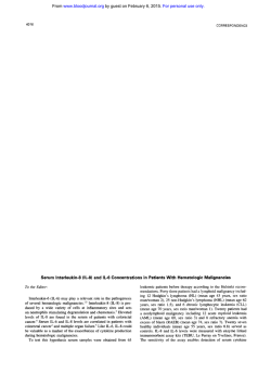
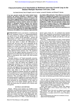
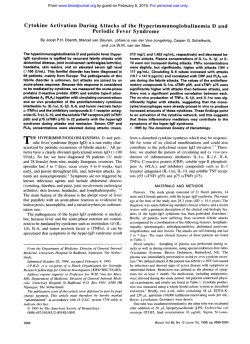
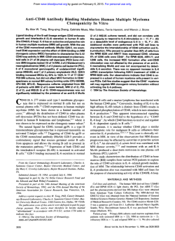
From www.bloodjournal.org by guest on February 6, 2015. For personal use only.
Expression of the Interleukin-6 (IL-6), IL-6 Receptor,
and gp130 Genes in Acute Leukemia
By Kazushi Inoue, Haruo Sugiyama, Hiroyasu Ogawa, Tamotsu Yamagami, Teruyuki Azuma, Yoshihiro Oka,
Hiroshi Miwa, Kenkichi Kita, Akira Hiraoka, Tohru Masaoka, Kaori Nasu, Taiichi Kyo, Hiroo Dohy, Junichi Hara,
Akihisa Kanamaru. and Tadamitsu Kishimoto
Expression patterns of interleukin-6 (IL-6). IL-6 receptor (IL6RI. and gp130 genes in 39 patients with acute myeloid
leukemia (AML), in 23 patients with acute lymphoblastic
leukemia (ALL), and in 7 patients with acute mixed lineage
leukemia (AMLL) were studied by quantitative reverse transcriptase-polymerasechain reaction. Significant levels of IL6 were expressed in 8 (21%) of 39 AML patients and in 2
(29%) of 7 AMLL patients, whereas in ALL, the expression
of IL-6 was almost negligible. IL-6R was expressed in all
patients with AML and AMLL, whereas only half of ALL patients expressed low levels of IL-6R as compared with those
with AML and AMLL. However, gp130 was ubiquitously expressed in all the leukemia patients, and there was no sig-
nificant difference in gp130 expression among AML, ALL,
and AMLL. Significant correlation was observed between
the expression of IL-6R and gp130 in AML. When tested for
in vitro response to IL-6, the leukemic cells from 3 of 7 AML,
none of 3 ALL, and both of 2 AMLL patients significantly
responded to IL-6, showing the correlation between the expression levels of IL-6R and gp130 and the responsiveness
of leukemic cells to IL-6. These results showed that quantitation of IL-6R and gp130 expression by reverse transcriptasepolymerase chain reaction is useful for the rapid prediction
of the responsiveness of leukemic cells to IL-6, especially in
cases of administration of IL-6.
0 1994 by The American Society of Hematology.
I
responsiveness of leukemic cellsto 1L-6 toelucidate the
roles of IL-6 in the pathogenesis of acute leukemia.
NTERLEUKIN-6 (IL-6) is a pleiotropic multifunctional
cytokine produced by various types of cells and plays
an important role in the pathogenesis of various disease.’
IL-6
is
a growth
factor
for
human
multiple
and
stimulates autocrine’ or paracrines.6 growth of myeloma
cells. Large quantities of IL-6 were shown to beproduced by
the hyperplastic lymph nodes frompatients with Castleman’s
disease,’ implicating the important roles of IL-6 in various
clinical manifestations of this syndrome. Concerning malignant lymphomas, it was shown that cultured supernatants of
neoplastic cells from a patient with advanced Ki-l-positive
lymphoma included high levels of IL-6’ and that Hodgkin’s
lymphoma cells expressed both IL-6 and IL-6 receptor (IL6R).9.’0Autocrinegrowth of IL-6 in twolymphoma cell
lines” and a Lennert’s lymphoma-derived T-cell line” was
shown. On the other hand, therehave been few reports concerning the expression of IL-6 and its related genes in acute
le~kemia.”.’~ The
purpose of this study is to clarify the patterns of expression of IL-6 and its relatedgenes in acute
leukemia and the relationship between the patterns and the
From the Departments of Medicine I l l and Pediatrics, Osaka
University Medical School, Osaka: the Third Department of Internal
Medicine, Kinki University School of Medicine, Osaka; the Second
Department of Internal Medicine, Mie University School of Medicine, Mie: The Center for Adult Diseases, Osaka: Osaka Red Cross
Hospital, Osaka; Hiroshima Red Cross Hospital and Atomic-Bomb
Survival Hospital, Hiroshima; and the Second Department of Internal Medicine, Hyogo College of Medicine, Hyogo. Japan.
Submitted February 7. 1994; accepted June 18, 1994.
Supported by Grants,from the Ministry of Education. Science, and
Culture .fJapan.
Address reprint request to Haruo Sugiyama, MD, Department of
Medicine Ill, Osaka University Medical School, 2-2, Yamada-Oka,
Suita City, Osaka 565, Japan.
The publication costs of this article were defrayed in part by page
charge payment. This article must therefore be hereby marked
“advertisement” in accordunce with 18 U.S.C. section 1734 solely to
indicate this fact.
0 1994 by The American Society of Hematology.
0006-497//94/8408-0037$3.00/0
2672
MATERIALS AND METHODS
Patients. At the onset of the disease, peripheral
blood (PB) or
bone marrow (BM) cells from 39 acute myeloid leukemia (AML),
23 acute lymphoblastic leukemia (ALL), and 7 acute mixed lineage
leukemia (AMLL) patients were obtained before therapy. The leukemic cells were classified according to the French-American-British
criteria. AMLL was defined as described previously.”
Purijication of leukemic ce1l.r. Heparinized PB or BM cells were
suspended in phosphate-buffered saline, put into a Ficoll-lsopaque
solution, and centrifuged. The leukemic cells were recovered from
theinterphase, washed twice with phosphate-bufferedsaline,and
used for experiments. In AML M3 cases, heparinized
blood samples
were suspended in a half volume of Haemacel (Boehringer Mannheim, Mannheim, Germany) and left to stand for 20 minutes to let
theerythrocytessettle.Thesupernatantcellswerethengathered
by centrifugation and examined. Microscopic examination showed
purity of leukemic cells to be more than 90% in all cases.
Cell lines. All celllinesexceptforIMS-MIand
TF-l were
kindly provided by the Japanese Cancer Research Resources Bank;
IMS-M I was a generous donation from DrS. Asano (Tokyo University, Tokyo, Japan). A human erythroleukemia cell line, TF-I, was
kindly donated by Dr T. Kitamura (DNAX Research Institute, Palo
Alto, CA). The cells were cultured and propagated in
RPM1 1640
medium containing 10% heat-inactivated fetal calf serum (FCS).
RNA purijication. Freshlyisolatedleukemiccells (5 X 10“ to 5
x IO’) were dissolved in 500 pL of 4 rnol/L guanidine thiocyanate
solution, and total RNA was isolated as described previously,Ih except that phenol-chloroform-isoamylalcoholextraction
was performed twice to improve thepurity of RNA. The RNA was dissolved
in diethylpyrocarbonate-treated waterandquantifiedspectrornetrically with absorbance at 260 nm.
Reverse rrunscription. Onemicrogram of‘ total RNA in 12.5
pL of diethylpyrocarbonate-treated water was heated at 65°C for S
minutes and then mixed with 17.5 WLof reverse transcription buffer
(50 mmollL Tris HCI [pH 8.31, 70 mmol/L KCI, 3 mmol/L MgC12.
I O mmoVL dithiothreitol)containing600U
of Moloneymurine
leukemiavirusreversetranscriptase(RT;GIBCO-BRL,Gaithersburg, MD), 500 pmoVL of eachdNTP(PharmaciaBiotech,AB,
Uppsala, Sweden), 750 ng of oligo-dT primer (Pharmacia), and 40
U ofRNaseinhibitor(BoehringerMannheim).Thesolution
was
Blood, Vol 84, No 8 (October 15). 1994: pp 2672-2680
From www.bloodjournal.org by guest on February 6, 2015. For personal use only.
IL-6 AND ITSRELATEDGENEEXPRESSION
2673
IN ACUTE LEUKEMIA
C
3
l*
1 05
1V
1 0’
1 0‘
L
0‘
24 2 1 30 33 36 39
” 0 1
2 ~ 2‘
~
2
5
4
2-1 2-2z - ,
Dilutions
it
24 21
30 33
10‘
Cycles
Cycles
20
2‘
10
r
-zol
- 201
Dilutions
p -actin
gpl30
IL-6R
IL-6
D
10
10
l0
1 5 182124
27
Cycles
-2ol
Dilutions
Dilutions
Fig 1. Detection of the exponential range by termination of PCR at sequential cycles orby serial dilutions of input cDNA. PCR was performed
at the indicated cycles (A through D) or on serial 1:2 dilutions (E through H) in the reverse transcription product prepared from 83 ng of total
RNA derived from U266 cells using the appropriate primers for IL-6 (A and E), IL-6 R (B and F), gp130 (C and G), or pactin (D and H).
incubated at 37°C for 90 minutes, then heated at 70°C for 20 minutes,
and stored at -20°C until use.
Polymerasechainreaction (PCR). The nucleotide sequence of
the upstream primer for IL-6 was 5’-ATGAACTCCTTCTCCACAAGCGC-3’ and that of the downstream primer 5‘-GAAGAGCCCTCAGGCTGGACTG-3’.” The sequence of the upstream primer
and of the
for IL-6R was 5’-CATTGCCATTGTI%TGAGGlTC-3’
of
downstream primer 5‘-AGTAGTCTGTATTGCTGATGTC-3”’;
the upstream primer for gp130 5’-ACAGATGAAGGTGGGAAGGAT-3‘ and of the downstream primer 5”AGATGACATGCATGAAGACCC-3”9; andof the upstream primer for @actin 5’GTGGGGCGCCCCAGGCACCA-3’ and of the downstream primer
5‘-GTCClTAATGTCACGCACGATITC-3’.*o
PCR wasperformed
in 50 pL of reaction solution containing cDNA derived from 83 ng
of the total RNA, 1.25 U of AmpliTaq polymerase, 250 pmolL of
each dNTP, and 0.5 pmol/L of sense and antisense primers. Each
PCR cycle included 1 minute of denaturation at 94°C. 1 minute of
primer annealing at 60”C, and 2 minutes of extensiodsynthesis at
72°C. PCR was performed with a DNA thermal cycler (Perkin Elmer-Cetus, Nonvalk, CT).
Quantitation of thePCR products. Quantitation of the PCR
products was performed as described previously.2’~2z
Briefly, the
products were subjected to electrophoresis on 1.3% agarose gels
containing 0.05 pglmL ethidium bromide, visualized by UV light,
and photographed with Polaroid 665 film (Polaroid, Cambridge,
MA). The negative film was developed for 5 minutes at 25°C and
washed with running water for 5 minutes. For quantitative analysis,
the film wassubjected to a densitometer CS-9OOO (Shimadzu, Kyoto,
Japan). PCR of the products of reverse transcription reaction without
addition of RT was used for negative controls.
P-actin is traditionally used to verify comparability of RNA loading between samples. In this study, weused P-actin to normalize
PCR products. The value of P-actin for the normalization of PCR
products is based on the concept that there will be no significant
differences in the amounts of P-actin per cell among various types
of cells. When P-actin was quantified in the leukemic samples, there
were no significant differences among them, confirming the propriety
of the use of P-actin for the normalization of PCR products.
Short-termproliferation assay. Proliferation of leukemic cells
to IL-6 was evaluated by measuring 3H-thymidine(3H-TdR)incorporation. Briefly, adherent cells were depleted from the purified leukemic cells by incubating the cell suspension (3 X lO“/mL) for 12
hours inRPM1 1640 medium containing 10% FCS. The leukemic
cells were cultured in the presence (100 ng/mL) or absence of IL6. Cultures were run in triplicate in 100 pL of a-minimal essential
medium containing 20% FCS in 96-well microtiter plates. After 96
hours of incubation, the cells were pulse-labeled with 0.5 pCi of
3H-TdR for 18 hours, and then ’H-TdR uptakes were determined by
a liquid scintillation counter.
RESULTS
Determination of the RT-PCR conditions for the quantitation of IL-6, IL-6R, and gp130 gene expression. First, the
From www.bloodjournal.org by guest on February 6, 2015. For personal use only.
INOUE ET AL
2674
optimal RT-PCR conditions to quantify mRNA expression
of IL-6, IL-6R, and gp130 genes were determined with the
cDNA from U266,23which is a human myeloma cell line
showing a high expression for IL-6,'4 IL-6R,24 and gp13O.l9
When the cDNA prepared from 83 ng of total RNA from
U266 cells was amplified with various numbers of PCR
cycles, exponential amplification for IL-6, IL-6R, and gp130
cDNA was observed at cycles 24 through 33, cycles 21
through 27, and cycles 21 through 30, respectively (Figs 1A
through C). When 32 cycles of PCR were used for IL-6,27
1 2 3 4 5 6
AML
--n
AMLL
ALL
Patient No. 80 79 122 17 38 49 64 81 90 20 82 U266
11-6-
IL-6R-
~.
IL-6gpl30-
IL-6R&actin-
-
--
Fig 3. Expression of IL-6.lL-6R. and gp130 genes in acute leukemic
cells. PCR products of representative cases (6 AML,3 ALL, and 2
AMLL cases) are presented.
~~~130-
B-actinFig 2. Spiking experiments. Ball-l cells (expression level: 11-6,
~ 0 . 0 1 ;IL-6R. ~ 0 . 0 1 gp130,
;
0.02) were spiked with increasing percentages of U266 cells (lane 1, 0%; lane 2, 5%; lane 3, 10%; lane 4,
50%; lane 5, 100%; and lanes 6, reaction buffer alone) and then RTPCR was performed with IL-6,IL-6R.gp130,or
p-actin primers as
described in Materials and Methods.
cycles for ILdR, and 30 cycles for gp130 at serial 1:2 dilutions of the cDNA prepared from 83 ng of total RNA from
U266 cells, exponential amplification was observed at 2-"
to 2" dilutions for IL-6, at 2-" to 2-' dilutions for IL-6R,
and at 2-' to 2" dilutions for gp130 (Figs IE through G ) .To
normalize the RNA loading for the RT-PCR, &actin gene"
was used as an internal standard. When the cDNA prepared
from 83 ng of total RNA from U266 cells was amplified
with various numbers ofPCR cycles using&actin gene
primers, exponential amplification was observed at cycles
16 through 21 (Fig ID). When 21 cycles of PCR were used
for serial 1 :2 dilutions of the cDNA prepared from 83 ng of
total RNA from U266 cells, exponential amplification was
observed at 2-' to 2' dilutions (Fig 1 H). As a result, patients'
samples were subjected to 32 cycles ofPCR for IL-6, 27
cycles for ILdR, 30 cycles for gp130, and 21 cycles for pactin.
Spiking experiments. To confirm that our assay of IL-6
and its related gene expression was quantitative, Ball-l cells
(expression level: IL-6, <0.01; IL-6R, <0.01; gp130, 0.02)
were spiked with increasing percentages of U266 cells, and
then RT-PCR was performed. As shown in Fig 2, the inten-
From www.bloodjournal.org by guest on February 6, 2015. For personal use only.
EXPRESSION
IL-6 ANDGENE
ITS RELATED
IN ACUTE LEUKEMIA
2675
Table 1. Clinical Features of Patients and Relative Gene Expression of 11-6, IL-6R. and gp130in Leukemic Cells
Relative Gene Expression
FAB
Classification
Relative Gene Expression
FAB
Classification
Patient No.
Age/Sex
IL-6
IL-6R
gp130
6
80
94
218
11
25
43
60
77
79
108
115
117
119
122
124
184
219
220
224
28
78
130
33
34
37
275
17
35
38
39
49
50
227
66
43/F
37/F
23/M
59/F
17/F
65/F
36/M
56lF
76lM
58lM
25lM
56lF
17/M
50/M
6OlF
60/M
56lM
<0.01
0.02
0.04
<0.01
<0.01
0.18
0.01
0.02
<0.01
0.13
<0.01
0.04
0.05
<0.01
3.41
0.14
0.10
0.43
0.34
0.12
0.60
0.12
0.12
0.21
0.17
0.38
0.50
0.20
0.22
0.11
0.20
0.74
0.20
0.31
0.25
0.08
0.31
0.03
1.27
<0.01
0.15
0.31
0.07
0.06
0.28
0.15
0.08
0.04
0.02
1.12
0.04
0.27
0.08
0.14
0.06
0.05
0.07
0.07
0.06
0.24
0.83
0.15
0.60
0.21
0.10
0.05
0.70
0.22
0.47
0.04
AM L
MI
M2
M3
M4
M5
M6
::;l}
0.16
<0.01
40/M
19/M
56/M 0.42<0.01
17/M
61/F
28/F
2O/F
65/F
15/M
25lF
<0.01
56/F
52/M
59/M
0.16
0.16
28/M
42/M
0.33
0.04
50/F
0.18
0.17
0.15
0.17
0.21
0.24
0.22
0.45
0.57
0.40
0.23
0.55
0.16
0.67
0.21
M7
MDS-AML
Patient No.
Age/Sex
IL-6
31/M
<0.01
IL-6R
gp130
45/F
0.27
0.23
0.21
0.09
0.04
0.03
0.13
0.12
81F
35/M
86lM
41/M
41/F
33/M
45/M
22/F
24/M
16lF
18lM
22/F
51/F
40/M
63lF
20/M
60/F
77/M
52/M
451M
15/F
<0.01
0.18
0.01
0.03
0.1 1
<0.01
<0.01
0.15
<0.01
0.12
0.16
0.13
0.14
0.07
<0.01
0.02
10.01
0.25
0.08
<0.01
0.10
0.28
<0.01
ND
0.05
0.02
0.20
0.08
0.60
0.02
0.13
0.40
0.18
0.40
c0.01
0.26
0.39
0.12
0.32
0.24
0.1 1
0.02
0.14
0.27
0.52
0.03
0.05
3.21
0.31
0.46
0.05
0.30
0.31
0.25
3.73
0.22
0.20
0.38
0.18
0.43
ALL
L1
L2
L3
AMLL
Cell Lines
Cell lines
Myeloma
IL-6
1.oo
Ball-l
HSB-2
0.05
<0.01
ALL
CML
blastic
MI
M3
AML
0.02
0.04
1.oo
0.05
0.11
KU812
KG-l
0.37
<0.01
HL-60 0.15 <0.01
0.20
0.60
IL-6R
gp130
431M
57/M
0.54
0.16
0.01
10.01
0.52
0.16
Abbreviations: FAB, French-American-British; ND, not determined.
10.01
C0.02
<0.01
0.10
}
1.68
3.54
0.03
0.02
0.10
<0.01
Relative Gene Expression
Origin
Cell Lines
tissues
1.oo
Donor BMNormal
0.03 0.17 0.01 no. 1
0.020.20
0.02
Donor no. 2
0.29no.3
0.13
Donor
0.09
0.07
Donor no. 4
0.02
Normal 0.17
PB 0.300.04Donor
1 no.
0.05
0.34
0.35
0.03
Donor no. 2
0.40
0.27
0.66
<0.01
34/M
53/F
47/F
18/F
461 M
Relative Gene Expression
Origin
0.09
IL-6
<0.01
IL-6R
gp130
From www.bloodjournal.org by guest on February 6, 2015. For personal use only.
2676
INOUE ET AL
A
4.0
.
(3.54)
(3.41)
3.0.
.
2.0
'
(1.68)
1.0.
.
0.5.
.
t
0
MI MzMs
M4 M5
Ms
L1 Lz L3
M7 MDS
+AML
AMLL PB BM AML ALL CML U266
cell Lines
4.0
(3.21)
3.0
2.0
1.o
O(1.00)
. .
..
..
.
-....
.
.
+ y f : t 3.
.
,f
__
0.5
:
.
L
V
-
0
. v
0
I
MI
I
"
0
I
I
M2 M3 M4
I
M5
I
I
M6 M7
.
I
Y
I
MDS L1
L2
+AML
L3
AMLL
sity of the bands of IL-6, IL-6R, gp130 became more intense
with the increase in the percentage of U266 cells, whereas
there were no significant differences in p-actin gene expression. The specificity of the bands wasconfirmedby
the
second-round PCR with the nested internal primers for IL6, IL-6R, and gp130 (data not shown). These results confirmed that IL-6, IL-6R, and gp130 gene expression can be
quantified under our RT-PCR conditions.
Expression of the IL-6, IL-6R, and gp130 genes in fresh
leukemic cells. Fresh leukemic cells were examined for the
-
:
I
PB BM AMLALL CML U266
Cell Lines
Fig 4. Relative expression of
IL-6, IL-6 in
R,
genes
gp130
and
the leukemic cells from patients
with acute leukemia and in human leukemic celllines: (A), IL6; (B),IL-6R; (C), gp130.
expression of IL-6, IL-6R, and gp130 genes. To normalize
the differences in RNA loading during RT-PCR and in RNA
degradation in individual samples, the values of IL-6, IL6R, and gp130 gene expression divided by @-actinexpression were used for comparing gene expressions, with the
value for U266 cells defined as 1.W. PCR products of representative cases are shown in Fig 3.
As shown in Table l and Fig 4A, significant expression
of IL-6 was observed in M2 and M5 forms of AML patients,
whereas, in ALL, the levels were almost negligible. Two
From www.bloodjournal.org by guest on February 6, 2015. For personal use only.
IL-6 AND ITS RELATEDGENEEXPRESSION
2677
IN K U T E LEUKEMIA
C
4.0.
0
(3.73)
&
X
W
.-E
0.5
+d
Q
Q,
Fig 4. (Cont'dl The levels of
11-6, IL-6 R, and gp130 gene expression in U266 cells were defined as 1.00. The average levels
ofexpression are indicated by
horizontal lines.
LT
0
AMLL patients (cases no. 7 and 20) expressed high levels
of IL-6 (> 1 .W). The levels of expression of IL-6 in PB and
BM cells was almost negligible.
As shown in Table l and Fig 4B, IL-6R was expressed
in all patients with AML, whereas only half of ALL patients
expressed lower levels of IL-6R as compared with those in
AML and AMLL patients. One AMLL patient (case no. 20)
expressed extremely high levels of ILdR. Among cell lines
from AML, ALL, and chronic myelogenous leukemia
(CML), only cell lines from AML expressed ILdR. These
cell lines appear to represent the characteristics of normal
counterparts.
In contrast to the expression of IL-6 and IL-6R, gp130
was ubiquitously expressed in all types of leukemias, as
shown in Table 1 and Fig 4C. One AMLL patient (case no.
20) expressed extremely high levels of gp130 as well as IL6 and IL-6R. There were no significant differences in gp130
expression among AML, ALL, and AMLL patients.
Correlation of expression between IL-6R and gpI30 in
AML. The correlation of expression was analyzed for two
genes in IL-6, IL-6R, and gp130 expression. Spearman's
correlation coefficient showed a significant correlation only
between IL-6R and gp130 in AML, as shown in Fig 5 (Spearman's correlation coefficient, .53; P = .0005).
Correlation between the levels of IL-6R and gp130 expression and the responsiveness of leukemic cells to IL-6. To
examine the biologic significance of the coexpression of IL6R with gp130, purified leukemic cells were studied for the
response to K-6 bymeasuring3H-TdR
incorporation. As
shown in Table 2, the leukemic cells from 2 of 7 AML, none
of 3 ALL, and both of 2 AMLL patients showed significant
proliferative reaction toE-6, whereas those from 1 of 7 AML
patientsshowedinhibitoryresponse.Thetendencywasobserved that the leukemic cells expressing higher levels of IL-
6R and gp130 showed response to IL-6. None
of the leukemic
cells from the 3 ALL patients responded to IL-6. The possibility
that no response to IL-6 orlow basal incorporation of 3H-TdR
in some leukemic samples was caused by low viability of the
leukemic cells is negligible, because only leukemic cells with
aviabilityof
greaterthan 80% by dyeexclusiontestwere
used for experiments, and the leukemic cells that did not show
response to IL-6 reacted to the other cytokines, including IL3 and granulocyte-macrophage colony-stimulating factor
(data
not shown). These results showed the correlation between the
levels of IL-6R and gp130 expression and the responsiveness
of leukemic cells to IL-6.
DISCUSSION
In this study, quantitative RT-PCR was used to determine
the patterns of expression of IL-6, IL-6R, and gp130 genes
E
4.01
3.0
2.0
S
1.0
;I
W
]
0
O
D
0
0 U266
0
*
U
0
0.5
1.0
2.0
3.0
4.0
Relatlve Expmsslon of ILdR
Fig 5. Correlation of the expression of IL-6R and gp130 genes in
AML (n = 391. Spearman's correlation coefficient between IL-6R and
gp130 expression in AML was .53 ( P = .ooo51.
From www.bloodjournal.org by guest on February 6, 2015. For personal use only.
INOUE ET AL
2678
Table 2. Response of Leukemic Cells to IL-6
Relative Gene Expression
IL-6R
Patient no.
20
218
49
38
139
219
115
224
220
44
140
90
Cell Lines
U 266
Tall-l
K 562
HL-60
~1~130
%TdR Incorporation (Mean t SE)
ILIL-6
3.21
0.60
0.55
0.40
0.30
0.25
0.22
0.18
0.16
0.16
0.10
0.07
3.37
1.27
0.70
0.10
0.18
0.08
0.08
0.06
0.14
0.40
0.27
0.39
3.54
<0.01
0.16
<0.01
10.01
<0.01
0.04
<0.01
<0.01
<0.01
10.01
<0.01
1.oo
10.01
<0.01
0.15
1.oo
0.09
0.40
<0.01
1.oo
10.01
0.52
<0.01
None
6
208 i- 11
1,639 t 289
6,007 i 259
4,946 f 403
1,262 i 27
5,885 i 846
70 -i 3.5
21,067 i- 3,025
32,614 2 5,010
121 i- 14
107 +- 23
110 t 6.0
1,301 f 184”
90 ? 34*
7,558 i 252*
5,787 i. 399
2,104 i 24t
7.759 t 2.241
183 t 52
31,319 i- 1.135$
41,296 1+ 2,359
156 ? 45
129 i 24
138 i 11
3,941
210,517
314,916
125,731
2 63
t 3,188
2 9,725
-t
6,875
7,076
203,610
311,475
144,921
L 149t
2 1,978
-t 4,499
t 6,945
Statistical analyses were performed for 3H-TdR incorporation.
Abbreviations: SE, standard error; FAB, French-American-British classification.
* P < .01.
t P i ,001.
P < .05.
*
in acute leukemia. Significant levels of IL-6 were expressed
in the M2 and M5 forms of AML. This result is fundamentally consistent with previous
showed
thatthat
IL-6 expression was observed in differentiated AML M4and
M5 forms as well as in less mature AML MI and M2 forms;
however, our 4 cases of M4 did not express IL-6. On the
other hand, the levels of the expression of IL-6 in ALL
patients were almost negligible. These results may represent
the characteristics of normal counterparts. A total of 1 case
of AML M2 (case no. 122) and 2 cases (cases no. 7 and 20)
of AMLL expressed high levels (>1.OO) of IL-6. Of these
3 cases, 1 (case no. 20) expressed high levels of IL-6R and
gp130 as well. A characteristic of this case is that, among 7
AMLL cases, only this case expressed B-lineage antigens
(CD19 and CD20) and cell surface IgG and K-chains thatare
expressed in mature B cells, together with myeloid-lineage
antigens (CD13 and CD33). The possibility of autocrine
growth of the leukemic cells by IL-6 might beraised in
this patient, because the leukemic cells showed apparent
proliferative reaction to IL-6 (Table 2).
In contrast to IL-6 expression, IL-6R expression was more
general. IL-6R was expressed in all patients with AML and
in half of the patients with ALL. The expression of IL-6R
was lower in both the frequencies and levels in ALL than
in AML.
gp130 is a membrane glycoprotein with a molecular
weight of 130 kD that associates with IL-6RI9. and transduces the signal only when the receptor is occupied by IL6. IL-6 signal transducer gp130 is shared in the formation of
high-affinity IL-6R,26 leukemia-inhibitory factor receptor,*’
Oncostatin M
and ciliary neurotrophic factor re-
~eptor.’~
gp130 was expressed inAML and AMLL cells
together with IL-6R, and a significant correlation was observed between IL-6R and gp130 expression in AML. Also
in AMLL, positive correlation appears to exist between IL6R and gp130 expression, although it is not statistically significant because of the small number of samples.
In this study, IL-6 was shown to act as growth-stimulatory
or -inhibitory stimulus in the leukemic cells from AML and
AMLL patients that expressed significant levels of IL-6R and
gp130.Theresultsareconsistentwithpreviousreportsthat
showed that IL-6 stimulates3’ or inhibits3’ thegrowth of leukemic cells, although the expression levels of L 6 R and gp130
were not examined in these studies. On the other hand, in ALL,
despite sufficient expression of gp130, ILdR expression was
infrequent and weak, and no correlation between
L-6R and
gp130 expression was observed. None oftheleukemic cells
from the 3 ALL patients responded to IL-6. This may reflect
the low expression of IL-6R in ALL
cells, although gp130 is
sufficiently expressed. Therefore, thegp130 expressed on ALL
cells might mediate the signals of cytokines other than IL-6.
Our present data showed the correlation between the levels of
ILdR and gp130 expression and the responsiveness to IL-6,
showing that the quantitation of IL-6R and gp130 expression
by RT-PCR is very useful for the rapid prediction
of the responsiveness of leukemiccells to IL-6, especially in cases of administration of IL-6.
Patients with refractory plasma cell leukemia3’ or Castleman’s disease33were effectively treated by the administration of anti-IL-6 antibodies. This suggests that patients expressing high levels of IL-6, IL-6R, and gp130, (eg, case
no. 20 of AMLL) might be effectively treated by anti-IL-6
From www.bloodjournal.org by guest on February 6, 2015. For personal use only.
IL-6 AND ITS RELATED GENEEXPRESSION
IN ACUTELEUKEMIA
or anti-IL-6Rantibodies. It has been reportedthat IL-6
promotesbothmurineandhumanmegakaryocyte
colony
formation." Clinical trials of IL-6 as a thrombopoietin have
justbegun. Ourdata showed thattheleukemic
cells that
showed proliferative reaction to IL-6 expressed significant
levels of IL-6R andgp130. Therefore, screening of leukemia
patients whose tumorcells show proliferative reaction to IL6 may become possible by quantitation of ILdR and gp 130
expression.
ACKNOWLEDGEMENT
We thank Masashi Nakagawa, Tatsuro Matsunashi, Koichi Sasaki,
Toyoshi Tatekawa, and Nohumasa Inoue for providing clinical samples; Hiromi Takeuchi for preparation of themanuscript; Kirin Brewery Company for synthesizing primers for RT-PCR; and the Japanese
Cancer Research Resources Bank (Tokyo, Japan) for donation of
various hematopoietic cell lines.
REFERENCES
1. Kishirnoto T: The biology of interleukin-6. Blood 74:1, 1989
2. Kawano M, Hirano T, Matsuda T, Taga T, Horii Y, Iwato K,
Asaoku H, Tang B, Tanabe 0, Tanaka H,Kuramoto A and Kishimoto T: Autocrine generation and requirement of BSF-2flL-6 for
human multiple myelomas. Nature 332:83, 1988
3. Nilsson K, Jemberg H, Pettersson M: IL-6 as a growth factor
for human multiple myeloma cells - A short overview. Curr Top
Microbiol Immunol 166:3, 1990
4. Zhang X-G, Klein B, Bataille R: Interleukin-6 is a potent myeloma-cell growth factor in patients with aggressive multiple myeloma. Blood 74:11, 1989
5.KleinB,
Zhang X-G, Jourdan M, Content J, Houssiau F,
Aarden L, Piechaczyk M, Bataille R: Paracrine rather than autocrine
regulation of myeloma-cell growth and differentiation by interleukin-6. Blood 73:517, 1989
6. Shimizu S, Yoshioka R, Hirose Y, Sugai S, Tachibana J, Honda
S: Establishment of two interleukin 6 (B cell stimulatory factor 2/
interferon P2-dependent human bone marrow-derived myeloma cell
lines. J Exp Med 169:339, 1989
7. Yoshizaki K. Matsuda T, Nishimoto N, Kuritani T, Taeho L,
Aozasa K, Nakahata T, Kawai H, Tagoh H, Komori T, Kishimoto
S, Hirano T, Kishimoto T: Pathogenic significance of interleukin-6
(IL-6/BSF-2) in Castleman's disease. Blood 74: 1360, 1989
8. Agematsu K, Takeuchi S, Ichikawa M, Yabuhara A, Uehara
Y, Kawai H, Miyagawa Y,IshiiK,
Katsuyama T, Nakahata T,
Komiyama A: Spontaneous production of interleukin-6 by Ki-lpositive large-cell anaplastic lymphoma with extensive bone destruction. Blood 77:2299, 1991
9. Freeman GJ, Freedman AS, Rabinowe SN, Segil JM, Horowitz
J, Rosen K, Whitman JF, Nadler LM: Interleukin 6 gene expression
in normal and neoplastic B cells. J Clin Invest 83: 1512, 1989
IO. Jucke M, Abts H, Li W, Schindler R, Merz H, Gunther A,
von Kalle C, Schaadt M, Diamantstein T, Feller AC, Krueger GRF,
Diehl V, Blankenstein T, Tesch H: Expression of interleukin-6 and
interleukin-6 receptor in Hodgkin's disease. Blood 77:2413, 1991
11. Yee C, Biondi A, Wang XH, Iscove NN, Sousa J, Aarden
LA, WangGG, Clark SC, Messner HA, Minden MD: A possible
autocrine role for interleukin-6 in two lymphoma cell lines. Blood
74:798, 1989
12. Shimizu S, Hirano T, Yoshioka R, Sugai S, Matsuda T, Taga
T, Kishimoto T, Konda S: Interleukin-6 (B cell stimulatory factor
2)-dependent growth of a Lennert's lymphoma-derived T-cell line
(KT-3). Blood7211826,1988
2679
13. Schoot CE, Jansen P, Poorter M, Wester MR. Borne AEG,
Aarden LA, Oers RHJ: Interleukin-6 and interleukin-] production
in acute leukemia with monocytoid differentiation: Blood 74:2081,
1989
14. Lopez M, Maroc N, Kerangueven F, Bardin F, Courcoul M,
Lavezzi C, Birg F, Mannoni P: Coexpression of the genes for interleukin 6 and its receptor without apparent involvement in the
proliferation of acute myeloid leukemia cells. Exp Hernatol 19:797,
1991
15. Yumura-Yagi K, Hara J, Kurahashi H, Okamura J, Koizumi
S, Toyoda Y, Murayama N, Inoue M, Ishihara S, Tawa A, Nishiura
T, Kaneyama Y, Okada S, Kawa-Ha K: Clinical significance of
CD7-positive stem cell leukemia. Cancer 68:2273, 1991
16. Chomczynski P, Sacchi N: Single-step method of RNA isolation by acid guanidinium thiocyanate-phenol-chloroform extraction.
Anal Biochem 162:156, 1987
17. Hirano T, Yasukawa K, Harada H, Taga T, Watanabe Y,
Matsuda T, Kashiwamura S, Nakajima K, Koyama K, Iwamatsu A,
Tsunasawa S, Sakiyama F, Matsui H, Takahara Y, Taniguchi T,
Kishimoto T: Complementary DNA for a novel human interleukin
(BSF-2) that induces B lymphocytes to produce immunoglobulin.
Nature 324:73, 1986
18. Yamasaki K, Taga T, Hirata Y, Yawata H, Kawanishi Y, Seed
B, Taniguchi T, Hirano T, Kishimoto T: Cloning and expression of
the human interleukin-6 (BSF-2flFN P2) receptor. Science 241 :825,
1988
19. Hibi M, Murakami M, Saito M, Hirano T, Taga T, Kishimoto
T: Molecular cloning and expression of an IL-6 signal transducer,
gp130. Cell 63:1149, 1990
20. Nakajima-Iijima S, Hamada H, Reddy P, Kakunaga T: Molecular structure of thehuman cytoplamic P-actin gene: Interspecies
homology of sequences in the introns. Proc NatlAcad Sci USA
82:6133, 1985
21. Becker S, Quay J, Soukup J: Cytokine (tumor necrosis factor,
IL-6, and IL-8) production by respiratory syncytial virus-infected
human alveolar macrophages. J Immunol 147:4307,1991
22. Murphy LD, Herzog CE, Rudick JB, Fojo AT, Bates SE: Use
of the polymerase chain reaction in the quantitation of mdr-l gene
expression. Biochemistry 29: 1035I , 1990
23. Nilsson K, Bennich H, Johansson SCO, Ponten J: Established
immunoglobulin producing myeloma (IgE) and lymphoblastoid
(IgG) cell line froman IgE myeloma patient. Clin Exp Immunol
7:477, 1970
24. Jernberg H, Pettersson M, Kishimoto T, Nilsson K: Heterogeneity in response to interleukin 6 (IL-6), expression of IL-6 and IL6 receptor mRNA in a panel of established human multiple myeloma
cell lines. Leukemia 5:255, 1991
25. Taga T, Hibi M, Hirata Y, Yamasaki K, Yasukawa K, Matsuda T, Hirano T, Kishimoto T: Interleukin-6 triggers the associationof its receptor with a possible signal transducer, gp130. Cell
58:573, 1989
26. Kishimoto T, Akira S, Taga T: Interleukin-6 and its receptor:
A paradigm for cytokines. Science 258593, 1992
27. Gearing DP, Thut CJ, VandenBos T, Gimpel SD, Delaney
PB, King J, Price V, Cosman D, Beckmann MP: Leukemia inhibitory
factor receptor is structurally related to the IL-6 signal transducer,
gp130. EMBO J 10:2839, 1991
28. Gearing DP, Comeau MR, Friend DJ, Gimpel SD, Thut CJ,
McGourty J, Brasher KK, King JA, Gillis S, Mosley B, Ziegler SF,
Cosman D: The 1L-6 signal transducer, gp130: An oncostatin M
receptor andaffinity
converter for the LIF receptor. Science
255:1434, 1992
29. Ip NY, Nye SH, Boulton TG, Davis S, Taga T, Li Y, Birren
SJ, Yasukawa K, Kishimoto T, Anderson DJ, Stahl N, Yancopoulos
From www.bloodjournal.org by guest on February 6, 2015. For personal use only.
2680
GD: CNTF and LIF act on neuronal cells via shared signaling pathways that involve the IL-6 signal transducing receptor component
gp130. Cell 69:1121, 1992
30. Akashi K, Hardda M, Shibuya T, Eto T, Takamatsu Y, Teshima T, Niho Y: Effects of interleukin-4 and interleukin-6 on the
proliferation of CD34' and CD34- blasts from acute myelogeneous
leukemia. Blood 78:197, 1991
3 1. Suzuki T, Bessho M, Hirashima K, Tohda S, Nagata K, Imai
Y,Nara N: Interleukin-6reducestheoptimalgrowth
in vitro of
leukemicblastprogenitorsfromacutemyeloblasticleukemiapatients. Acta Haematol 8753, 1992
INOUE ET AL
32. Klein B, WijdenesJ,Zhang
XG, JourdanM,BoironJM,
BrochierJ,LiautardJ,Merlin
M, Clement C, Morel-Fournier B,
Lu ZY, Mannoni P. Sany J, Bataille R: Murine anti-interleukin-6
monoclonal antibody therapy for a patientwith plasma cell leukemia.
Blood, 78: 1198, 1991
33. Beck JT, Hsu S-M, Wijdenes J, Bataille R, Klein B, Vesole
D, Hayden K, Jagannath S. BarlogieB:Alleviation
of systemic
manifestations of Castleman's disease by monoclonal anti-interleukin 6 antibody. N Engl J Med, 330:602, 1994
34. Yang Y-C, Yin T: Interleukin-l I and its receptor. Biofactors
4: 14, 1992
From www.bloodjournal.org by guest on February 6, 2015. For personal use only.
1994 84: 2672-2680
Expression of the interleukin-6 (IL-6), IL-6 receptor, and gp130 genes
in acute leukemia
K Inoue, H Sugiyama, H Ogawa, T Yamagami, T Azuma, Y Oka, H Miwa, K Kita, A Hiraoka and T
Masaoka
Updated information and services can be found at:
http://www.bloodjournal.org/content/84/8/2672.full.html
Articles on similar topics can be found in the following Blood collections
Information about reproducing this article in parts or in its entirety may be found online at:
http://www.bloodjournal.org/site/misc/rights.xhtml#repub_requests
Information about ordering reprints may be found online at:
http://www.bloodjournal.org/site/misc/rights.xhtml#reprints
Information about subscriptions and ASH membership may be found online at:
http://www.bloodjournal.org/site/subscriptions/index.xhtml
Blood (print ISSN 0006-4971, online ISSN 1528-0020), is published weekly by the American
Society of Hematology, 2021 L St, NW, Suite 900, Washington DC 20036.
Copyright 2011 by The American Society of Hematology; all rights reserved.


© Copyright 2026