
Blood 93/6
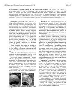
From www.bloodjournal.org by guest on February 6, 2015. For personal use only.
CORRESPONDENCE
2131
Hereditary Spherocytosis Due to a Novel Frameshift Mutation in AE1 Cytoplasmic COOH
Terminal Tail: Band 3 Vesuvio
To the Editor:
Band 3 (anion exchanger 1, AE1) is the most abundant integral
protein of the red blood cell membrane. It is composed of 911 amino
acids encoded by the EPB3 gene. Several mutations in the EPB3 gene
have been found associated to different diseases, eg, hereditary spherocytosis, Southeast Asian ovalocytosis, chorea-acanthocytosis, and familial distal renal tubular acidosis, thus demonstrating that the effects of
EPB3 mutation are dependent on type and location.1,2 The function of
the band 3 carboxyl terminus tail that protrudes on the cytoplasmic side
of the erythrocyte membrane is unknown.3 We describe the first
example of a disease-causing mutation located in the extreme 3Ј end of
the AE1 coding sequence.
Hereditary spherocytosis (HS) is a common hemolytic anemia in
which the spheroidal, osmotically fragile erythrocytes are destroyed in
the spleen. HS due to band 3 deficiency has an autosomal dominant
mode of transmission and is characterized by mild to moderate anemia.1
Six children, belonging to unrelated Italian families from the
Vesuvian area (Southern Italy), presented a homogeneous clinical
picture showing moderate hemolytic anemia and splenomegaly. HS was
diagnosed on the basis of clinical findings, familial history, and routine
hematological investigations. The protein phenotype, performed by
sodium dodecyl sulfate-polyacrylamide gel electrophoresis (SDSPAGE), was typically that of band 3-deficient HS (band 3/spectrin:
Fig 1. Heteroduplex analysis of exon 20 of band 3
genomic DNA. In all 11 HS patients, heteroduplexes
were visible (they were generated on PCR between
normal fragments and fragments carrying the C
deletion). *Band shows heteroduplex produced by
silent polymorphism occurring in trans.
Ϫ21% Ϯ 2% compared with that of the controls).4 In all patients,
single/strand conformation polymorphism (SSCP) screening of band 3
gene showed an alteration in exon 20 (not shown).4 Direct sequencing
of the abnormal fragments constantly showed a deletional frameshift
mutation in codon 894 (ACC = AC; not shown). We designated this
mutation with the area of origin as Vesuvio. Heteroduplex analysis
confirmed this mutation in 5 additional patients in the 6 families. This
analysis failed to find the mutation in 4 healthy members (Fig 1). In 2
brothers of family 3 we also detected a previously described silent
polymorphism (codon 904, TAC = TAT)5 occurring in trans (Fig 1 and
not shown). Analysis of the PstI polymorphic site of the EPB3 gene
indicated that HS was invariably associated with a PstI (ϩ) allele (not
shown). Semiquantitative reverse transcription-polymerase chain reaction (RT-PCR) amplification with primers flanking the deletion (primer
sense in exon 19 and primer antisense in exon 20) and with lower
numbers of cycles produced heteroduplexes, suggesting that mRNA
corresponding to allele Vesuvio was expressed (Fig 2).
Urinary pH and acid excretion were in the normal range in all
patients.
Band 3 Vesuvio is the first mutation located in the cytoplasmic
COOH terminal tail. The singularity of this mutation is the opening of
the reading frame for an extra 133 codons (instead of 18), before the
new stop codon at position 1027. The presence of the corresponding
mRNA suggests that neither the transcription of the mutant gene nor the
From www.bloodjournal.org by guest on February 6, 2015. For personal use only.
2132
CORRESPONDENCE
Fig 2. Mutant band 3 mRNA is detected in patient
II1 of family 1. The presence of the cDNA Vesuvio was
verified by PCR amplification of reverse-transcribed
total reticulocyte RNA. Aliquots of the PCR reaction
after 14, 16, 18, and 20 cycles were screened using
heteroduplex analysis.
stability of the mutant mRNA is affected. Nevertheless, because no
elongated additional band 3 was seen by Western blotting (not shown),
we offer one of the following possibilities to explain the band 3
deficiency: (1) abnormal band 3 insertion in the endoplasmic reticulum;
(2) defective translocation of mutant band 3 from the endoplasmic
reticulum to the plasma membrane; and (3) premature band 3 degradation in the bone marrow erythroblasts. The COOH-terminal 18
amino acids of the normal band 3 are predicted to be replaced
by an abnormal sequence in band 3 Vesuvio. Alignment of the 13
known sequences pertaining to different members of the three anion
exchanger families (AE1, AE2, and AE3) shows that the amino acids in
positions 897, 901, 905, and 906 are highly conserved through
evolution. As reported by Anderson and von Heijne,6 these affected
residues could act as stop transfer signals during cotranslational
insertion of band 3 protein into the endoplasmic reticulum membrane of
erythroid precursor cells. Furthermore, alternatively and/or additionally,
the presence of a much longer COOH terminus could be the hindrance
to the band 3 insertion. Band 3 Vesuvio mutation shows that the
cytoplasmic COOH terminal tail is also very critical for the assembly
and/or maintaining of band 3 in the mature erythrocyte membrane.
Finally, we did not evaluate the unlikely possibility of a mutant band 3
insertion into the plasma with proteolytically truncation to a size similar
to normal band 3.
We found band 3 Vesuvio in 6 unrelated families from the same
geographic area. Because this mutation is located on the same Pst1
allele and does not occur in a hot spot area, the presence of a founder
effect is strongly suspected.
Silverio Perrotta
Raffaella Polito
Maria Luisa Conte
Bruno Nobili
Stefano Cutillo
Emanuele Miraglia del Giudice
Dipartimento di Pediatria
Seconda Universita` degli Studi di Napoli
Napoli, Italy
Vincenzo Nigro
Istituto di Patologia Generali ed Oncologia
Seconda Universita` degli Studi di Napoli
Napoli, Italy
Achille Iolascon
Dipartimento di Biomedicina dell’Eta` Evolutiva
Universita` di Bari
Bari, Italy
Giovanni Amendola
Dipartimento di Pediatria
Ospedale San Leonardo
Castellammare di Stabia
Italy
REFERENCES
1. Gallagher PG, Forget BG, Lux SE: Disorders of the erythrocyte
membrane, in Nathan DG, Orkin SH (eds): Nathan and Oski’s Hematology
of Infancy and Childhood, vol 1. Philadelphia, PA, Saunders, 1998, p 544
2. Bruce LJ, Cope DL, Jones GK, Schofield AE, Burley M, Povey S,
Unwin RJ, Wrong O, Tanner MJA: Familial distal renal tubular acidosis
is associated with mutations in the red cell anion exchanger (Band 3,
AE1) gene. J Clin Invest 100:1693, 1997
3. Lieberman DM, Reithmeier RAF: Localization of the carboxyl
terminus of band 3 to the cytoplasmic side of the erythrocyte membrane
using antibodies raised against a synthetic peptide. J Biol Chem
263:10022, 1988
4. Miraglia del Giudice E, Vallier A, Maillet P, Perrotta S, Cutillo S,
Iolascon A, Tanner MJA, Delaunay J, Alloisio N: Novel band 3 variants
(bands Foggia, Napoli I and Napoli II) associated with hereditary
spherocytosis and band 3 deficiency: Status of the D38A polymorphism
within the EPB3 locus. Br J Haematol 96:70, 1997
5. Eber SW, Gonzales JM, Lux ML, Scarpa AL, Tse WT,
Dornwell M, Herbers J, Kugler W, Ozcan R, Pekrun A, Gallagher PG,
Schroter W, Forget BG, Lux SE: Ankyrin-1 mutations are a major cause of
dominant and recessive hereditary spherocytosis. Nat Genet 13:214, 1996
6. Anderson H, von Heijne G: A 30-residue-long ‘‘export initiation
domain’’ adjacent to the signal sequence is critical for protein translocation across the inner membrane of Escherichia coli. Proc Natl Acad Sci
USA 88:9751, 1991
Splenectomy in Agnogenic Myeloid Metaplasia
To the Editor:
Agnogenic myeloid metaplasia (AMM) is a clonal myeloproliferative
disorder characterized by progressive bone marrow fibrosis and extramedullary hematopoiesis.1 Because of the lack of curative options,
treatment is mostly palliative and is essentially aimed at improving
severe cytopenia and at relieving symptomatic organomegaly.2 In a
substantial number of patients this can only be achieved with splenectomy.3 It
was therefore with the greatest interest that we read the report by Barosi et al4
reporting the association of splenectomy with a higher than expected
incidence of acute leukemic transformation in patients with AMM. Although
the observations and the conclusions reported in the study are interesting, we
believe that alternative explanations should also be considered. This has
important clinical implications, because the hypothesis proposed by Barosi et
From www.bloodjournal.org by guest on February 6, 2015. For personal use only.
1999 93: 2131-2132
Hereditary Spherocytosis Due to a Novel Frameshift Mutation in AE1
Cytoplasmic COOH Terminal Tail: Band 3 Vesuvio
Silverio Perrotta, Raffaella Polito, Maria Luisa Conte, Bruno Nobili, Stefano Cutillo, Emanuele Miraglia del
Giudice, Vincenzo Nigro, Achille Iolascon and Giovanni Amendola
Updated information and services can be found at:
http://www.bloodjournal.org/content/93/6/2131.full.html
Articles on similar topics can be found in the following Blood collections
Information about reproducing this article in parts or in its entirety may be found online at:
http://www.bloodjournal.org/site/misc/rights.xhtml#repub_requests
Information about ordering reprints may be found online at:
http://www.bloodjournal.org/site/misc/rights.xhtml#reprints
Information about subscriptions and ASH membership may be found online at:
http://www.bloodjournal.org/site/subscriptions/index.xhtml
Blood (print ISSN 0006-4971, online ISSN 1528-0020), is published weekly by the American Society of
Hematology, 2021 L St, NW, Suite 900, Washington DC 20036.
Copyright 2011 by The American Society of Hematology; all rights reserved.


© Copyright 2026