
Antibacterial activity of different crude extracts of Dodonaeaviscosa
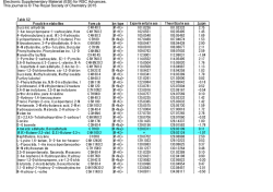
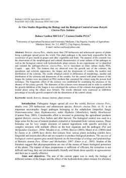
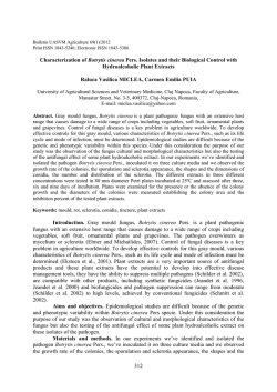
Available online www.jocpr.com
Journal of Chemical and Pharmaceutical Research, 2015, 7(1):350-355
Research Article
ISSN : 0975-7384
CODEN(USA) : JCPRC5
Antibacterial activity of different crude extracts of Dodonaea viscosa
R. Mrutyunjaya Rao1, K. Ramakrishna1 and M. Ashapriya2
1
Department of Chemistry V. S. M. College, Ramachandrapuram, India
Department of Pharmacy, B. V. C. College of Pharmacy, Ramachandrapuram, India
2
_____________________________________________________________________________________________
ABSTRACT
The present study of antimicrobial activities of various crude solvent extracts of Dodonea viscosa were determined
against a wide variety of pathogenic bacteria. Crude extracts of Dodonea viscosa shows mild to significant activities
for most of the treated bacteria. Crude extracts of n-hexane, dichloromethane, ethyl acetate and methanol showed
antibacterial effect against most of the tested organisms. It has been expected that the present work on antimicrobial
screening of the plant materials will help researchers who wish to work in designing clinical drugs concerning the
killer diseases.
Key words: Antibacterial activity, Mueller-Hinton Agar (MHA), Dodonea viscosa
_____________________________________________________________________________________________
INTRUDUCTION
Many organisms can cause several diseases and now, in this world of modern science, man can face any challenge
against any disease. But in spite of the tremendous advancement of medical science and technology, diseases are the
leading health problem particularly in the under privileged population in the remote rural areas in the developing
countries. Nature has been a source of medicinal agents for thousands of years and an impressive number of modern
drugs have been isolated from natural sources, many based on their use in traditional medicine. Various medicinal
plants have been used for years in daily life to treat disease all over the world. They have been used as a source of
medicine. The wide spread use of herbal remedies and health care preparations, such as those described in ancient
texts like Vedas and the Bible, has been traced to the occurrence of natural products with medicinal properties. In
fact, plants produce a diverse range of bioactive molecules, making them a rich source of different types of
medicines. Plants with possible antibacterial activities should be tested against an appropriate microbial model.
The plant Dodonaea viscosa belonging to the family Sapindaceae is distributed as a weed from coast to the elevation
of more than 2000 meters. The weed is distributed in tropical as well as subtropical regions of the world.
Dodonaea viscosa has many medicinal properties and has been used by native peoples from all regions where it is
found. It is a traditional medicine worldwide, administered orally or as poultice to treat a great variety of ailments.
Stem or leaf infusions are used to treat sore throats; root infusions to treat colds. The stems and leaves are used to
treat fever, and seeds (in combination with those of other plants and coated in honey) to treat malaria. The stems are
used as fumigants to treat rheumatism. The leaves are used to relieve itching, fevers swellings, aches and can be
used as a antispasmodic agent leaves and roots as a painkiller to soothe toothaches and headaches and a lotion made
from unspecified plant parts to treat sprains, bruises, burns and wounds.
350
R. Mrutyunjaya Rao et al
J. Chem. Pharm. Res., 2015, 7(1):350-355
______________________________________________________________________________
EXPERIMENTAL SECTION
PLANT MATERIAL:
Pradesh, India.
The plant material Dodonia viscosa was obtained from Maredumelli forest area, Andhra
PREPARATION OF EXTRACT:
The dried powdered leaf was defatted using petroleum ether later the defatted material was subjected to maceration
using distilled water as a solvent. By using Methanol, N-hexane, Di- chloromethane and Ethyl acetate soxhlet
extraction had been performed. Each extract was concentrated at 37° C temperature. Those obtained extracts were
screened to identify the chemical constituents present; they are stored in Desiccator for further uses.
PREPARATION OF THE TESTED ORGANISMS:
The lyophilized forms of different strains of microorganisms like Escherichia coli [MTCC-2126], Staphylococcus
aureus [MTCC-3160], were obtained from the Microbial Type Culture Collection and Gene bank (MTCC),
Chandigarh, India and the bacterial Strains Streptococcus faecalis [NCIM-2603] and Streptococcus pyogens ,
Bacillus subtilis [NCIM-2655] were obtained from National Collection of Industrial Microorganisms (NCIM), Pune
India. The bacterial cultures were maintained on Mueller-Hinton Agar (MHA) and were sub cultured periodically.
The average number of viable organisms per ml organ stock suspension was about 109 colony forming units (CFU)
per ml which was maintained by following McFarland Standardization[6]. Each time fresh stock suspension had been
prepared; constant experimental conditions were maintained to obtain close viable counts.
INOCULATION:
Single loopful of an overnight grown nutrient broth culture of each test organism served as inoculum for the
antimicrobial activity determination. The average size of inoculum was about 1×10⁶ cells contained in 3mm
diameter of standard loop.
DETERMINATION OF MINIMUM INHIBITORY CONCENTRATION [MIC]:
The solution of nutrient agar medium [250 mL] was prepared and sterilized. 25mL of media was dispersed in each
of 5 conical flask, and they were autoclaved after plugged with cotton. Stock solution of Dodonaea viscosa bearing
concentration of 4 mg/ml in Dimethyl sulphoxide [DMSO] was prepared. Each Petri dish was equally filled with
nutrient agar about 20 mL/petri dish. The petri dishes were marked. One sterile nutrient agar plate without extract
but with equal volumes of solvent served as the control plate. All plates were allowed to refrigerate overnight for
uniform diffusion of the extract through the media. Those plates were dried at 37° C. A loop of an overnight grown
peptone water culture of each test organism was placed in petri dish and they were marked. The spot inoculated
plate was also incubated at 37° C, for 24h and the MIC values were obtained [7-8]. The experiment was repeated and
the values were given in table number 01.
DETERMINATION OF ZONE OF INHIBITION:
Agar Well Assay Method: In Agar Well Assay Method 20 ml nutrient agar medium was poured in sterilized Petri
plates (100 X15 mm) and allowed to solidify at room temperature. 24 h broth culture of test bacteria was used as
inoculums under sterile conditions. The freshly prepared 100µl or 0.1ml (1×10⁹ cells/ml) of organisms was set to 0.5
optical density spread with a sterile L shaped. Using cork borer several wells of 6mm in diameter were punched. To
each well 100µl extract three sets of two dilutions (2mg/ml, 4mg/ml) of C. Dodonaea viscosa extracts of methanol,
n-hexane, dichloro methane and ethyl acetate extracts prepared in double distilled water ,were poured into wells.
The Petri dishes were incubated at 37 ºC for 24hrs and the diameter of the zone of inhibition were measured in mm.
Similar procedure was adopted for the pure ciprofloxacin and the corresponding zone diameter were compared
accordingly. The experiment was repeated in triplicate and average values were written in the Table no 01.
RESULTS AND DISCUSSION
Results for the antibacterial activity of Dodonaea viscosa extracts of n-.hexane Di- chloromethane and ethyl acetate
extracts are shown in table given before. MIC and zone of inhibition of both extracts are carried out by using five
bacterial strains the MIC of test compound have been compared with standard drug. From the results zone of
inhibition values all the extracts have their activity on Gram positive bacteria when compared with Gram negative
bacteria. According to the zone of inhibition Bacillus subtilis showed very less sensitivity to the aqueous extracts.
The remaining four bacterial strains effectively inhibited by the ethanolic and aqueous extracts at various
351
R. Mrutyunjaya Rao et al
J. Chem. Pharm. Res., 2015, 7(1):350-355
______________________________________________________________________________
concentration levels. From the above observations various extracts of Dodonaea viscosa can be selected for the
further antibacterial studies against gram positive bacteria and eve pathogenic strains can be studied to know the
potency of these extracts. Fascinatingly E.coli and Bacillus subtilis shown slight resistant to the standard drug
ciprofloxacin. Streptococcus faecalis [NCIM-2603] shows similar sensitivity to all extracts extracts of Dodonaea
viscosa at the same concentration levels.
Table number 01: Determination of MIC of various extracts of Dodonaea viscosa
S.No.
1.
2.
3.
4.
5.
Name of the bacteria
Staphylococcus aureus
Bacilli subtilis
Staphylococcus faecalis
Streptococcus pyogens
E.coli
Conc. of methanolic
2mg/mL
4mg/mL
+
+
+
+
+
+
+
+
-
Conc.of n-hexane
2mg/mL
4mg/mL
+
+
+
+
+
+
+
+
-
Conc. of Dichloro methane
2mg/mL
4mg/mL
+
+
+
+
+
+
+
+
-
Conc.of ethyl acetate
2mg/mL
4mg/mL
+
+
+
+
+
+
+
+
-
Table number 02: Determination of zone of inhibition of methanolic and n-hexane extracts of Dodonaea viscosa
S.No.
1.
2.
3.
4.
5.
Concentration of
Concentration of
Ciprofloxacin
methanolic extract
n-Hexan extract
(µg/mL)
2mg/mL 4mg/mL 2mg/mL 4mg/mL
10
20
30
Staphylococcus aureus
10±0.11 13±0.15 13±1.23
16±1.2
20±0.14 23±0.24 25±0.35
Bacilli subtilis
07±0.34 10±0.16 10±1.25 13±1.43
Staphylococcus faecalis 10±0.26 13±0.24 12±1.26 13±1.24 10±0.41 15±0.25 18±0.34
Streptococcus pyogens
15±0.31 20±0.31 10±1.24 15±1.24 13±0.26 15±0.24 20±0.24
E.coli
1±0.12
1±0.24
1±0.24
1±0.38
09±0.24 11±0.26 13±0.34
*All values are mean of triplicate readings; - Absent; values ± Standard deviation
Name of the bacteria
40
28±0.25
20±0.35
23±0.42
15±0.41
Table number 03: Determination of zone of inhibition of Dichloro methane and ethyl acetate of Dodonaea viscosa
S.No.
Name of the bacteria
1.
2.
3.
4.
5.
Staphylococcus aureus
Bacilli subtilis
Staphylococcus faecalis
Streptococcus pyogens
E.coli
Concentration of
di chloro methane extract
2mg/mL
4mg/mL
10±0.11
13±0.15
09±0.34
11±0.16
11±0.26
13±0.24
16±0.31
22±0.31
1±0.12
1±0.24
Concentration of
ethyl acetate extract
2mg/mL 4mg/mL
14±1.23
18±1.2
10±1.25 13±1.43
13±1.26 14±1.24
10±1.24 15±1.24
1±0.24
1±0.38
10
20±0.14
10±0.41
13±0.26
09±0.24
Ciprofloxacin
(µg/mL)
20
30
23±0.24 25±0.35
15±0.25 18±0.34
15±0.24 20±0.24
11±0.26 13±0.34
Fig. no. 1: Graphical representation for the obtained results of methanolic extract of Dodonaea viscosa
352
40
28±0.25
20±0.35
23±0.42
15±0.41
R. Mrutyunjaya Rao et al
J. Chem. Pharm. Res., 2015, 7(1):350-355
______________________________________________________________________________
Fig. no. 2: Graphical representation for the obtained results of n-hexane extract of Dodonaea viscosa
Fig. no. 3: Graphical representation for the obtained results of dichloro methane extract of Dodonaea viscosa
353
R. Mrutyunjaya Rao et al
J. Chem. Pharm. Res., 2015, 7(1):350-355
______________________________________________________________________________
Fig. no. 4: Graphical representation for the obtained results of Ethyl acetate extract of Dodonaea viscosa
Fig. no. 5: Graphical representation for the obtained results of Ciprofloxacin
CONCLUSION
Plants are the natural sources to promote health, from the present study it can be concluded that the antibacterial
activity of Dodonaea viscosa leaf extracts of methanolic, n-hexane, dichloro methane and ethyl acetate are effective
against various gram positive organisms. And extensive studies are to be carried out to find out which of the
chemical constituent might be responsible for antibacterial activity. Finally Dodonaea viscosa may be useful to
various other diseases also; further investigation need to be done for this.
354
R. Mrutyunjaya Rao et al
J. Chem. Pharm. Res., 2015, 7(1):350-355
______________________________________________________________________________
Acknowledgements
The author Dr. Mrutyunjaya Rao and K. Ramkrishna are grateful to University Grants Commission, New Delhi for
awarding Major Research Project.
REFERENCES
[1] C.K. Atal, K.L. Dhar and J. Singh, Lloydia, 38, 256 (1975).
[2] Cribb AB and CribbJW,Wild medicine in Australia,collins,Sydney, 1981, 228.
[3] Dixon RA (2001). Nature. 411: 843 - 847.
[4] Forbes AB, Sahm FD, Weissfeld SA (2007). Mycology. In: Bailey & Scott’s Diagnostic Microbiology. Mosby,
Elsevier, St. Louis. pp. 696 -697.
[5] Herborn, J.B. 1973. Phytochemical methods, A Guide to Modern Techniques of Plant Analysis, pp. 5-11, 2nd
edition, Hall, New York.
[6] Joshi, S.D., Badiger, A.M., Ashok, K., Veerapur,V.P. and Shastry, C.S. 2003. Indian drugs, 40: 549-552.
[7] Kefale, T., Tsige, G.M., Asres K. and Engidawork, E. 2009. Phytotherapy Res.,13:60-69. doi: 10.1002/ptr.2869.
[8] Kirtikar, K.R., and Basu, B.D., 1993. Indian Medicinal Plants, 2nd ed., Vol. I, Dehradun:International Book
Publisher; p. 641.
[9] M.A. Ali, N.M. Alam, M.S. Yeasmin, A.M. Khan and M.A. Sayeed, Res. J. Agric. Biol. Sci., 3, 852 (2007).
[10] Rojas AS, Cruz H, Ponce-Monter, and Mata R Planta medica. 1996 ,62;154-159.
[11] Subashini, HD, Malarvannan S, Renjith RP. Current Science 2004; 86 (1): 26-28
[12] Suresh, K., Saravana Babu, S., and Harisaranraj,R., 2008. Ethnobotanical Leaflets, 12: 586-590.
355
© Copyright 2026