
Evaluation of in vitro antioxidative, cytotoxic and apoptotic activities
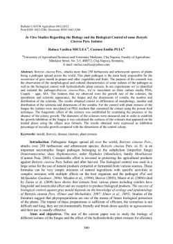
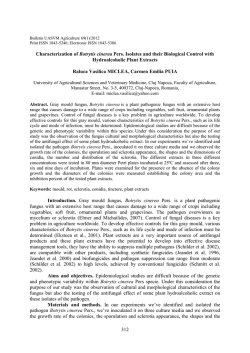
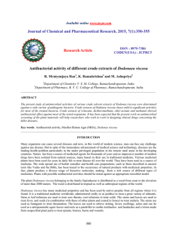
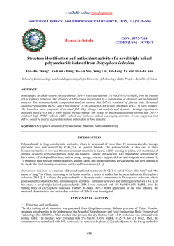
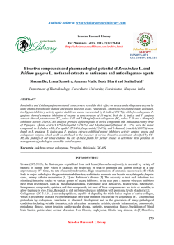
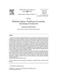
Journal of Plant Sciences
2014; 2(6): 339-346
Published online January 28, 2015 (http://www.sciencepublishinggroup.com/j/jps)
doi: 10.11648/j.jps.20140206.22
ISSN: 2331-0723 (Print); ISSN: 2331-0731 (Online)
Evaluation of in vitro antioxidative, cytotoxic and apoptotic
activities of Rheum ribes ethyl acetate extracts
Pembegul Uyar1, Nursen Coruh2, Mesude İscan3
1
Department of Biology, Selçuk Üniversity, Konya, Turkey
Department of Chemistry, Middle East Technical University, Ankara, Turkey
3
Department of Biology, Middle East Technical University, Ankara, Turkey
2
Email address:
[email protected] (P. Uyar)
To cite this article:
Pembegul Uyar, Nursen Coruh, Mesude İscan. Evaluation of in Vitro Antioxidative, Cytotoxic and Apoptotic Activities of Rheum ribes Ethyl
Acetate Extracts. Journal of Plant Sciences. Vol. 2, No. 6, 2014, pp. 339-346. doi: 10.11648/j.jps.20140206.22
Abstract: Rheum species are medicinally important plants due to the presence of anthracene derivatives. This study was
designed to determine the antioxidative, cytotoxic and apoptotic properties of Rheum ribes shoot and root ethyl acetate extracts
using human promyelocytic leukemia (HL-60) cell line as a model system. R. ribes shoot and root dry powder samples were
prepared and extracted with ethyl acetate. The extracts were revealed to be a potential scavenger of DPPH radicals (IC50 value
of 206.28 µg/ml for shoot and 10.92 µg/ml for root) and the chemical composition of the extracts was quantified by
colorimetric determination of total phenol (GAE) and flavonoid (CAE) contents. HL–60 cells were cultured in the presence of
various concentrations of extracts up to 72 h. R. ribes inhibited the survival of HL-60 cells in a concentration- and timedependent manner, shown by XTT assay. R. ribes caused HL-60 cells apoptosis via formation of phosphatidylserine
externalization, as evidenced by flow cytometry. Exposure of HL-60 cells to higher concentrations of extracts for 72 h resulted
in a shift of 87% of the cell population from normal to the early/late apoptotic stage. These findings suggest that Rheum ribes
ethyl acetate root extracts exhibits potential antioxidant and cytotoxic properties against HL-60 cells better than shoot extracts
and exert their toxicity via induction of apoptosis.
Keywords: Rheum ribes, Antioxidant, HL-60, Cytotoxicity, Apoptosis
1. Introduction
Natural products are of great importance in the drug
discovery process, particularly in the areas of cancer. The
screening with innovative biological assays allows the
verification of their active principle. These improvements
provided the basis for rational drug discovery and have
fundamentally contributed to the development of important
pharmaceuticals of natural origin.
Rheum species has anthracene derivatives found in the
subterranean parts of the plant which makes them
medicinally important. Rheum ribes L. (Polygonaceae) is
used to obtain a component of most important crude drugs in
the Middle East (Kashiwada, 1988). Rheum ribes L.
(Polygonaceae) is grown mostly in Iran, Lebanon and
Eastern Turkey (Shokravi, 1997). Rheum ribes L. is
cultivated in some temperate countries for its edible red leaf
stalks and a perennial. ‘‘Işkın, uşgun or, uçgun’’ are the local
names of R. ribes. (Shokravi, 1997).
R. ribes (young shoots and petioles) is used against
diarrhea and also stomachic and antiemetic as well as against
hemorrhoids, measles, smallpox and cholagogue (Baytop,
1999). Fresh stems and petioles of Rheum ribes are
consumed as vegetable, and used as digestive and appetizer
in Eastern Turkey, while the roots are used to treat diabetes,
hypertension, obesity, ulcer, diarrhea and as antihelmintic and
expectorant (Abu-Irmaileh, 2003; Tabata, 1994). R. ribes
decoction root extracts showed significant blood sugar
lowering activity in alloxan-induced diabetic mice, although
this extract did not show hypoglycemic action in healthy
mice. The antioxidant activity of chloroform and methanol
extract of roots and stems of R. ribes L. were also studied.
(Ozturk, 2007)
Flavonoids, stilbenes and anthraquinones are the major
phenolic constituents of Rheum ribes to provide a potential
source of antioxidants. The pharmaceutically relevant
Journal of Plant Sciences 2014; 2(6): 339-346
compounds in rhubarb are mainly anthraquinone derivatives,
including
emodin,
rhein,
aloe-emodin,
physcion,
chrysophanol and their glucosides. Among these
anthraquinones, emodin has been under intensive
investigations since last decade, and it has been shown that
emodin possesses a number of biological activities. Some
chemicals that occur naturally in plants have begun to receive
much attention as safe antioxidants, as they have been
consumed by people and animals for years (Namiki, 1990).
Therefore, considering the phenolic constituent profile of R.
ribes, they appeared to provide a potential source of
antioxidants, which have well known pharmacological
actions such as free radical scavenging, chemoprevention and
tumor suppression.
Currently, Rheum ribes its toxicity, pharmaceutical
potentials and molecular mechanisms have not been well
investigated. We report here the potential of Rheum ribes
antioxidant, cytotoxic and apoptotic effects against human
promyelotic leukemia (HL-60) cells.
2. Materials and Methods
2.1. Plant Material
Rheum ribes roots and young shoots were collected from
Van, Turkey. The plants were placed on filter papers, and
dried under airflow at room temperature in the dark prior to
use and analysis. Dry plants were powdered by Waring
(model 32BL80) commercial blender at a high speed for at
least 2 min and stored in dark bottles, at room temperature,
25 °C until use.
2.2. Preparation of Plant Extracts
Rheum ribes shoot and root dry powder samples (30 g)
were separately dissolved in ethyl acetate (EtOAc) at a ratio
of 1:12 (w/v). The samples were extracted by sonicating for 1
h in a bath sonicator (Bandelin Sonorex Model RK 100H,
Berlin, Germany) followed by incubation at 50 °C on a
rotational incubator at 150 rpm for 24 h. Then extracts were
filtered through a coarse filter paper on a Büchner funnel and
dried in a rotary evaporator at around 40 °C (Heidolph
Laborota 4000) and lyophilizator (Maxi Dry Lyo HetoHolten, Allerod, Denmark). The freeze-dried extracts were
kept at -20 °C in the dark.
Freeze-dried extracts were named as follows; ethyl acetate
shoot extract (ASE) and root extract (ARE).
2.3. Determination of in vitro Antioxidant Activity, DPPH
Method
DPPH (2,2-Diphenyl-1-picrylhydrazyl) method was
applied as proposed by Blois (1958) for determining the free
radical scavenging activities of extracts and adapted for 96well microplate reader measurements. DPPH is the purplecolored stable free radical that is reduced into the yellow
colored diphenylpicryl hydrazine compound by subtracting
hydrogen from the phenolic compounds found in extracts.
340
Rheum ribes extracts prepared in ethyl acetate were
prepared as serial dilutions, ranging from 1333.33 µg/ml to
2.60 µg/ml. The assay was conducted in triplicate. Aliquots
of 20 µl of plant extract prepared in methanol were plated out,
to which 280 µl of DPPH (1.5x10⁻⁴ M), prepared in methanol
was added to the wells. Plates were then tightly covered with
lid and aluminum foil to prevent evaporation and were
shaken for 2 min, after which it was stored in the dark for 30
min at room temperature. The percentage decolourisation
was measured spectrophotometrically at 517 nm using the
SPECTRAmax 340PC microtiter plate reader. The negative
controls contained 20 µl of plant extract to which 280 µl of
methanol was added, in the absence of DPPH and the
positive controls were prepared using quercetin and emodin,
to which 280 µl of DPPH was added. The percentage
decolourisation was then determined for each of the test
samples (using equation 2.1), as a measure of the DPPH free
radical scavenging activity.
The antioxidant activity is expressed as 50 % effective
concentration (EC50), which is defined as the concentration
(in µg/ml) of extract required to scavenge 50 % of DPPH in
reaction mixture. Percentage DPPH Scavenging Effect was
plotted against the concentration of the sample and the EC50
values were determined.
Equation 2.1
DPPH Scavenging Effect (%) = [[Av controls – (Av
sampleDPPH – Av samplemethanol)] / Av controls] * 100
Where:
Av controls = average absorbance of all DPPH control
wells – average absorbance of all methanol control wells
Av sampleDPPH = average absorbance of sample wells with
DPPH
Av samplemethanol = average absorbance of sample wells
with methanol
2.4. Determination of Total Phenolic Contents
Total phenolic constituents of the extracts were analyzed
by Folin–Ciocalteu method using gallic acid as standard as
described by Singleton, Orthofer & Lamuela-Raventos (1999)
with some modifications.
For the preparation of gallic acid stock solution, 50 mg of
dry gallic acid was dissolved in 1mL of ethanol and diluted to
10 ml with ddH2O. Then the gallic acid stock solution was
diluted with ddH2O at different concentrations, prepared as
serial dilutions, ranging from 500 to 50 µg/ml. The extracts
(3.5 µl) from a 1 mg/ml methanol stock solution or standard
solutions of gallic acid (3.5 µl) or ddH2O as blank were
added to separate test tubes and mixed thoroughly with 276.5
µl ddH2O and 17.5 µl of Folin-Ciocalteu reagent (1N). After
8 min 52.5 µl of 7% Na2CO3 (0.66 M) solution was added,
and mixed thoroughly by pipetting. The final concentration
of the extracts in each well was 10 µg/ml. The solutions were
incubated at room temperature for 2 h and the absorbance
versus blank (0 µg/ml gallic acid) was read at 765 nm using
the SPECTRAmax 340PC microtiter plate reader.
The total phenol content of the extracts was determined by
comparing with a calibration curve of the gallic acid standard
341
Pembegul Uyar et al.: Evaluation of in Vitro Antioxidative, Cytotoxic and Apoptotic Activities of Rheum ribes
Ethyl Acetate Extracts
and represented as mg gallic acid equivalents (GAE) /g of
dried samples according to the equation obtained from the
standard gallic acid graph.
2.5. Determination of Total Flavonoid Contents
The total flavonoid content in the extracts was determined
by aluminium colorimetric assay (Zhishen, Mengcheng &
Jianming, 1999) with some modifications.
A standard solution of (+) catechin at different
concentrations (20, 40, 60, 80 and 100 µg/ml) was prepared
by dissolving (+) catechin in ddH2O. The extracts (35 µl)
from a 1 mg/ml stock solution or standard (+) catechin
solutions (35 µl) or ddH2O (as blank) were mixed thoroughly
by pipetting with 140 µl ddH2O in a 96 well plate. Then 10.5
µl of 5% NaNO2 was added. The mixture was incubated for 5
min at 25 ˚C and 10.5 µl of 10 % AlCl3 was added to and
then 6 min later, 70 µl 1 M NaOH was added to the wells.
The total volume was made up to 350 µl by adding ddH2O.
The absorbance was read at 490 nm using the SPECTRAmax
340PC microtiter plate reader. ddH2O was used as a blank.
The total flavonoid content was expressed as mg of
catechin equivalents (CE)/g of dried samples according to the
equation obtained from the standard catechin graph.
2.6. Cell Line and Culture Conditions
HL-60 cells, a human promyeloblastic leukemia cell line,
were grown in RPMI-1640 supplemented with 10% heatinactivated fetal bovine serum (FBS), 2mM l- glutamine and
0.2 % (50 mg/ml) gentamicine in the vessels appropriate for
the designed experiment. The cells were incubated at 37 0C
with 95 % air and 5% CO2 in a Hepa filtered Heraeus Hera
Cell 150 incubator. They were manipulated in a HERAsafe
Class II Biological Safety (laminar flow) cabinet by using
appropriate cell culture techniques. The medium was
collected and refreshed every 2-3 days simulating usual cell
culture conditions to maintain a constant cell to growth
medium ratio.
2.7. Measuring Viability of Cells using XTT Assay
The effects of the Rheum ribes shoots and roots ethyl
acetate extracts on the cytotoxicity of HL-60 cell line were
evaluated by means of the Cell Proliferation Kit (Biological
Industries, Israel) in 96 well flat bottomed microtiter plates.
Aliquots of 50 µl of the 2x10³ cells/ml cell suspension
were seeded into the 96-well plate. The plates were then
incubated at 37 °C in 5 % CO2 for 12 hours to achieve the
cell maintenance. No cells were seeded into the blank wells
(instead, 100 µl of experimental media was added). Plant
extracts (50 µl) were added in triplicate to the wells already
containing 50 µl of cell suspension. The controls comprised
of: (i) DMSO in experimental media, <0.1 % (negative
control), (ii) plant extract with experimental media in the
absence of HL-60 cells (color control), (iii) experimental
media in absence of both plant extract and HL-60 cells (blank
control). Control wells aided in the determination of any
background extract absorbance, especially in the event of
colour interferences or interaction of the extract with the
XTT solution. Each plate contained four wells for the blank
cell-free control. At the end of the incubation time, 100 µL of
phenazine metho-sulfate is added to 5 ml of XTT reagent,
and 50 µl of this XTT solution was added to each well before
being incubated for a further 5 hours. Then, XTT reagent was
applied to form a soluble dye. After incubation at 37 ºC for 5
h, the dissolution of formazan crystals that were produced by
mitochondrial enzymes of the living cells occurred, the
optical density of chromogenic product was measured at 415
nm with a Spectromax 340 96-well plate reader (Molecular
Devices, Sunnyvale, CA, USA). The results were expressed
in terms of percentage cellular viability, calculated using
equation 2.2, taking the relevant controls into account. The
IC50 value for each sample was determined from a log
sigmoid dose response curve generated. The assay was
performed in triplicate.
% Cell Viability =
Where;
Abss (withcell)
extracts
Abss (cell free)
Abs s ( with cell ) − Abs s (cell free )
Abs c ( with cell ) − Abs c (cell free )
x100
= Average absorbance of cells treated with
= Average absorbance of extracts in cell
free medium
Abs c ( with cell ) = Average absorbance of untreated cells,
control
Absc(cell free) = Average absorbance of cell free medium
Abs = Absorbance at 415 nm
2.8. Cell Observation using an Inverted Microscope
Cell shrinkage, membrane blebbing and the formation of
apoptotic bodies are characteristic events during apoptosis,
which can be easily detected by light microscopy. HL-60
cells (2 x 10⁵ cells/ml; 1 ml/well; 24-well plate) were either
left untreated or stimulated with 100 µg/ml ASE and ARE,
for 72 h. Cells were viewed at a 400-fold magnification with
an inverted microscope. (Olympus CKX 41).
2.9. Flow Cytometry Analysis
The two modes of cell death, apoptosis and accidental cell
death (necrosis), differ fundamentally in their morphology,
biochemistry and biological relevance. In this part, to
characterize and differentiate between two different
mechanisms of cell death, apoptosis and necrosis flow
cytometry was used.
Detection of apoptosis using Annexin V after Rheum ribes
treatment was performed using an APC- labeled
Recombinant Human Annexin V and 7AAD antibody.
Annexin V-Apc (λ ex: 633, λ em: 660 nm) is detected in FL4
on dual laser instruments and 7AAD (λ ex: 488, λ em: 647 nm)
is detected in FL3 channel on most instruments.
All measurements were performed on a FACScalibur (BD
Journal of Plant Sciences 2014; 2(6): 339-346
concentration dependent manner up to a certain extract
concentration, and then reached a plateau.
The DPPH radical scavenging capacity which was
expressed as EC50 was obtained in ARE with 10.92±0.21
µg/ml and 96.67±0.98 is higher than obtained in ASE with
206.28±10.21 µg/ml EC50 value, which was also exhibited
maximum 80.34±0.98 % scavenging potential as shown in
the Figure 3.1 and Table 3.1. This difference obtained
between the shoot and root residue with ethyl acetate
extraction could be due to presence of less phenolic
constitutes in the ASE than ARE.
% Radical Scavenging Activity
Biosciences, Palo Alto, CA, USA), equipped with a 488 nm
argon-ion laser, using CellQuestTM software. Analysis was
investigated using FlowJo software (Treestar, Ashland, OR,
USA).
HL-60 cells (2.5 x 10⁵ cells/ml; 1 ml; 24-well plate) were
either left untreated or treated with a concentration range of
Rheum ribes freeze-dried extracts for 24, 48 and 72 h. Cells
were harvested by centrifugation (500 x g, 5 min, RT),
washed with cold PBS, resuspended in a volume of 500 µl
cold PBS and incubated with 1X Annexin V-apc (50x)
solution and 1X 7AAD (100x) solution for 15 min at room
temperature, 25 °C in dark room. After centrifugation (500 x
g, 5 min, RT) the cells was washed twice with 1000 µl PBS
(Biochrom L 1825) and then obtained cell pellet was
resuspended with 500 µl PBS. The samples were
immediately analyzed by flow cytometry.
It was important to have separate and distinctive
populations. In these populations; (i) cells that stained
positive for Annexin V-apc and negative for 7-AAD were
undergoing apoptosis, (ii) cells that stain positive for both
Annexin V-apc and 7-AAD were either in the end stage of
apoptosis which were undergoing necrosis, or were already
dead, (iii) cells that stain negative for both Annexin V-apc
and 7-AAD were alive and not undergoing measurable
apoptosis.
342
100
75
50
ASE
ARE
Quercetin
Emodin
25
0
0
500
1000
1500
Extract Concentration (µg/ml)
2.10. Statistical Analysis
All experiments were performed in triplicate unless
otherwise noted; results are expressed as mean ± standard
deviation. Data analysis and graphing was performed using
the GraphPad Prism version 5 (GraphPad Software,
San Diego, California, USA). For all the measurements,
oneway ANOVA followed by Tukey's Multiple Comparison
Test was used to assess the statistical significance of
difference between control and Rheum ribes extract-treated
groups in vitro. A statistically significant difference was
considered to be at p < 0.05.
3. Results and Discussion
3.1. Determination of Antioxidant Capacities of Rheum
ribes
Relatively stable radicals such as DPPH are often preferred
in the assessment of radical scavenging activity. This radical
has been widely used in various studies of plant extracts and
foods (Koleva 2002; Lee, 2003). The popularity of using the
DPPH free radical method for estimating free radical
scavenging activity may be due to its simple, rapid and
economic properties. In our study, DPPH radical scavenging
activity of extracts prepared by ethyl acetate extraction was
monitored at 517 nm for 30 minutes.
The Radical Scavenging Activity (RSA) of each extract
was calculated and used for determination of EC50 values,
which indicated the amount of the extract scavenging 50 %
of DPPH radical. As shown in Figure 3.1 the antioxidant
capacities (% RSA) in both samples increased in a
Figure 3.1. Percent DPPH scavenging activities (% RSA) of Rheum ribes
shoot and root extracts prepared in ethyl acetate (EtOAc) and the reference
materials. Each point is the mean of quadruple measurements from three
different sets of experiments (n=3) (* p<0.05 compared to and analyzed by
one way ANOVA).
As compared to result of the present study, Ozturk et al.,
2007 observed higher antioxidant activity with methanol
extract of Rheum ribes shoots, however they obtained lower
antioxidant activity with methanol root, chloroform shoot and
root extracts. This could be due to the difference at applied
time and temperature during extraction (Ozturk, 2007).
DPPH radical scavenging capacities of reference materials
such as quercetin and emodin were also determined since
they were quite often used in the literature as phenolic
standards, and displayed in the Figure 3.1. Besides, emodin
was one of the anthraquinones found in Rheum ribes (Tosun,
2003). ARE showed greater antioxidant activity than emodin
and almost the same activity to that of quercetin.
3.2. Determination of Total Phenol and Total Flavonoids
Total phenolic and flavonoid contents of Rheum ribes
extracts were determined by the method of Singleton and
Rossi (1965), the Folin-Ciocalteu reagent assay (FC) and the
aluminium colorimetric assay (Whiten, 1999), respectively.
Total phenolic compounds were determined as mg
equivalents of gallic acid per g of crude extracts and total
flavonoid compounds were determined as mg equivalents of
catechin per g of crude extracts given in the Table 3.1
Ethyl acetate extraction of root presented the higher total
phenolic and flavonoid contents than shoot.
343
Pembegul Uyar et al.: Evaluation of in Vitro Antioxidative, Cytotoxic and Apoptotic Activities of Rheum ribes
Ethyl Acetate Extracts
Table 3.1. Summary of antioxidant activity, contents of Total phenol and flavonoid in the ethyl acetate extracts of Rheum ribes GAE: Gallic Acid Equivalents
CAE: Catechin Equivalents.
Samples
Antioxidant Activity
(EC50 µg/ml± SD℘)
Maximum % Radical Scavenging
Activity (%RSA± SD℘)
Total Phenol (mg GAE/g dried
extract± SD℘)
Total Flavonoid (mg CAE/g
dried extract± SD℘)
ASE
206.28±10.21
80.34±0.98
21.11±1.11
2.29±1.04
ARE
10.92±0.21
96.67±0.98
207.22±6.96.72
50.49±2.03
Emodin
164.30±23.13
85.98±1.47
NA
NA
Quercetin
4.49±2.54
95.39±1.96
NA
NA
TP GAE : Total phenolic contents mg equivalents of gallic acid/g of plant extract, TF CAE : Total flavonid contents mg equivalents of catechin/g of plant
extract, ND: not detected, NA: not applicable,ϒSD : was derived from three independent experiments.
As known, there is a significant linear correlation between
phenolic concentration and free radical scavenging activity
and flavonoids are natural phenolic compounds and well
known antioxidants. In this study, antioxidant activity of root
extracts extracted in ethyl acetate was found to be fairly
higher which are also rich in flavonoids.
From the chemical analysis of Rheum ribes extracts; ASE
was the lower effective extract. Shoot extract with lower
polarity had lower antioxidative activity with lower phenolic
flavonoid content.
3.3. Cytotoxicity of Rheum ribes Shoot and Root Extracts in
HL-60 Cells
3.3.1. Light Microscopic Analysis of Cell Morphology
For the investigation of cytotoxic effects of Rheum ribes
extracts, morphological criteria were combined with
commonly accepted biochemical methods. The effects of
Rheum ribes on morphological changes and viability in HL60 cells treated for 48h were analyzed under light inverted
microscope (magnification, 400X) as shown in Figure 3.2.
HL-60 cells (2 x 10⁵ cells/ml; 1 ml/well; 24-well plate) were
either left untreated or treated with 100 µg/ml ASE and ARE
for 48 h.
Figure 3.2. Light Microscopic Analysis of effects of Rheum ribes Extracts on
morphological changes in HL-60 cells for 48 h. (a) Un-treated cell; (b)
DMSO % 0.1, 100 µg/ml of (c) ASE and (d) ARE; Arrow (d) indicates a
typical apoptotic cell with apoptotic body. The cells were photographed
under inverted light microscopy (magnification, 400X).
The HL-60 cells grow in single cell suspension without
any tendency to clump or to adhere to plastic or glass (Fig
3.2 a) with a round or ovoid shape, and occasional cells have
blunt pseudo pods. After incubation with ASE and ARE
alterations and cell growth inhibition in HL-60 cells were
illustrated comparing with control and DMSO treated cells
(Fig 3.2 a and b). Control or DMSO treated cells were round
in normal shape. Exposure of HL-60 cells to those ARE for
48 h led to the membrane blebbing and apoptotic body
formation (Fig 3.2 d ) which were observed by light inverted
microscope (400X).
At 100 µg/ml concentration, cell growth ratio was
decreased significantly higher by ARE than ASE for 48 hin
microscopic analysis.
3.3.2. XTT Assay
Figure 3.3. Effects of Rheum ribes shoot (A) and root (B) ethyl acetate
extract on cell survival in HL-60 cells. Cytotoxicity was measured by the
XTT assay.. The percentage of cell growth in the control group was
designated as 100%.
Journal of Plant Sciences 2014; 2(6): 339-346
Metabolically active HL-60 cells were detected using XTT
assay upon treatment with Rheum ribes extracts for 24, 48
and 72 hours. Mitochondrial enzymes of the metabolically
active cells reduced tetrazolium salt; XTT to orange colored
formazan product was measured using ELISA plate reader.
Cell viability measurements obtained from XTT assay were
converted to percent cell viability by setting control (0.1 %
DMSO) results as 100 % cell viable. HL-60 cells were
precultured in 96-well microplates for 12 h and then
incubated with 0- 300 µg/ml of Rheum ribes shoot and root
ethyl acetate extract for 24, 48 and 72 h. % cell viability for
different time points (0, 24, 48 and 72 h) versus different
Rheum ribes extracts concentrations 3D column graph were
constructed (Figure 3.3). The IC50 values were determined
344
using GraphPad Prism version 5 for Mac OS X as shown in
Table 3.2.
IC50 values that were the Rheum ribes extracts
concentrations at which 50% of cells are viable were
calculated as 288 µg/ml, 252 µg/ml and 192 µg/ml after ASE
treatment for 24, 48 and 72 h, respectively. The results were
not statistically significant for all time points. ASE had
growth inhibitory effects; however ASE cytotoxicity was not
occurred at a time dependent manner in HL-60 cells.
IC50 values were calculated as 149 µg/ml, 135 µg/ml and
74 µg/ml after ARE treatment for 24, 48 and 72 h,
respectively. The results were not statistically significant for
24 and 48 h, however for 72 h cells viability dropped
dramatically.
Table 3.2. Comparison of IC50 values in HL-60 cells obtained by XTT Assay after treatment of Rheum ribes for 24, 48 and 72 h.
Incubation time (h)
Parameter
24
Extracts
48
72
IC50 (µg/ml)
% viability*
IC50 (µg/ml)
% viability*
IC50 (µg/ml)
% viability*
ASE
288
53
252
42
192
34
ARE
149
<1
135
<1
74
<1
*% viability was calculated considering the viable cell count at highest extract concentration (500 µg/mL) compared to control viable cell count at every
incubation time point NA: not applicable
These results showed that the difference between the IC50
values of ASE and ARE in HL-60 cells was statistically
significant, p<0.05 and the growth inhibitory effect of ARE
was higher than ASE in HL-60 cells for all time points,
because the amount of extract concentration required to
inhibit 50 % metabolic activity of the cells were significantly
low.
According to the criteria established by the U.S. National
Cancer Institute (NCI), the compounds with IC50 < 30 µg/ml,
30 µg/ml <IC50 < 100 µg/ml and IC50 > 100 µg/ml are judged
as active, moderately active and inactive, respectively
(Suffness, 1990). ARE exhibited the highest activity and
showed moderate cytotoxicity with IC50 (calculated from
TBA assay)< 100 µg/ml that fall within the NCI criteria. ASE
shows some cytotoxicity, however those were judged as
inactive according to NCI criterion for 72 h.
Biological activity studies were carried out using HL-60
cells. Rheum ribes extract prepared in ethyl acetate was
applied at different concentrations for 24, 48 and 72 h to the
feeding environment of the cells. As a conclusion, ARE had
the highest potential with lower IC50 for all time points from
the obtained results of XTT assay.
3.4. Detection of Phosphatidylserine Translocated to the
Cell Surface
Rheum ribes treatment led to the exposure of
phosphatidylserine (PS) on the outside of the plasma
membrane, a characteristic event in early stages of apoptosis,
detected by Annexin V-apc staining and FACS analysis . Costaining with 7AAD allowed discrimination between
apoptotic and necrotic cells.
Cells were treated with ASE and ARE at concentrations of
100 and 250 µg/ml for 72 h. After treatment, cells were
stained with annexin V/7AAD and analyzed by flow
cytometry. Cells treated with 0.1 % DMSO was used as
control.
345
Pembegul Uyar et al.: Evaluation of in Vitro Antioxidative, Cytotoxic and Apoptotic Activities of Rheum ribes
Ethyl Acetate Extracts
Figure 3.4. Cell apoptosis, percentage of HL-60 cells after treatment with ASE and ARE by flow cytometric analysis for 72 h.
These results demonstrated that there was a dosedependent fashion in annexin V/7ADD double positive cells
induced by ASE and ARE as shown in Figure 3.4 and Table
3.3. At 72 h, the observed maximal apoptosis was about 42%
for ASE and 81% for ARE at the concentration of 250 µg/ml.
Apoptosis in response to ARE was early, whereas apoptosis
in response to ASE was significantly late.
In apoptosis induced by ASE for 72 h, PS externalization
occurred as the same time as disruption of membrane
integrity. This was a result of presence of early and late
apoptotic components in the ASE.
The apoptosis induced by ARE of 100 µg/ml for 72 h was
dominantly PS externalization, however by ARE of 250
µg/ml, dominant cell death was late apoptosis or necrosis
which was a result of disruption of membrane integrity. This
result showed that the components found in ARE were
generally shown characteristics of early apoptotic agents.
There are other reports indicating the effects of Rheum
species on apoptosis in several cell lines. In human cervical
Bu 25 TK cancer cells and human lung squamous carcinoma
CH 27 cells, emodin induced apoptosis through
mitochondrial activation of caspase 3 and 9 and with Bax
upregulation (Srinivas et al., 2003; Lee, 2001).
to catch on to the cutting-edge technologies.
4. Conclusions
Considering all the results obtained in the present study,
Rheum ribes could be considered as a dietary antioxidant and
anticancer agent. Our investigations for the pursuit of these
interactions may not result in hundred percent prevention or
in total cure of leukemia, but this was a rational way forward,
Acknowledgements
This study was supported by the Research Fund of METU
ÖYP Grant No: BAP-08-11-DPT-2002K120510. The authors
are also grateful to botanist Assoc. Prof. Dr. Fevzi Özgökçe
for collecting plant samples and his collaboration.
References
[1]
Abu-Irmaileh, B. E., & Afifi, F. U. (2003). Herbal medicine in
Jordan with special emphasis on commonly used herbs.
Journal of Ethnopharmacology, 89, 193–197.
[2]
Awika, J. M., Rooney, L. W., & Waniska, R. D. (2005).
Anthocyanins from black sorghum and their antioxidant
properties. Food Chemistry, 90, 293–301.
[3]
Baydar, N. G., Ozkan, G., & Sagdic, O. (2004). Total phenolic
contents and antibacterial activities of grape (Vitis vinifera L.)
extracts. Food Chemistry, 15, 335–339.
[4]
Baytop, T. (1999). Rheum ribes L. In T. Baytop (Ed.). Therapy
with medicinal plants in Turkey, Nobel Tıp Publication Press,
1, pp. 319–320).
[5]
Blois, M.S., (1958). Antioxidant determinations by use of a
stable free radical. Nature, 181, 1199-1200 .
[6]
Cai, Y., Sun, M., Xing, J., & Corke, H. (2004). Antioxidant
phenolic constituents in roots of Rheum officinale and Rubia
cordifolia: structure-radical scavenging activity relationships.
Journal of Agricultural Food Chemistry, 52, 7884–7890.
Journal of Plant Sciences 2014; 2(6): 339-346
[7]
Castañeda-Ovando, A., de Lourdes Pacheco-Hernández, Ma,
Elena, Ma, Páez- Hernández, J. A., RodríguezGalán-Vidal, J.
A., & Galán-Vidal, C. A. (2009). Chemical studies of
anthocyanins: A review. Food Chemistry, 113, 859–871.
[8]
Collins, S.J., (1987). “The HL-60 promyelocytic leukemia cell
line: proliferation, differentiation, and cellular oncogene
expression”, Blood, Vol 70, 1233-1244.
[9]
Cullen, J. (1966). Rheum L. In PH. Davis (Ed.). Flora of
Turkey and the East Aegean Islands. Edinburgh: Edinburgh
University Press, 2, 268–269.
[10] Enayat, S,. Banerjee., S., 2009. Comparative antioxidant
activity of extracts from leaves, bark and catkins of Salix
aegyptiaca sp. Food Chemistry, Vol 116, 23-28
[11] Fadok, V. A., Voelker, D.R., Campbell, P.A., Cohen, J.J.,
Bratton, D.L., Henson, P.M., (1992). “Exposure of
phosphatidylserine on the surface of apoptotic lymphocytes
triggers specific recognition and removal by macrophages”,
Journal of Immunology, 148, 2207-2216.
346
[19] Mericli, A. H., & Tuzlacı, E. (1990). Constituents of Rheum
ribes. Fitoterapia, 61, 375.
[20] Özbek, H., Ceylan, E., Kara, M., Özgökce, F., & Koyuncu, M.
(2004). Hypoglycemic effect of Rheum ribes roots in alloxan
induced diabetic and normal mice. Scandinavian Journal of
Laboratory Animal Science, 31, 113–115.
[21] Ozgokce F, Ozcelik H (2004). Ethnobotanical aspects of some
taxa in East Anatolia (Turkey), Econ. Bot. 58: 697-704.
[22] Ozturk M., F. Aydogmus-Ozturk, M.E. Duru and G. Topcu,
Antioxidant activity of stem and root extracts of Rhubarb
(Rheum ribes) An edible medicinal plant, Food Chem 103
(2007), 623–630
[23] Shokravi, A., & Agha Nasiri, K. (1997). Synthesis of
1,2,3,4,5,6,7,8Octahydro-9-ethoxy-10-hydroxy-1anthracenone (OEHA). Iranian Journal of Chemistry and
Chemical Engineering, 16, 10–15.
[12] Freshney, R. I., (2005). “Culture of Animal Cell: A manual of
Basic Technique”, (5th Edition).
[24] Singleton, R. Orthofer and R.M. Lamuela-Raventos, Analysis
of total phenols and other oxidation substrates and
antioxidants by means of Folin–Ciocalteu reagent, Oxidants
and Antioxidants
[13] Gulcin, I., Oktay, M., Kirecci, E., & Kurevioglu, O. _I. (2003).
Screening of antioxidant and antimicrobial activities of anise
(Pimpinella anisum L.) seed extracts. Food Chemistry, 83,
371–382.
[25] Tabata, M., Sezik, E., Honda, G., Yesilada, E., Fuki, H., Goto,
K., et al. (1994). Traditional medicine in Turkey III. Folk
Medicine in East Anatolica, Van and Bitlis provinces.
International Journal of Pharmacognosy, 32, 3–12.
[14] Hollman, P. C. H., & Katan, M. B. (1999). Dietary flavonoids:
Intake, health effects and bioavailability. Food and Chemical
Toxicology, 37, 937–942.
[26] Tosun, F., & Akyuz-Kızılay, C. (2003). Anthraquinones and
flavonoids from Rheum ribes. Journal of Faculty of Pharmacy
Ankara, 32, 31–35.
[15] Kashiwada, Y., Nonaka, G., Nishioka, I., & Yamagishi, T.
(1988). Galloyl and hydroxycinnamoyl glucoses from
Rhubarb. Phytochemistry, 27, 1473–1477.
[27] Tsao, R., & Yang, R. (2003). Optimization of a new mobile
phase to know the complex and real polyphenolic composition:
Towards a total phenolic index using high-performance liquid
chromatography. Journal of Chromatography A, 1018, 29–40.
[16] Lapornik, B., Prosek, M., & Golc, Wondra. A. (2005).
Comparison of extracts prepared from plant by-products us
ing different solvents and extraction time. Journal of Food
Engineering, 71, 214–222.
[17] Li, P., Nijhawan, D., Budihardjo, I., Srinivasula, S.M., Ahmad,
M., Alnemri, E.S. and Wang, X., 1997. “Cytochrome c and
dATP-dependent formation of Apaf-1/caspase-9 complex
initiates an apoptotic protease cascade”, Cell, Vol 91, 479–489.
[18] Liu, Q., Cai, W., & Shao, X. (2008). Determination of seven
polyphenols in water by high performance liquid
chromatography combined with preconcentration. Talanta, 77,
679–683.
[28] Tuzlaci, E., & Mericli, A. H. (1992). Some studies on Is kın
(Rheum ribes) and its distribution in Turkey. Proceedings of
the 9th symposium on plant drugs (Pub No. 641, pp. 336–341).
Eskis ehir, Turkey: Anatolia University Press.
[29] Yesilada E, Sezik E, Honda G, Takaishi Y, Takeda Y, Tanaka T
(1999). Traditional medicine in Turkey IX. Folk medicine in
north-west Anatolia. J. Ethnopharmacol. 64: 195-210.
[30] Zhishen, T. Mengcheng and W. Jianming, The determination
of flavonoid contents in mulberry and their scavenging effects
on superoxide radicals, Food Chemistry 64 (4) (1999), 555–
559.
© Copyright 2026