
The “Normandy” Variant of von Willebrand Disease
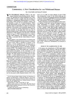
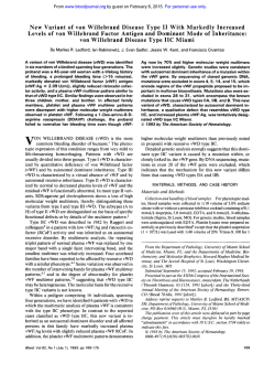
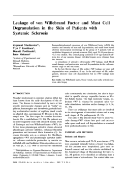
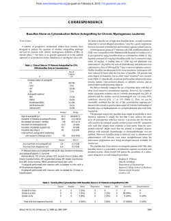
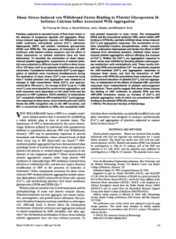
From www.bloodjournal.org by guest on February 6, 2015. For personal use only.
The “Normandy” Variant of von Willebrand Disease: Characterization of a Point
Mutation in the von Willebrand Factor Gene
By C. Gaucher, S. Jorieux, B. Mercier, D. Oufkir, and C. Mazurier
We previously reported a functional defect of von Willebrand
factor (vWF) in a new variant of von Willebrand disease
(vWD) tentatively named vWD ”Normandy.” The present
work has attempted to characterize the molecular abnormality of this vWF that fails to bind factor Vlll (FVIII). The
immunopurifiedvWF from normal and patient’s plasma were
digested by trypsin and the resulting peptides were compared. The electrophoresis of “vWF Normandy” showed a
shift in the band corresponding to a polypeptidefrom amino
acid 1 to 272. Consequently, we performed the molecular
analysis of the portion of the vWF gene of this patient
encoding this amino acid sequence. Exons 18-24 were amplified by the use of polymerase chain reaction and their
nucleotide sequences corresponding to 1.8 kb were deter-
mined. Our analysis showed a point mutation C to T at codon
791, resulting in the substitutionof Methionine for Threonine
at position 28 of the mature vWF subunit. Because this
nucleotide substitution destroyed a Mae II restriction site,
this mutation was conveniently sought in various individual
DNAs. The patterns obtained were consistent with the
homozygous and heterozygous state of this mutation in the
patient and in her son, respectively, and with its absence in
28 normal individuals. We conclude that Threonine at position 28 in plasma vWF may be crucial for the conformation
and FVIII-binding capacity of its cystine-rich N-terminal domain.
o 1991 by The American Society of Hematology.
V
patient with FVIII deficiency but normal primary hemostasis. Her plasma vWF, which did not present any quantitative or multimerization abnormality, was shown to be
unable to bind FVIII. This report details the study we have
performed to characterize the underlying molecular pathology of this patient.
ON WILLEBRAND FACTOR (vWF) is a large multimeric glycoprotein found in plasma and in platelets
that is synthetized by endothelial cells and megakaryocytes.
Besides its major role as a mediator of initial platelet
adhesion to vascular subendothelium, vWF is also the
The association
carrier of factor VI11 (FVIII) in plasma.132
of vWF with FVIII has been shown in vitro to stabilize the
coagulant activity of both human3and recombinant435FVIII.
Furthermore, clinical observations in patients confirm the
importance of normal vWF in prolonging FVIII
The human vWF gene has been studied in some detail
and is located on chromosome 12.8,9It is = 178 kb in length
and contains 52 exons, the intron boundaries of which were
recently determined, = 19% of the gene being sequenced.”
The 9-kb vWF mRNA encodes a 2,813-amino acid (AA)
precursor consisting of a 22-AA signal peptide, a 741-AA
propeptide, and a 2,050-AA mature subunit. Functional
domains involved in binding to platelet membrane glycoproteins, collagen, and heparin have been localized on the vWF
subunit.“ More recently, a major FVIII-binding domain of
vWF was characterized on the NHz-terminal region of
mature vWF. FVIII binds to Sp fragment 111‘ (AA 1-910),
but not to Sp fragments I (AA 911-1,365) or I1 (AA
1,366-2,050),obtained by digestion of vWF with Staphylococcus aureus V8 p r ~ t e a s e . ’ ~Furthermore,
-’~
this function is
maintained on a tryptic fragment (SpIII-T4) containing the
amino-terminal 272 AA of vWF.’~
von Willebrand disease (vWD), the most common inherited bleeding disorder, is heterogenous and originates from
either quantitative (vWD types I11 and I) or qualitative
(vWD type 11) alterations of vWF.” Many phenotypic
subtypes of vWD have been distinguished, generally according to the multimeric profile of plasma and platelet vWF.I6
Depending on the disease type or subtype, the inheritance
has been found either dominant (type I, subtypes IIA and
IIB) or recessive (type 111 and subtype IIC). The cause of
vWD at the level of gene structure is known in only a few
families. Total or partial gene deletions for vWD type 11117-19
as well as single point mutations for vWD types IIAz0,21
and
IIBZ2have been reported.
In an earlier article,z3we described a new variant form of
vWD, tentatively named vWD “Normandy,” in a female
Blood, Vol77, No 9 (May 1). 1991: pp 1937-1941
PATIENT AND METHODS
Case report. For a complete data report refer to our earlier
p ~ b l i c a t i o nA
. ~50-year-old French woman with a lifelong history
of bleeding was referred to us in August 1988. Investigations
confirmed FVIII deficiency (5 to 8 IU/dL) but showed normal
levels of vWF antigen and ristocetin cofactor activity and normal
vWF multimeric pattern. FVIII-binding assays showed that the
patient’s vWF, in contrast to normal vWF, was unable to bind
FVIII. Her two children, a son and a daughter, as well as her
parents, who were third cousins, were said to have no bleeding
history. However, plasma from the patient’s son showed both
moderate FVIII deficiency and FVIII-binding values intermediate
between the normal values and the patient’s values (data not
shown).
Antibodies. Monoclonal antibody (MoAb)-239 to human vWF,
prepared in collaboration with Immunotech (Marseilles, France),
recognizes all the multimeric forms of vWF.‘~MoAb-418 was a
generous gift of D. Meyer (INSERM U143, Paris, France); it
specifically inhibits the binding of FVIII to vWF and reacts with the
unreduced N-terminal tryptic (AA 1-272) or plasmic (AA 1-298)
fragment of the vWF subunit.’* Its epitope was further localized to
the first 106 AA of the mature vWF subunit.25Two other MoAbs,
obtained by immunization of Balb/c mice with SpIII-T4 fragment,”
were also used. MoAb-175-35A8 inhibits the FVIII binding tovWF
and recognizes both reduced and unreduced SpIII-T4 fragment;
From the Laboratoire de Recherche sur I’HCmostase, Centre RCgional de Transfusion Sanguine, Lille, France.
Submitted October 25, 1990; accepted December 20, 1990.
Address reprint requests to Claudine Mazurier, PhD, C.R.T.S., 21
rue C. Gudrin, 59012 Lille C C d q France.
The publication costs of this article were defrayed in part by page
charge payment. This article must therefore be hereby marked
“advertisement” in accordance with 18 U.S.C.section 1734 solely to
indicate this fact.
0 I991 by The American Society of Hematology.
0006-497I/91/7709-0005$3.00/0
1937
From www.bloodjournal.org by guest on February 6, 2015. For personal use only.
GAUCHER ET AL
1938
MoAb-175-31H3 does not inhibit FVIIlhrWF interaction and
reacts only with unreduced SpIII-T4 fragment.
v WFpunJcationand analysis of trypsin digest. vWF was purified
from the patient’s and normal plasma, using immunoaffinity
chromatography on anti-vWF MoAb-239 as previously reported.=
Purified vWF was submitted to a trypsin treatment according to a
previously published methodn with some minor modifications:
immunopurified vWF diluted in Tris 50 mmol/L, NaCll50 mmolL,
pH 7.35, was incubated at 37°C for 10 minutes with L-(tosylamido
2-phenyl) ethyl chloro methyl ketone (TPCK)-treated trypsin
(Worthington Biomedical Corp, Freehold, NJ) and an enzyme to
substrate ratio of 1:25 (wtht).
Polymerase chain reaction (PCR) amplification ofgenomic DNA.
All PCR amplifications were performed on genomic DNA extracted from peripheral blood leucocytes according to the method
described by Miller et aI.% Amplifications were performed using
Taq polymerase (Amersham, Buckinghamshire, England) in a
reaction mixture containing 0.5 Fg genomic DNA, 100 pmol of
each primer, 200 Fmol/L of each deoxynucleotide triphosphate
(dNTP), and the enzyme buffer ( l o x ) provided by the manufacturer. Before addition of the enzyme (2 to 2.5 Ull00 pL reaction),
the reaction mixture was heated at 94°C for 5 minutes to allow
proper genomic DNA denaturation. Amplification cycles were
subsequently performed under standard conditions: 94°C for 1
minute, 55°C for 1 minute, and 72°C for 2 minutes. After 30 cycles,
the PCR products were purified on Centricon 30 (Amicon Corp,
Danvers, MA) to remove unincorporated primers and dNTPs.
Each set of primers was designed to allow amplification of
introns sequences adjacent to the exon coding sequences.
Restriction endonuclease analysis. PCR-amplified exon 18 fragments were digested with Mae I1 restriction endonuclease (Boehringer, Mannheim, Germany) according to the manufacturer’s
conditions and separated on a 12% polyacrylamide gel. After
electrophoresis, the gel was stained with ethidium bromide for
direct visualization under UV light.
PCR products direct sequencing. Single-stranded DNA suitable
for sequencing was obtained by performing a second PCR amplification of 25 to 30 cycles on double-stranded PCR products as a
template with only one of the two primers used in the first PCR
reaction. Before sequencing, single-stranded products were purified on Centricon 100 or on diethyl aminoethyl (DEAE) cellulose
paper during agarose gel electrophoresis” to remove doublestranded template, unincorporated primers, and dNTPs. Sequencing was performed with a sequenase kit, (USB, Cleveland, OH)
using PCR primers and deoxyadenosine 5’-(a-”S) thiotriphosphate
(a-”S dATP) (Amersham) as a label. The samples were analyzed
on a 6% denaturing sequencing gel.
RESULTS
vWtrypticfragmentsanalysis. Fragments resulting from
trypsin digestion of purified vWF were separated with
sodium dodecyl sulfate polyacrylamide gel electrophoresis
(SDS-PAGE) (Fig 1). They displayed a different pattern in
the patient compared with the control fragments. The
smaller peptide characterized migrated faster than the
corresponding 31-Kd peptide obtained with normal vWF,
with an apparent molecular weight of approximately 29 Kd.
However, under reducing conditions, no mobility shift was
observed, the two peptides migrating with an apparent
molecular weight of 34 Kd. Western blot analysis performed on both tryptic digests showed that MoAb-418,
which recognizes normal unreduced 31-Kd peptide, was
A
N
P
B
N
-
’
P
kDa
I
I
-.200-
&a&
w
0 .
-’
-97 . 68
-
.43 I
t
. 26
u
NR
u
R
-
rr
- +
- +
’
Fig 1. Analysis by SDS-PAGE (3.5% to 16% gradlent) of normal (N)
and patient’s (P) vWF. The molecular mass of marker proteins (in
kilodaltons)is indicated. (A) Coomassie blue-stained gel run in either
nonreducing (NR) or reducing (R) conditions. All lanes correspond to
limited trypsin digestion samples. The arrows indicate Splll-T4 fragments identified separately with radiolabeled polyclonal anti-vWF
antibodies (data not shown). (B) Autoradiography of a gel run under
nonreducing conditions and transferred onto nitrocellulose before
incubation with radiolabeledMoAb-418. Samples are analyzed either
before (-1 or after (+) limited trypsin digestion.
unable to recognize the patient’s peptide. On the other
hand, both the patient’s unreduced 29-Kd peptide as well as
normal 31-Kd peptide, were recognized by MoAbs-17535A8 and 175-31H3 (data not shown).
DNA sequencing. Direct sequencing of PCR-amplified
exons 18 to 24, covering the N-terminal region of mature
vWF up to AA 311, was performed on both sense and
antisense strands. DNA sequence analysis showed a single
base mutation C to T in exon 18 at nucleotide 2372.’”ThisT
for C base replacement changes an ACG codon to an ATG
codon and predicts the substitution of Threonine (Thr) by
Methionine (Met) at position 28 in the mature vWF. The
patient was found to be homozygous for the substitution
while her son was found to be heterozygous (Fig 2).
Restriction endonucleaseanalysis. The mutation ACG +
ATG destroyed a Mae I1 restriction site ACGT. PCR-
From www.bloodjournal.org by guest on February 6, 2015. For personal use only.
A MISSENSE MUTATION IN vWD ''NORMANDY''
a
b
T A G C
T A G C
1939
DISCUSSION
Jq
27 Lys
Lys n
CGl T i
28Met
T h r l M e t 28
c y s 29
29 Cys
Fig 2. Part of the nucleotide sequence gels of amplified vWF exon
18 in the patient (a) and the patient's son (b). The asterisk indicates
the point mutation. The numbers indicatethe position of corresponding amino acids in the mature protein.
amplified exon 18 fragments of the patient, her son, and the
controls were digested to confirm the sequencing data and
to search for this nucleotide substitution in normal individuals. The undigested pattern (one single band of 281 bp)
found in the patient confirmed her homozygous state for
the mutation. Because her son exhibited both digested
(113 + 168 bp) and undigested (281 bp) restriction fragments, he was confirmed as heterozygous. On the other
hand, all the 28 control DNAs from normal individuals
tested presented the homozygous digested pattern with two
bands of 113 and 168 bp (Fig 3).
M1 234
bp
bP
I
I
-281
2131 9 2 F
-168
1841
123/124-
Because the vWF defect of the patient tentatively named
"vWD Normandy" resulted from a specific FVIII-binding
alteration, as shown in our previous report,2' we decided to
direct our investigations toward the vWF N-terminal region
containing a major FVIII-binding d ~ m a i n . ' ~More
. ' ~ precisely, we focused on the tryptic fragment lying between AA
1 and 272 of mature vWF and able to inhibit FVIII binding
to vWF,14although another potential FVIII-binding site has
been reported in the C-terminal region of the vWF subunit
but without precise localization.'" Comparison of normal
and the patient's vWF tryptic digests confirmed that a
defect was indeed located in the N-terminal region of the
vWF molecule, as shown by the mobility shift observed on
the patient's peptide, migrating in SDS-PAGE with an
apparent lower molecular weight than normal. This mobility alteration could originate from a small deletion or from
a point mutation in the vWF gene. Indeed, a single AA
substitution may induce either a charge modification creating a different SDS-peptide stochiometry," the disappearance of a glycosylation site, or a conformational alteration
of the vWF N-terminal peptide. However, the identical
behavior of reduced normal and patient's tryptic peptides
suggests that a conformational change is the most likely
explanation for the change in electrophoretic mobility. This
structure alteration hypothesis was further supported by
our immunoblotting analysis because MoAb-418, a conformational MoAb unable to recognize normal reduced SpIIIT4, failed to detect the patient's unreduced corresponding
peptide.
Having verified and more precisely localized the vWF
region altered in vWD "Normandy", the molecular analysis
of the 7 exons of vWF gene encoding the 311 N-terminal
AA of mature vWF was performed. In the 1.8-kb region
sequenced, corresponding to exons 18 to 24, we found a
single base mutation in exon 18, changing the Thr at
position 28 of mature vWF into a Met. This mutation,
substituting codon ATG for ACG, may result from a
methylation-induced C to T transition at a CpG dinucleotide, well known for being mutational hotspots in mammals." The base substitution, destroying a Mae I1 ACGT
restriction site, was conveniently confirmed by restriction
digest analysis of PCR-amplified exon 18. The patient was
homozygous for this mutation and her son was heterozygous. In a previous report," we discussed the possible mode
of inheritance of this defect, which could be in theory either
dominant, with only one gene affected in the patient and
not transmitted in this case to her children, or recessive,
with either double heterozygosity for one mutated allele
and one silent allele, or homozygosity for a defective gene.
The present genotypic data for the patient and her son
confirm the hypothesis of a recessive gene abnormality,
which appeared the most likely in light of the consanguinity
of the patient's parents.
The failure to find this mutation among normal individuals indicates that it is not a common (non-pathogenic)
amino acid sequence polymorphism and suggests that the C
to T transition is probably responsible for the FVIII-
-113
Fig 3. Pattern of the Mae II restriction digests of amplified vWF
exon 18 run on a 12% polyacrylamide gel. M, molecular weight
marker; 1, patient with undigested 281-bp fragment (the mutation
destroys a Mae II restriction site); 2, patient's son with both digested
and undigested fragments; 3 and 4, two normal individuals with
digested fragments (two bands, 168 and 113 bp).
From www.bloodjournal.org by guest on February 6, 2015. For personal use only.
GAUCHER ET AL
1940
binding defect in this patient. It is worth noting that the
altered AA 28 is localized on the 13-Kd polypeptide (AA 1
to 106) containing the epitope of MoAb-418, which totally
inhibits FVIII binding to v W F , ~but not on the nonadecapeptide sequence (AA 78 to 96) shown to be the epitope
of another anti-vWF MoAb, MoAb W5-6A, also inhibiting
FVIIIIvWF interaction.” Whether Thr2*of vWF directly
participates in the FVIII/vWF interaction, or its substitution by Met2* alters the conformation of the N-terminal
portion of mature vWF, which seems to be crucial for
FVIII/vWF i n t e r a ~ t i o nis, ~an
~ open question. It would be
particularly interesting to determine if this substitution of
Met for Thr may induce a change in the secondary structure
of this cystine-rich region. The identification of the gene
defect in other patients whose vWF fails to bind FVII17334
will be useful in providing an answer. Nevertheless, to prove
that this mutation is responsible for the FVIII-binding
defect characterized, the insertion of this mutation in a
eukaryotic expression vector of vWF-cDNA is required.
Studying the FVIII-binding ability of the corresponding
recombinant mutant vWF should help us to elucidate the
origin of the FVIII-binding deficiency observed in the vWD
“Normandy” variants and provide valuable information on
the vWF/FVIII interaction.
ACKNOWLEDGMENT
We thank Dr J. Dieval from Compiegne Hospital for the blood
samples. We are indebted to Dr E. Sadler (St Louis, MO) for
providing partial vWF gene sequence” before publication and for
his advice. We thank Drs D. Meyer and J.P. Girma (U143, Hopital
Bicttre) for the generous gift of MoAb-418. We are indebted to Dr
H. Broly (Laboratoire IngBnierie Cellulaire, CRTS Lille) for
preparing MoAbs-175-35A8 and 175-31H3. We thank Prof M.
Goudemand for his encouragement. We are grateful to D. Hoguet
and V. Duretz for their excellent technical assistance. We thank V.
Ditval for typing the manuscript.
REFERENCES
1. Hoyer LW: The factor VI11 complex: Structure and function.
Blood 58:1, 1981
2. Owen W, Wagner R: Antihemophilic factor: Separation of an
active fragment following dissociation by salts or detergents.
Thromb Diath Haemorr 27:502,1972
3. Weiss HJ, Sussman 11, Hoyer LW: Stabilization of factor VI11
in plasma by the von Willebrand factor. Studies on post transfusion
and dissociated factor VI11 and in patients with von Willebrand’s
disease. J Clin Invest 60390,1977
4. Pavirani A, Meulien P, Harrer H, Schanber F, Dott K,
Villeval D, Cordier Y, Wiesel ML, Mazurier C, Van de Pol H,
Piquet Y, Cazenave JP, Lecocq JP: Choosing a host cell for active
recombinant factor VI11 production using vaccinia virus. Biotechnology 5:389, 1987
5. Kaufman RJ, Wasley LC, Dorner AJ: Synthesis, processing,
and secretion of recombinant human factor VI11 expressed in
mammalian cells. J Biol Chem 13:6352,1988
6. Tuddenham EG, Lane RS, Rotblat F, Johnson AJ,Snape TJ,
Middleton SM, Kernoff PB: Response to infusions of polyelectrolyte fractionated human factor VI11 concentrate in human haemophilia A and von Willebrand‘s disease. Br J Haematol52:259,1982
7. Mazurier C, Gaucher C, Jorieux S, Parquet-Gernez A,
Goudemand M: Evidence for a von Willebrand factor defect in
factor VI11 binding in three members of a family previously
misdiagnosed mild haemophilia A and haemophilia A carriers.
Consequences for therapy and genetic counselling. Br J Haematol
76:372, 1990
8. Ginsburg D, Handin RI, Bonthron DT, Donlon DT, Bruns
GA, Latt SA, Orkin SH: Human von Willebrand factor (vWF):
Isolation of complementary DNA (cDNA) clones and chromosomal localisation. Science 228:1401,1985
9. Bonthron DT, Orkin SH: The human von Willebrand factor
gene: Structure of the 5’ region. Eur J Biochem 171:51,1988
10. Mancuso DJ, Tuley EA, Westfield LA, Worrall NK, SheltonInloes BB, Sorace JM, Alevy YG, Sadler JE: Structure of the gene
for human von Willebrand factor. J Biol Chem 264:19514,1989
11. Mazurier C: Purification, biochemistry and structure/
fonction relationships of vWf, in Seghatchian MJ, Savidge G F
(eds): Factor VIII-von Willebrand Factor (vol 1). Boca Raton, FL,
CRC, 1989, p 41
12. Takahashi Y, Kalafatis M, Girma JP, Sewerin K, Anderson
LO, Meyer D: Localization of a factor VI11 binding domain on a 34
kilodalton fragment of the N-terminal portion of von Willebrand
factor. Blood 70:1679,1987
13. Jorieux S, Magallon T, Mazurier C: Evidence that NH,terminal but not COOH-terminal moiety of plasma von Willebrand
factor binds to factor VIII. Thromb Res 48:205, 1987
14. Foster PA, Fulcher CA, Marti T, Titani K, Zmmerman TS:
A major factor VI11 binding domain resides within the aminoterminal 272 amino acid residues of von Willebrand factor. J Biol
Chem 262:8443,1987
15. Berkowitz SD, Ruggeri ZM, Zimmerman TS: Von Willebrand disease, in Zimmerman TS, Ruggeri ZM (eds): Coagulation
and Bleeding Disorders. The Role of Factor VI11 and von
Willebrand Factor. New York, NY,Dekker, 1989, p 215
16. Ruggeri ZM, Zimmerman TS: Von Willebrand factor and
von Willebrand disease. Blood 702395,1987
17. Ngo KV, Glotz VT, Koziol JA, Lynch DC, Gitschier J,
Ranieri P, Ciavarella N, Ruggeri ZM, Zimmerman TS: Homozygous and heterozygous deletions of the von Willebrand factor gene
in patients and carriers of severe Willebrand disease. Proc Nat
Acad Sci USA 85:2753,1988
18. Shelton-Inloes BB, Chehab FF, Mannucci PM, Federici AB,
Sadler JE: Gene deletions correlate with the development of
alloantibodies in von Willebrand disease. J Clin Invest 79:1459,
1987
19. Peake IR, Liddell MB, Moodie P, Standen G, Mancuso DJ,
Tuley EA, Westfield LA, Sorace JM, Sadler JE, Verweij CL,
Bloom AL: Severe type 111 von Willebrand’s disease caused by
deletion of exon 42 of the von Willebrand factor gene: Family
studies that identify carriers of the condition and a compound
heterozygous individual. Blood 75:654, 1990
20. Ginsburg D, Konkle BA, Gill JC, Montgomery RR, Bockenstedt P, Johnson TA, Yang AY: Molecular basis of human von
Willebrand disease: Analysis of platelet von Willebrand factor
mRNA. Proc Nat Acad Sci USA 86:3723,1989
21. Lyons S, Bruck ME, Bowie EJ, Montgomery RR, Gill JC,
Yang AY, Ginsburg D: Molecular basis of type IIA von Willebrand
disease. Blood 74:54a, 1989 (abstr, suppl)
22. Vink T, de Groot PG, Federici AB, Mannucci PM, Sixma JJ:
The molecular defect in a von Willebrand disease type IIB patient.
Thromb Haemost 62:472,1989 (abst 1476)
23. Mazurier C, Dieval J, Jorieux S, Delobel J, Goudemand M:
A new von Willebrand factor defect in a patient with factor VI11
deficiency but with normal levels and multimeric patterns of both
From www.bloodjournal.org by guest on February 6, 2015. For personal use only.
A MISSENSE MUTATION IN vWD “NORMANDY”
plasma and platelet vWf. Characterization of abnormal vWf/FVIII
interaction. Blood 7520,1990
24. Mejan 0, Fert V, Delezay M, Delaage M, Cheballah R,
Bourgois A Immunopurification of human factor VIII/vWF complex from plasma. Thromb Haemost 59:364,1988
25. Pietu G, Ribba AS, Meulien P, Meyer D: Localization within
the 106 N-terminal amino acids of von Willebrand factor (vWF) of
the epitope corresponding to a monoclonal antibody which inhibits
vWF binding to factor VIII. Biochem Biophys Res Commun
163:618,1989
26. Marti T, Rosselet SJ, Titani K, Walsh KA: Identification of
disulfide-bridged substructures within human von Willebrand factor. Biochemistry 2623099, 1987
27. Houdijk WP, Girma J-P, Van Mourik JA, Sixma J, Meyer D:
Comparison of tryptic fragments of von Willebrand factor involved
in binding to thrombin-activated platelets with fragments involved
in Ristocetin-induced binding and binding to collagen. Thromb
Haemost 56:391,1986
28. Miller SA, Dykes DD, Polesky H F A simple salting out
procedure for extracting DNA from human nucleated cells. Nucleic Acids Res 16:215,1988
29. Mack D, Kwon OS, Faloona F: Novel viruses, in Innis MA,
1941
Gelfand DH, Sninsky JJ, White TJ (eds): PCR Protocols. A Guide
to Methods and Applications, San Diego, CA, Academic, 1990, p
378
30. Koedam JA: Interaction between factor VI11 and von
Willebrand factor. Doctoral thesis, University of Utrecht, Netherlands, 1989
31. Dejong WW, Zweers A, Cohen LH: Influence of single
amino acids substitutions on electrophoretic mobility of sodium
dodecyl sulfate-protein complexes. Biochem Biophys Res Commun 82532,1978
32. Barker D, Schafer M, White R: Restriction sites containing
CpG show a higher frequency of polymorphism in human DNA.
Cell 36:131, 1988
33. Bahou WF, Ginsburg D, Sikkink R, Litwiller R, Fass DN: A
monoclonal antibody to von Willebrand factor (vWF) inhibits
factor VI11 binding. Localization of its antigenic determinant to a
nonadecapeptide at the amino terminus of the mature vWF
polypeptide. J Clin Invest 84:56,1989
34. Nishino M, Girma J-P, Rothschild C, Fressinaud E, Meyer
D: New variant of von Willebrand disease with defective binding to
factor VIII. Blood 74:1591, 1989
From www.bloodjournal.org by guest on February 6, 2015. For personal use only.
1991 77: 1937-1941
The "Normandy" variant of von Willebrand disease: characterization
of a point mutation in the von Willebrand factor gene
C Gaucher, S Jorieux, B Mercier, D Oufkir and C Mazurier
Updated information and services can be found at:
http://www.bloodjournal.org/content/77/9/1937.full.html
Articles on similar topics can be found in the following Blood collections
Information about reproducing this article in parts or in its entirety may be found online at:
http://www.bloodjournal.org/site/misc/rights.xhtml#repub_requests
Information about ordering reprints may be found online at:
http://www.bloodjournal.org/site/misc/rights.xhtml#reprints
Information about subscriptions and ASH membership may be found online at:
http://www.bloodjournal.org/site/subscriptions/index.xhtml
Blood (print ISSN 0006-4971, online ISSN 1528-0020), is published weekly by the American
Society of Hematology, 2021 L St, NW, Suite 900, Washington DC 20036.
Copyright 2011 by The American Society of Hematology; all rights reserved.


© Copyright 2026