
New Variant of von Willebrand Disease Type I1 With
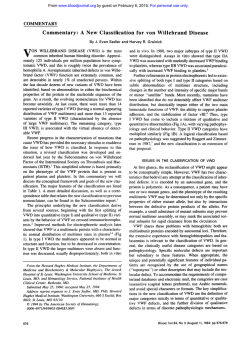
From www.bloodjournal.org by guest on February 6, 2015. For personal use only.
New Variant of von Willebrand Disease Type I1 With Markedly Increased
Levels of von Willebrand Factor Antigen and Dominant Mode of Inheritance:
von Willebrand Disease Type IIC Miami
By Marlies R. Ledford, Ian Rabinowitz, J. Evan Sadler, Jessie W. Kent, and Francisco Civantos
A variant of von Willebrand disease (vWD) was identified
in six members of a kindred spanning four generations.The
proband was a 46-year-old woman with a lifelong history
of bleeding, a prolonged bleeding time (>I5 minutes),
markedly elevated von Willebrand factor (vWF) antigen
(vWF:Ag = 2.09 U/mL), slightly reduced ristocetin cofactor activity, and a plasma vWF multimer pattern similar to
that of vWD type IIC. Similar findings were observed in her
three children, mother, and brother. In affected family
members, platelet and plasma vWF multimer patterns
were discrepant with higher molecular weight multimers
observed in platelet vWF. Following a 1 -Des-amino-8-Darginine vasopressin (DDAVP) challenge, the proband
failed to normalize her bleeding time even though vWF:
Ag rose by 70% and higher molecular weight multimers
were increased slightly. Genetic studies were consistent
with autosomal dominant inheritance of a mutation within
the vWF gene. By sequencing of cloned genomic DNA,
mutations were excluded in exons 4 , 5 , 14,and 15,which
encode regions of the vWF propeptide proposed to be important in multimer biosynthesis. Mutations also were excluded in exons 28 to 31,which encompass the known
mutations that cause vWD types HA, IIB, and B. This new
variant of vWD, characterized by autosomal dominant inheritance, a qualitative defect that resembles vWD type
IIC, and increased plasma vWF:Ag, was tentatively designated vWD type IIC Miami.
0 1993 by The American Society of Hematology.
V
higher molecular weight multimers than previously noted
in propositi with recessive vWD type IIC.
Detailed genetic analysis strongly suggests that this dominant type IIC variant is caused by a mutation within, or
closely linked to, the ~ W F g e n eBy
. DNA sequencing, mutations in exon 28 of the vWF gene were excluded, which
indicates that the mechanism for this new variant differs
from that causing vWD types IIA and IIB.
ON WILLEBRAND DISEASE (vWD) is the most
common bleeding disorder of humans.’ The phenotypic expression of this condition ranges from very mild to
life-threatening hemorrhaging. Patients with vWD are
usually divided into three groups. Type I vWD is characterized by quantitative deficiency of von Willebrand factor
(vWF) and by autosomal dominant inheritance. Type 111
vWD is characterized by a virtual absence of vWF and by
autosomal recessive inheritance. Type I1 vWD is characterized by normal to decreased plasma levels of vWF and the
residual vWF is functionally abnormal. In most type I1 vanants, SDS-agarose gel electrophoresis shows a loss of high
molecular weight multimers, thereby distinguishing these
variants from type I and type 111 vWD. The subtypes (A to
H) of type I1 vWD are distinguished on the basis of specific
functional defects or by details of the multimer pattern.’
Type IIC vWD was described in 1982 by Ruggeri and
colleagues3in a patient with low vWFAg and ristocetin cofactor (RCoF) activity and was inherited as an autosomal
recessive disorder. By multimeric analysis, the repeating
triplet pattern of normal plasma vWF was replaced by one
major band with a single faint intervening band, and the
smallest multimer was relatively increased. Four unrelated
families have been reported to be affected by recessive vWD
with a similar phenotype:-’Some variation was observed in
the number of intervening bands for plasma vWF multimer
patterns,’-’ and in the degree of abnormality for platelet
vWF multimer pattern^,',^ suggesting that vWD type IIC
may be heterogeneous. The molecular basis for the recessive
type IIC variants is not known.
Within a pedigree comprising 16 individuals, spanning
four generations, we have identified 6 patients with vWD in
which the multimeric analysis of plasma vWF is consistent
with the type IIC phenotype. In contrast to the reported
cases classified as vWD type IIC, this new variant is inherited as an autosomal dominant disorder and all affected
persons in this family have markedly increased plasma
vWFAg levels with slightly reduced plasma vWF RCoF. In
addition, the platelet vWF multimeric pattern demonstrates
Blood, VOI 82, NO 1 (July 1). 1993: pp 169-175
MATERIALS, METHODS, AND CASE HISTORY
Materials and Methods
Collection and handling of blood samples. For phenotypic studies, blood samples were collected in 1/10 volume of 3.8% sodium
citrate with or without a protease inhibitor cocktail consisting of 0.1
mmol/L leupeptin, 5.0 mmol/L EDTA, and 6 mmol/L N-ethylmaleimide (Sigma, St Louis, MO). For genetic studies, blood samples
were anticoagulated with EDTA. Platelet lysates were prepared essentially as previously described’ except that the platelet suspension
(1 X 10⁹/L)was lysed with 1/40 volume of 20% Triton-X 100 for 1
From the Department of Pathology. University of Miami School
of Medicine, Miami, FL; and the Departments of Medicine, Biochemistry, and Molecular Biophysics, Howard Hughes Medical Institute, and The Jewish Hospital of St Louis, Washington University, St Louis, MO.
Submitted September 15, 1992; accepted February 19, 1993.
Presented in part at the XIIIth Congress ofthe International Society on Thrombosis and Haemostasis, Amsterdam, The Netherlands
(Thromb Haemost; 65.1124, 1991 [abstr])and the Thirty-third
Annual Meeting of the American Society of Hematology, Denver,
CO (Blood 78:68a, 1991 [abstr]).
Address reprint requests to Marlies R. Ledford, BS, MT(ASCP)
SH, Department ofPathology, University ofMiami School of Medicine, PO Box 016960 (R-5), Miami, FL 33101.
The publication costs of this article were defrayed in part by page
charge payment. This article must therefore be hereby marked
“advertisement” in accordance with 18 U.S.C. section 1734 solely to
indicate this fact.
0 1993 by The American Society of Hematology.
0006-4971/93/8201-003 7$3.00/0
169
From www.bloodjournal.org by guest on February 6, 2015. For personal use only.
LEDFORD ET AL
170
hour at 37°C. Platelet counts and mean platelet volume (MPV)
were determined with a Coulter STKR (Coulter Corp, Hialeah,
FL).
A.w2j6 of'vWF.function and concentration. Ivy bleeding times
(BT) were performed using a Surgicutt device (International Technidyne Corp, Edison, NJ). Ristocetin-induced platelet agglutination (RIPA) and RCoF activity assays were performed by standard
aggregometric methods using ristocetin concentrations from 0.4 to
1.5 mg/mL and 1 .O mg/mL, respectively. For the RCoF assay, lyophilized platelets were from Bio/Data Corp, and a CAP (College of
American Pathologists, Chicago, IL) reference vWF preparation
was used for establishing a standard curve. Botrocetin and lyophilized platelets for botrocetin cofactor assay were kindly provided by
American Diagnostica, Inc (Greenwich, CT). The assay was performed on a PAP-4 aggregometer (Bio/Data Corp, Horsham, PA)
using botrocetin at a final concentration of 5 U/mL, lyophilized
platelets adjusted to 8 X 10⁸/L, and normal pooled plasma (30
donors) for calibration. vWF:Ag was determined with an ELISA kit
(Diagnostica Stago, American Bioproducts Co, Parsippany, NJ)
and also by the method of Laurell" using a polyclonal rabbit antihuman vWF antibody (Diagnostica Stago).
Electrophoretic analysis of v WF structiire. Multimeric analysis
of plasma and platelet vWF was performed by two methods': (1)
autoradiography, using an F(ab'), rabbit anti-human vWF antibody (Diagnostica Stago) and (2) electroblotting, using the same
polyclonal antibody as for the Laurell method and a horseradish
peroxidase-conjugated goat anti-rabbit affinity-purified IgG (CalBiochem, La Jolla, CA) as the secondary antibody. LGT VI1 agarose (Sigma) was used at concentrations of 1.4% or 2.0% as indicated.
Subunit composition of plasma vWF was determined in a gel
system similar to that used for multimer analysis except that samples contained 5% 2mercaptoethanol and were heated for I minute
at 100°C. 5.0%NuSieve GTG agarose was used for the running gel
and I .O% IsoGel was used for the stacking gel (FMC Corp, Rockland, ME).
Other assays. Activated partial thromboplastin time (APTT)
and one-stage factor VIII:C assays were performed on a Coag-AMate X2 (Organon Teknika, Durham, NC) using Organon Teknika reagents and reference plasmas. Factor VII1:C was also assayed
by a chromogenic substrate method using a commercially prepared
kit with substrate CBS 48.03 (Diagnostica Stago).
Analysis of PCR-amplified DNA. Genomic DNA was prepared
from peripheral blood leukocytes.'' Polymerase chain reaction
(PCR)**was performed using a Perkin Elmer-Cetus (Norwalk, CT)
DNA Thermal Cycler as previously described." Primers used for
amplification of exon 28 were No. 226, TGC GAA TAT GGA
ACT CAT TG; No. 227a, CCG ATC CTT CCA GGA CGA AC;
and No. 375a, TCT TGG CAG ATG CAT GTA GC. PCR-amplified fragments of exon 28 using primers 226 and 227a were digested
with BstEII (New England Biolabs, Beverly, MA), electrophoresed
on a 1 % agarose gel, and visualized by staining with ethidium bromide. Analysis of intron 40 variable number of tandem repeat
(VNTR) polymorphism was performed as previously des~ribed.'~.'~
Primers used for intron 40 were No. 474, AGC TAT ATA TCT
ATT TAT CAT and No. 475a, AGA TAC ATA CAT AGA TAT
AGG. DNA subcloning and sequencing were performed as previously described.I6
Case History
The proband of family M, a 46-year-old white woman. was referred in March 1990 to evaluate a lifelong bleeding diathesis before
elective minor surgery. Initial studies were: BT > 15 minutes (normal [N] < 8 minutes), APTT = 30 seconds (upper limit of N =
33.4), VIII:C = 0.79 U/mL (N = 0.5 to I S ) , platelets = 2.76 X
108/L (N = 1.50 to 3.90 x lo8),vWFAg = 2.09 U/mL (N = 0.5 to
1.7), RCoF = 0.46 U/mL (N = 0.5 to 1.5), and plasma vWF multimeric pattern apparently consistent with the type IIC phenotype. A
preliminary diagnosis of vWD type IIC was made. In August 1990,
the proband underwent a 1-Des-amino-8-D-arginine
vasopressin
(DDAVP) challenge showing near normalization of her BT (8.5
minutes), and maximum values for vWF:Ag (3.35 U/mL) at 30
minutes post-DDAVP, RCoF (1.62 U/mL) at 1 hour post-DDAVP,
and VIII:C(2.57 U/mL)at 2 hourspost-DDAVP. Multimericanalysis of plasma vWF demonstrated a slight increase in the higher
molecular weight forms between 30 and 120 minutes postDDAVP. In November 1990, a fine-needle aspiration ofthe thyroid
was successfully performed with cryoprecipitate coverage. The patient has occasional gingival bleeding, menorrhagia, and prolonged
bleeding from minor skin lacerations. Since puberty she has had
epistaxis requiring nasal packing on numerous (>IO) occasions.
Transfusions of unknown type were required for bleeding following
both a tonsillectomy and appendectomy at age 5. The patient has
had several teeth extracted uneventfully; however, removal of a
wisdom tooth at age 25 resulted in the formation of a large facial
hematoma. The patient has had seven pregnancies, of which three
ended in miscarriage. Two vaginal deliveries were uneventful. Two
cesarian sections were complicated by bleeding that required blood
transfusions. One month following the delivery ofher last child, she
experienced excessive uterine bleeding requiring blood transfusions. No transfusions were needed for a subsequent hysterectomy.
The pedigree of family M is shown in Fig 1. The proband (11-3)
and five other affected family members have vWFAg levels greater
than 1 S O U/mL. The proband's mother (I- I ) had a history of menorrhagia and of bleeding after each of five vaginal deliveries but
never received blood products. The proband has three brothers with
uneventful histories. A sister has a history of menorrhagia, occasional epistaxis that was particularly severe after an automobile
accident, and bleeding after vaginal delivery treated with platelet
transfusion. The proband has three surviving children; each has
gingival bleeding and both daughters have menorrhagia.
RESULTS
Values for BT, VIII:C, vWFAg, and RCoF are summarized in Table 1. The six affected family members had prolonged bleeding times (> 15 minutes) and normal platelet
counts. These individuals had markedly elevated plasma
vWF:Ag levels by ELISA and also by the Laurell technique
(data not shown). Plasma RCoF activity was variable
among the six affected members ranging from low to within
normal limits; however, the ratio of vWFAg to RCoF was
-4.6: 1 for the affected members compared with I .2: 1 for
the unaffected family members. All individuals were A B 0
blood group type 0 except 11-4, 111-1, and 111-4, who were
type A. The six affected family members had normal V1II:C
levels, although the ratio ofvWFAg to VII1:C was increased
(-2.3:l). RIPA with concentrations of 0.8 to 1.5 mg/mL
ristocetin was normal in all tested individuals, with no enhanced responsiveness to low-dose ristocetin (0.4 mg/mL).
Plasma botrocetin cofactor activity was 58% and 78% for
11-3 and 111- 1, respectively. Representative data for platelet
vWF from three affected members (11-3,III-l, 111-3) showed
platelet vWF:Ag levels above the normal range and normal
platelet RCoF activity. Platelet vWF from 111-1 demonstrated 35% botrocetin cofactor activity.
For the six affected individuals, analysis of plasma vWF
-
From www.bloodjournal.org by guest on February 6, 2015. For personal use only.
171
VON WILLEBRAND DISEASE TYPE IIC MIAMI
I
Fig 1. Pedigree and genetic studies for dominant vWD type IIC family M. Roman numerals denote generations, Arabic numbers indicate individuals within each generation, and the arrow denotes
the proband. Filled symbols identify family
members with elevated levels of vWF:Ag and a
type IIC multimer pattern. Letters a, b, c, d correspond to alleles of a VNTR marker system (for deT identifies a polytails see legend to Fig 3). C
morphism at nucleotide 4641 of the vWF exon 28.
PCR amplification of exon 28 was performed using
primers 2 2 6 and 227a. Product fragments were digested with BstEll to produce a fragment of 1 ,I90
bp (T-) or 1,045bp (C +). The aC haplotype appears to be linked to the vWD type IIC phenotype in
family M.
Ibb
2ab
C+T-
3ab
C+T-
C+T-
I
4bb
T-T-
5ab
C+T-
I
6 bc
1
C+C+
8
7
+, -
1 ab
5C+Tab
6C+Tbb 7c+c+ %+c+
C+T-
IV
bd
'T-T-
The subunit composition of plasma vWF from the proband (11-3) and her affected daughter (111-1) was compared
with that of normal plasma vWF and vWF from an individual with type IIA vWD. Both affected patients demonstrated proteolytic fragments comparable to those found in
normal plasma vWF and type IIA vWF (data not shown).
Two marker systems were used to investigate linkage of
the vWD phenotype to the vWF gene in this family. One
system exploits a highly polymorphic tetranucleotide
(ATCT), repeat within intron 40.15 In Fig 3, the observed
PCR product sizes are designated a to d. The second system
is a C/T polymorphism in exon 28 at nucleotide 464 1 numbered from the initiation codon. This polymorphism is detected by digestion with the enzyme BstEIII7 that cuts the
DNA (indicated by +)if C is present but does not (indicated
by -) if T is present. For 111-4, plasma samples were available for phenotypic characterization (Table I), but blood
cells for DNA isolation could not be obtained. All analyzed
multimers in 1.4% low gelling temperature (LGT) agarose
gels (Fig 2A) revealed loss of the highest molecular weight
multimers, no intervening bands, and an increase in the
smallest multimer, with the latter two findings also observed
using 2% LGT agarose gels (data not shown). In Fig 2B,
platelet vWF from representative affected family members,
in an intermediate resolution gel, clearly demonstrates the
presence of high molecular weight multimers that are comparable to normal plasma but not to normal platelet vWF.
Densitometric scans (data not shown) showed high molecular weight multimers totaling 58%and 50%for normal platelet vWF and IIC platelet vWF, respectively. Gels of lower
poiosity (2.0% agarose, data not shown) consistently disclosed an intervening band between multimers 1 and 2 as
well as an increase in staining intensity of lower multimers
relative to normal platelet vWF. These data suggest either
that limited proteolysis occurs in vivo or that multimer assembly is abnormal in this vWD variant.
Table 1. Laboratory Data From Family M
Platelet vWF
(U/lOs plt/mL)
Plasma vWF
Family
Member
I- 1
11-1
11-2
11-3
11-4
11-5
11-6
111-1
111-2
111-3
111-4
111-5
111-6
111-7
111-8
F VII1:C
BT (min)
215
8
>15
>I5
4
6
6.5
>15
5
>I5
>15
8.5
6.5
9
A
IV-l
Normal
<8
(U/mL)
1.15
1.40
1.21
1.09
0.86
0.47
0.50
0.61
0.47
0.78
1.17
0.94
0.41
0.50
0.50
1.29
0.5-1.5
vWF:Ag
(ELISA) (U/mL)
2.90
0.90
1.50
2.44
0.90
0.66
0.90
2.17
0.73
2.24
2.38
1.03
0.67
0.80
0.50
0.98
0.5-1.7
RCoF
(UlmL)
0.77
0.88
0.52
0.48
0.67
0.46
0.77
0.29
0.42
0.39
0.54
0.68
0.59
0.54
0.83
0.84
0.5-1.5
vWF:Ag
(ELISA) (U/mL)
RCoF
(U/mL)
0.47
0.38
0.30
0.43
0.16
0.39
0.14
0.20
0.40
0.28
0.14-0.34
0.10-0.44
Abbreviations: BT, bleeding time; vWF: Ag, von Willebrand factor antigen; ELISA. enzyme-linked immunosorbent assay; RCoF, ristocetin cofactor
activity of von Willebrand factor.
From www.bloodjournal.org by guest on February 6, 2015. For personal use only.
LEDFORD ET AL
172
A PlasmavWF
Platelet vWF
B
-=
11-3
CKT
111-1
CKT
Ill4
CIT
111-3
CIT
NP
11-3
11-4
NPLT
111-3
NP
Fig 2. SDS-LGT agarose gel electrophoresis of plasma and platelet vWF antigen using gels of intermediate (1.4%) porosity. vWF multimers were detected by "'I-labeled rabbit antihuman vWF antibody and visualized by autoradiography. Arrow indicates stacking and
running gel interface. Plasma samples for 111-4 and 111-3 were collected in 3.8% sodium citrate (CIT), whereas those from the proband (11-3)
and her daughter (111-1)were collected in the presenceof a protease inhibitor cocktail (CKT). All samples for platelet analysis were collected
in the presence of a protease inhibitor cocktail. Platelet suspensions were adjusted to 1 X 10⁹/L and treated with Triton X-100. Plasma
samples and platelet lysates were diluted 1:10 with sample buffer and 40 r L was loaded onto each lane. A normal pooled plasma (NP) from
each run is shown for reference. Side A: Plasma vWF from the proband (11-3) and her three children. Mobility of individual bands from
affected members corresponds to the central band of each triplet from NP (each band of the first triplet is indicated by lines). Likewise, the
gel shows an absence of subbands in vWF from affected family members as compared with NP. The characteristic accentuated fastest
moving multimer of vWD type IIC is denoted by an asterisk. Side B: Platelet vWF from the proband (ll-3),her normal husband (ll-4),normal
individual (NPLT), and the proband's son (111-3). Platelet vWF from affected members shows the presence of high molecular weight multimers but not the extra large multimers found in NPLT vWF. In comparisonwith NPLT, 11-3and 111-3show increasedstaining intensity of the
lower molecular weight multimers.
matings were informative and the results are consistent with
linkage between the aC haplotype (Fig I ) and the type IIC
phenotype (lod = 2. I , 0 = 0.00). The genotype of 1-2. who
was not available for study. is inferred to be bT/cC.
In an attempt to identify the causative mutation, regions
of the vWF gene from patient 111-3 were subcloned and
sequenced. Clones for both alleles were identified on the
basis of heterozygosity for known DNA sequence polymorphisms. N o candidate mutations were found for either allele
in exons 4 to 5. exons 14 to IS. or exons 28 to 3 I. In particular. mutations in exon 28 known to cause vWD types IIA,
IIB. and Bt8were not present (data not shown).
DISCUSSION
The laboratory studies of the proband showed a disparity
between vWFAgand RCoFand an absence ofhigh molecular weight vWF multimers. The normal triplet structure of
the plasma vWF multimer pattern was replaced by a single
band and the smallest multimer was accentuated (Fig 2h).
This pattern resembles that reported for vWD types IIC.F7
and IIE"? it is distinguished from type IIE by the prominence ofthe smallest multimer."On the basis ofthe plasma
vWF multimer pattern. the disorder in family M is most
similar to vWD type IIC.
The cases of vWD type IIC reported to date** show some
phenotypic heterogeneity (Table 2). Common features include low levels of vWFAg. even lower RCoF activity, a h
sent RIPA, and apparently autosomal recessive inheritance.
Howevcr. differences are present among the vWF multimer
patterns. Plasma vWF multimers consistently show an increase in the smallest multimer. but some patients show an
absence of intervening bands5-' and others show a single
faint intervening
The platelet vWF multimer pattern is more variable. For some patients, it appears to be
identical to plasma vWF,'.~but in others. there are differ-
From www.bloodjournal.org by guest on February 6, 2015. For personal use only.
VON WILLEBRAND DISEASE TYPE IIC MIAMI
173
MW lI-4 11-3 III-I ID-3 II-2 1-1It-6 11-5 III-5 II-6
118-
Fig 3. Analysis by PCR of the vWF intron40 tetranucleotide (ATCT). repeat polymorphism. Following extraction with chloroform, amplified DNA products were electrophoresed on an 8 % SDS-PAGE
gel and stained with ethidium bromide. The molecular weight standard (MW) of 118 bp is Haelll-digested 6x1 74 DNA. The four observed PCR product sizes are arbitrarily designated a t o d: "a" allele
(99 bp) corresponds t o 6 ATCT repeats. "b" allele
(103 bp) corresponds t o 7 ATCT repeats, "c" allele
(119 bp) corresponds t o 11 ATCT repeats, and "d"
allele (123 bp) corresponds t o 12 ATCT repeats.
The "a" allele is linked t o the type IIC phenotype in
this pedigree.
1
-b
\a
MW
enccs in the intensity of intervening bands' or o f the first
multimcr.'
The plasma vWF multimeric structure of the six family
M patients more closely rexmblcs that of the Spanish.6 Italian.s and French' type IIC patients. In common with the
E-l II-6 II-2 III-2
Spanish patients?' affected members of family M show a
disparity hetween plasma and platelet vWF multimer structure: however. platelet vWF from family M patientsdemonstrates normal lcvcls of vWFAg and the presence of high
molecularweight multimcrs. Asscen in Fig I.clevated vWF
Table 2. Comparison of Dominant and Recessive v W D Type IIC
Pallems
Affected
individuals
Inheritance
Clinical history
Bleeding time
Factor VI1I:C
vWF:Ag (EIA)
vWF:Ag
(ELlSA/lRMA)
RCoF
RIPA
Multimer pattem
Plasma
Platelet
Famlly M'
Spanish"
Swedtsh't
Italian"
French'
Engllsh'
6
2
1
1
1
1
Autosomal
dominant
Mild to severe
>15
1.oo
2.41
2.27
0.50
Autosomal
recessive
Mild
>20
0.14
0.14
0.03
Normal
<0.01
Absent
iHMWM
Single bands
No subbands
ttMultimer 1
JHMWM
Single bands
No subbands
ttMultimer 1
Presence of HMWM
tMultimer 1
Intervening band
Between multimer 1
and 2
JHMWM
tMultimer 1
Intervening band
Between multimer
1 and 2
Autosomal
recessive
Moderate
>30
0.67
0.50
-
0.10
-
JHMWM
Single bands
Faint intervening
bands
ttMultimer 1
Same as plasma
Autosomal
recessive
Moderate
130
0.85
1.25
0.45
Autosomal
recessive
Severe
>20
0.24
0.14
Absent
0.03
Absent
JHMWM
Single bands
No subbands
ftMultimer 1
iHMWM
Single bands
No subbands
ttMultimer 1
Same as plasma
Faint band for
Multimer
1
-
0.16
Autosomal
recessive
Moderate
212
0.31
0.50
0.18
0.03
-
iHMWM
Single bands
Faint intervening
bands
ttMultimer 1
-
Abbreviations: vWF: Ag. von Willebrand factor antigen; EIA, electroimmunoassay: ELISA, enzyme-linkedimmunosorbent assay: IRMA, immunoradiometric assay: RCoF, ristocetin cofactor activity of von Willebrand factor; RIPA, ristocetin-induced platelet agglutination: HMWM, high molecular
weight multimers.
* Mean values for 6 affected persons.
t Original patient.
From www.bloodjournal.org by guest on February 6, 2015. For personal use only.
LEDFORD ET AL
174
antigen and type IIC multimer pattern are transmitted in an
autosomal dominant fashion in family M that differs from
the autosomal recessive transmission described previously
for vWD type IIC.
In contrast to other patients with vWD type IIC, affected
members of family M have markedly elevated levels of vWF
antigen (Table 2). To exclude the possibility that the high
antigen levels were not representative of the phenotype, several affected individuals were tested on multiple occasions
and consistently demonstrated these findings. For affected
members of family M, vWF function as determined by the
ristocetin cofactor assay was borderline normal to low but
significantly higher than values reported for other type IIC
propositi. Although V1II:C levels were normal in family M,
the ratio of VII1:C to vWFAg was low; administration of
DDAVP to the proband increased this ratio into the normal
range.
In the plasma of normal individuals or of patients with
vWD types IIA or IIB, vWF consists mostly of the intact
250-Kd subunit but also contains proteolytic fragments of
189,176, and 140 Kd.I9 Several other variants with aberrant
structure of individual multimers show markedly decreased
amounts of these proteolytic fragments, particularly vWD
types IIC (Swedish proband), IID, and IIE.19 In contrast,
plasma vWF from two affected members of family M demonstrates the presence of fragments similar to those found in
normal plasma vWF (data not shown). Thus, a marked decrease in intervening or satellite multimer species (Fig 2A)
does not necessarily provide evidence for decreased subunit
proteolysis.
Genetic analysis of family M showed complete concordance between intragenic vWF polymorphisms and the
vWD phenotype, and this is consistent with a mutation in
the vWF gene. If there is a single intragenic mutation, the
mechanism by which it causes both defective multimer
structure and high vWF:Ag levels is not obvious. In principle, the decrease in large multimers might be caused by
impaired biosynthesis or by enhanced proteolysis. Our studies of plasma and platelet vWF do not provide clear evidence for either mechanism, and no explanation for the
high vWF:Ag levels is apparent. One possibility is that the
mutation could facilitate the transport of vWF through the
endoplasmic reticulum, a process that is unusually prolonged for normal vWF.~'By reducing intracellular degradation or storage, such an effect could thereby increase the rate
of vWF secretion.
The vWF gene of the proband was examined for mutations that might disrupt multimer biosynthesis or increase
multimer degradation. The vWF propeptide is proposed to
catalyze vWF multimer
Segments of the
vWF propeptide that are encoded by exons 4 and 5 (D1
domain) and exons 14 and 15 (D2 domain)16show similarity to sequences in protein disulfide isomerase, and mutagenesis of these regions abolishes multimer formation.23By
DNA sequencing of PCR products, mutations within these
sequences were excluded (data not shown). Similarly, of the
mutations that cause vWD type IIA, some impair multimer
formation and others increase sensitivity to proteolytic degradation.' By DNA sequencing, mutations were excluded in
exons 28 to 3 1 (data not shown); these regions that encode
for domains A 1 and A2 of the mature protein include all of
the currently known mutations that cause vWD type IIA, as
well as those that cause vWD types IIB and B.l* Thus, the
molecular basis of vWD in family M is different from these
previously characterized variants, which suggests that the
pathophysiologic mechanism also is different.
To our knowledge, this is the first report of a qualitative
defect of vWF that is associated with elevated levels of
vWFAg. The unique combination of autosomal dominant
inheritance, type IIC-like multimer pattern, and high
vWF:Ag appears to represent a new variant of type I1 vWD.
Until classification can be based on either pathophysiologic
mechanism or genetic defect, we propose to designate this
new variant as vWD type IIC Miami to avoid the addition
of new terms to the present nomenclature.
ACKNOWLEDGMENT
We acknowledge and thank Dr Robert Glasser for refemng this
interesting patient, the family for their unlimited cooperation,
Doug Roach and Pam Little from the Biomedical Communications
Department of the University of Miami School of Medicine for
preparation of the figures, and Patricia M. Wright and Ann F.
Wright for their secretarial assistance.
REFERENCES
1. Ginsburg D, Bowie EJW: Molecular genetics of von Wille-
brand disease. Blood 79:2507, 1992
2. Ruggeri ZM, Zimmerman TS: von Willebrand factor and von
Willebrand disease. Blood 705395. 1987
3. Ruggeri ZM, Nilsson IM, Lombardi R, Holmberg L, Zimmerman TS: Aberrant multimeric structure of von Willebrand factor in
a new variant of von Willebrand's disease (type IIC). J Clin Invest
70:1124, 1982
4. Armitage H, Rizza CR: Two populations of factor VIII-related antigen in a family with von Willebrand's disease. Br J Haematol 41:279, 1979
5. Mannucci PM, Lombardi R, Pareti FI, Solinas S, Mazzucconi
MG, Mariani G: A variant of von Willebrand's disease characterized by recessive inheritance and missing triplet structure of von
Willebrand factor multimers. Blood 62: 1000, 1983
6. Batlle J, Lopez Fernandez MF, Lasierra J, Fernandez Villamor A, Lopez Berges C, Lopez Borrasca A, Ruggeri ZM, Zimmerman TS: von Willebrand disease type IIC with different abnormalities of von Willebrand factor in the same sibship. Am J Hematol
21:177, 1986
7. Mazurier C, Mannucci PM, Parquet-Gernez A, Goudemand
M, Meyer D: Investigation of a case of subtype IIC von Willebrand
disease: Characterization of the variability of this subtype. Am J
Hematol22:301, 1986
8. Batlle J, Lopez-Fernandez MF, Fernandez Villamor A, Lopez
Berges C, Zimmerman TS: Multimeric pattern discrepancy between platelet and plasma von Willebrand factor in type IIC von
Willebrand disease. Am J Hematol 22237, 1986
9. Ledford MR, Kent J, Civantos F: A comparative study of
three methods for the visualization of von Willebrand factor (vWF)
multimers: Thromb Haemost 64569, I990
10. Laurel1 CB: Quantitative estimation of proteins by electrophoresis in agarose gel containing antibodies. Anal Biochem 15:45,
1966
11. Grunebaum L, Cazenave JP, Camerino G, Kloepfer C,
Mandel JL, Tolstochev P, Jaye M, DeLasalle H, Lecocq J P Carrier
detection of hemophilia B by using restriction site polymorphism
From www.bloodjournal.org by guest on February 6, 2015. For personal use only.
VON WILLEBRAND DISEASE TYPE IIC MIAMI
associated with the coagulation factor IX gene. J Clin Invest
73:1491, 1984
12. Saiki RK, Gelfand DH, Stoffel S, ScharfSJ, Higuchi R, Horn
GT, Mullis KB, Erlich HA: Primer-directed enzymatic amplifications of DNA with a thermostable DNA polymerase. Science
239:487, 1988
13. Randi AM, Rabinowitz I, Mancuso DJ, Mannucci PM,
Sadler JE: Molecular basis of von Willebrand disease type IIB. Candidate mutations cluster in one disulfide loop between proposed
platelet glycoprotein Ib binding sequences. J Clin Invest 87: 1220,
1991
14. Peake IR, Liddell MB, Moodie P, Standen G, Mancuso DJ,
Tuley A, Westfield LA, Sorace JM, Sadler JE, Venveij CL, Bloom
AL: Severe type 111 von Willebrand’s disease caused by deletion of
exon 42 of the von Willebrand factor gene: Family studies that
identify camers of the condition and a compound heterozygous
individual. Blood 75:654, 1990
15. Peake IR, Bowen D, Bignell P, Liddell MB, Sadler JE, Standen G, Bloom AL: Family studies and prenatal diagnosis in severe
von Willebrand disease by polymerase chain reaction amplification
of a variable number tandem repeat region of the von Willebrand
factor gene. Blood 76555, 1990
16. Mancuso DJ, Tuley EA, Westfield LA, Worrall NK, Shelton-Inloes BB, Sorace JM, Alevy YG, Sadler JE: Structure of the
gene for human von Willebrand factor. J Biol Chem 264:19514,
1989
175
17. Nichols WC, Lyons SE, Harrison JS, Cody RL, Ginsburg D:
Severe von Willebrand disease due to a defect at the level of von
Willebrand factor mRNA expression: Detection by exonic PCRrestriction fragment length polymorphism analysis. Proc Natl Acad
Sci USA 88:3857, 1991
18. Ginsburg D, Sadler JE: von Willebrand disease: A database
of point mutations, insertions, and deletions. Thromb Haemost
69:177, 1993
19. Zimmerman TS, Dent JA, Ruggeri ZM, Nannini LH: Subunit composition of plasma von Willebrand factor. Cleavage is present in normal individuals, increased in IIA and IIB von Willebrand
disease, but minimal in variants with aberrant structure of individual oligomers (types IIC, IID, and IIE). J Clin Invest 77:947, 1986
20. Wagner DD, Marder VJ: Biosynthesis of von Willebrand
protein by human endothelial cells. Identification of a large precursor polypeptide chain. J Biol Chem 258:2065, 1983
2 I . Venveij CL, Hart M, Pannekoek H: Expression of variant
von Willebrand factor (vWF) cDNA in heterologous cells: Requirement of the pro-polypeptide in vWF multimer formation. EMBO J
6:2885, 1987
22. Wise RJ, Pittman DD, Handin RI, Kaufman RJ, Orkin SH:
The propeptide of von Willebrand factor independently mediates
the assembly of von Willebrand multimers. Cell 52:229, 1988
23. Mayadas TN, Wagner DD: Vicinal cysteines in the prosequence play a role in von Willebrand factor multimer assembly.
Proc Natl Acad Sci USA 89:3531, 1992
From www.bloodjournal.org by guest on February 6, 2015. For personal use only.
1993 82: 169-175
New variant of von Willebrand disease type II with markedly increased
levels of von Willebrand factor antigen and dominant mode of
inheritance: von Willebrand disease type IIC Miami
MR Ledford, I Rabinowitz, JE Sadler, JW Kent and F Civantos
Updated information and services can be found at:
http://www.bloodjournal.org/content/82/1/169.full.html
Articles on similar topics can be found in the following Blood collections
Information about reproducing this article in parts or in its entirety may be found online at:
http://www.bloodjournal.org/site/misc/rights.xhtml#repub_requests
Information about ordering reprints may be found online at:
http://www.bloodjournal.org/site/misc/rights.xhtml#reprints
Information about subscriptions and ASH membership may be found online at:
http://www.bloodjournal.org/site/subscriptions/index.xhtml
Blood (print ISSN 0006-4971, online ISSN 1528-0020), is published weekly by the American
Society of Hematology, 2021 L St, NW, Suite 900, Washington DC 20036.
Copyright 2011 by The American Society of Hematology; all rights reserved.

© Copyright 2026