
BRIEF COMMUNICATION
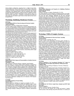
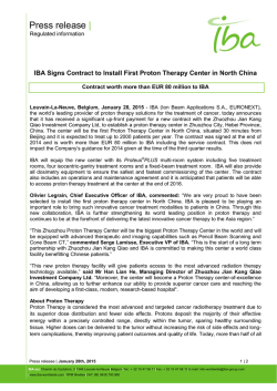
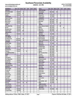
BRIEF COMMUNICATION
ORDER OF PROTON UPTAKE AND RELEASE BY
BACTERIORHODOPSIN AT LOW PH
DRAKE MITCHELL AND G. W. RAYFIELD
Physics Department, University ofOregon, Eugene, Oregon 97403
ABSTRACr The order of proton uptake and release in an aqueous suspension of purple membrane in response to a light
flash has been investigated at lowered pH. pH indicator dyes and a flash spectrophotometer were used for the study. At
pH 6.6 it was found that the release of protons from the purple membrane precedes uptake, as reported by other
investigators. At pH 5.9, 4.9, and 4.1 it was also found that release precedes uptake. These results are not in agreement
with those of previous investigators.
INTRODUCTION
Bacteriorhodopsin (bR) is a light activated proton pump
that transports protons across the plasma membrane of
Halobacterium halobium. This protein is found in large,
ordered, two dimensional arrays known as purple membrane sheets. Recent reviews cover these and other aspects
of bacteriorhodopsin in detail (1-4).
When an aqueous suspension of purple membrane sheets
is illuminated, a transient change in the pH of the bathing
solution is observed. This uptake and release of protons into
the bathing medium has been investigated in some detail.
An optical method employing pH sensitive dyes (5-7), a
volumetric technique (8), and a novel technique based on
changes in the ohmic conductance of the bathing solution
(9) have been employed for these studies. Near pH 7 it was
found that protons are first released into the bathing
solution from the photo-stimulated protein and then taken
up (5-9) by it. However, at lowered pH (pH 4 to 5) single
flash measurements have been reported in which the
sequence is reversed, that is uptake precedes release (5, 9).
Steady-state measurements at low pH using pH electrodes
and an aqueous suspension of purple membrane sheets
have been reported (10) that show alkalization of the
bathing medium under illumination. This was interpreted
as demonstrating that uptake of protons occurs more
quickly than release at low pH.
The retinal chromophore of bR is connected to a lysine
residue of the opsin by a Schiff base, which is protonated in
bR568 (11, 12). Studies have tested the notion that protonation of the Schiff base is correlated with the photoreaction
cycle of the chromophore and with proton release and
uptake from the bathing medium (6). In particular,
detailed studies at pH 7 indicate that, in the M412
intermediate of the photoreaction cycle, the Schiff base is
deprotonated (12, 13). Measurements of the K (11) and L
BIOPHYS. J.©BiophysicalSociety
Volume 49 February 1986 563-566
*
(14) intermediates show that they are protonated, and
M412 is therefore the first deprotonated intermediate
(14). The formation of 0640, the principle intermediate in
the M412 to bR568 conversion, has been kinetically associated with uptake of protons by bR (15), and at pH 7 it is
found to be protonated (16). To our knowledge the protonation state of M412 intermediate has not been studied at
low pH (pH 4 to 5).
The decay of M412 has been correlated with the uptake
of protons from the bathing solution (17, 7), and from pH 7
to 4.5 the time constant of this decay has been shown to
increase (18). Over this pH range the time constant of
M412 formation remains constant (19). If the sequence of
release and uptake is reversed at lowered pH and the
photoreaction cycle is essentially unaltered, then the correlation between the two becomes obscure.
We have repeated measurements of the uptake and
release sequence at low pH and find that release precedes
uptake in agreement with the order of events near neutral
pH.
MATERIALS AND METHODS
Bacteriorhodopsin in the form of purple membrane sheets was derived
from strain JW 3 of Halobacterium halobium following the procedures of
Becher and Cassim (20) with some minor modifications. Samples were
stored in 40% sucrose (wt/wt) at 40C. Small aliquots were removed from
this stock solution and used within 10 d. The sample was dialyzed for 24 h
to remove sucrose.
The flash spectrophotometer used for this investigation was constructed in our laboratory. The actinic light flash was provided by a
flashlamp-pumped dye laser (model CMX-4; Chromatix Inc., Sunnyvale,
CA) using rhodamine 590 dye and operating at a wavelength of 598 nm.
The flash had a pulse width of 1 js and an energy of 3 to 4 mJ as
measured by a calibrated bolometer (Scientech Instruments, Inc., Boulder, CO). A beam splitter in the laser beam sent a small fraction of the
actinic beam to a PIN photodiode. The output of this photodiode was fed
to the B channel of a Nicolet 4094 digital oscilloscope (Nicolet Instru-
0006-3495/86/02/563/04 $1.00
563
ment Corp., Madison, WI), and served as an external trigger. The
amplitude of this signal was also used to monitor the laser intensity.
The light source for the measuring beam was a quartz halogen lamp
(I150 W). A stabilized, 24 V DC power supply was used for the lamp. The
measuring beam passed through a high intensity, grating monochromator
(Bausch & Lomb Inc., Rochester, NY) (1,200 grooves per millimeter)
and was collimated by quartz lenses through the bottom third of a 1 x 1
cm quartz cuvette containing the sample. The volume of interaction
between the two beams was maximized by using a quartz lens to spread
the laser beam to approximately the size of the sample (1 x 1 cm). The
measuring beam, after passing through the sample, was then focused by a
quartz lens on a UV enhanced, inverted channel, PIN photodiode
(UDT-UV005; United Technologies Corp., Hartford, CT). Appropriate
interference filters were used to isolate the detector from the actinic laser
flash. After amplification by an op-amp (OPACM 104 cm; Analog
Devices Inc., Norwood, MA) in a current-to-voltage configuration, the
signal from the photodiode was stored by the Nicolet oscilloscope. Signal
averaging was used when it was found desirable to improve the signal to
noise ratio.
The proton uptake and release to the bathing medium of the photoactivated bR sample was monitored by using a pH indicator dye. To observe
the time dependent absorbance change of the dye it was necessary to
perform a difference measurement that removes the absorbance changes
of the photocycling bacteriorhodopsin. This may be accomplished in
either of two ways: (a) by comparing the absorbance changes of two
samples with and without dye, or (b) by comparing two samples containing dye, with one containing an appropriate buffer. We chose the first
method because of possible problems associated with an ionic strength
effect on the photocycle kinetics (21). A second reason for not choosing
the buffer method was a recent observation of a flash induced absorbance
change due to proton release in a buffered solution of purple membrane
and p-nitro-phenol (22).
The data was transferred from the Nicolet 4094 (Nicolet Instrument
Corp.) to an IBM PC (IBM Instruments, Inc., Danbury, CT) for further
analysis. Along with the flash-induced kinetic absorbance data we
included the voltages corresponding to the absolute baseline and the
magnitude of the laser flash intensity in each computer data file. The
absolute base line was used to calculate the absolute absorbance change
from the observed intensity change. This is a necessary step for comparing
changes in two separate samples with different initial absorbances. Both
traces were normalized to the same actinic flash intensity. Following these
procedures a computer program performed the appropriate subtraction
and plotted the absorbance change of the dye.
The pH indicating dyes used in this study were either those used by
other investigators (p-nitrophenol, bromocresol green) (5-7) or were
selected according to a modified protocol of Lozier (21). The final choice
of a dye had to satisfy several experimental considerations: (a) The dye
should be soluble in water and have little affinity for the purple
membrane. (b) The dye should not affect the photocycle kinetics of the
bacteriorhodopsin. (c) The absorbance of the dye must be sensitive to
changes in pH at the pH of interest. (d) The dye should show little or no
absorption at the actinic wavelength (598 nm). This last requirement is
essential if one is to compare samples with and without dye.
P-nitrophenol was satisfactory near pH 6.6 and pH 5.9. A search of
other pH indicating dyes showed that 2-5 dinitrophenol is suitable for
measurements near pH 5 while 2-4 dinitrophenol can be used near pH 4.
The static absorption spectra of these dyes at different pH were obtained
using a Hewlett-Packard spectrophotometer (model no. 8450 UV/vis).
No dye was detected when a pellet of bR from a dye-bR sample was
resuspended in distilled water. The static absorption spectra of the dyes
was unchanged in the presense of bR. The time-dependent absorption at
660 nm of light-activated bR was unaffected by the presence of any of the
three nitrophenol pH indicator dyes.
Bacteriorhodopsin has significant buffering capacity in the full pH
range of interest. This buffering effect was taken into account by
measuring the change in static absorbance of a mixture of purple
membrane and dye as a function of H+ added. The buffering capacity of
564
the purple membrane alone was also measured independently, using a pH
electrode and titrating against HCI.
Table I shows the type of indicator dye used for each pH along with the
dye concentration and measuring wavelength for each sample. All
measurements were made at a bR concentration of 3 IAM in 250 mM KC1.
This salt concentration was used to maximize proton pump activity (7, 8).
All measurements were carried out at room temperature, 19-210C.
RESULTS
A typical set of time dependent absorption curves used to
determine the proton concentration in the bathing medium
after a light flash is shown in Fig. 1 for 2-5 dinitrophenol at
pH 4.9. Curve 1 a (without dye) is subtracted from curve 1
b (with dye) to yield AA, the dye response. AA is linearly
related to the number of protons released to the bathing
medium when the buffering of the bR sample is taken into
account (see Methods above). AA can be converted to ApH
which measures the number of free protons in solution not
the number released to solution. AA/ApH in Fig. 1 is
0.51.
Fig. 2 shows a similar set of absorption curves taken with
and without 2-4 dinitrophenol at pH 4.1. The dye response
is shown in Fig. 2 c and is consistent with release preceding
uptake at pH 4.1. The results shown in Figs. 1 and 2
contradict the order of uptake and release reported by
Dencher and Wilms at pH 5 (5) as well as the results
reported by Marinetti and Mauzerall at pH 4.2 (9).
Further studies at pH 5.9 and pH 6.6 using the indicator
dye p-nitrophenol were consistent with results reported by
Lozier et al. at pH 6.5 (6) and Govindjee et al. at pH 6.6
(7), which showed that release precedes uptake.
An attempt was made to repeat the results of Dencher
and Wilms using bromocresol green. The high absorbance
of this dye at the actinic wave-length (OD = 0.7 at 598 nm
for a 4 ,uM solution at pH 5) means that the two samples to
be compared must both contain dye, and one must be
buffered. Dencher and Wilms used 6 ,uM bR and 46 ,M
bromocresol green in distilled water as the sample, and the
same concentrations of dye and bR in 98 mM citric
acid/phosphate buffer as the control, both at pH 5. It was
necessary to reduce the dye concentration to 4 ,uM since 46
,uM bromocresol green has an OD of -10 at 620 nm and
pH 5, which was too high for our system. We found that
the buffer had a significant effect on the bR photocycle at
620 nm. This was found to be consistent with an observed
ionic strength effect on the photocycle (unpublished
results). To negate the ionic strength effect an experiment
TABLE I
Dye
concentration
Measuring
wavelength
p-nitrophenol
bromocresol green
170,uM
400 nm
3 ,M
2-5 dinitrophenol
2-4 dinitrophenol
190,uM
70MM
620 nm
435 nm
400 nm
pH
Dye
5.90
5.00
4.90
4.10
BIOPHYSICAL JOURNAL VOLUME 49 1986
was performed using 4 ,M bromocresol green in a solution
of 3 MM bR and 250 mM KCl for both the sample and
control. The control was buffered at pH 5.0 by 10 mM
malic acid. Measurements at 620 nm on this pair yielded a
dye signal that showed release of protons occurring first, in
agreement with our other measurements.
CONCLUSIONS
0
1~~~~A
Ib
d
LOG TIM-E.(
acid
FIGURE 1 The flash-induced absorbance change of bacteriorhodopsin
(a) bR without pH indicator dye. (b) bR in the
presence of 190 AiM 2-5 dinitrophenol. (c) b-a, the flash-induced
absorbance change of 2-5 dinitrophenol. The curves (a, b) are the average
of 100 flashes, 5 s apart. Both of the solutions contained 3gM bR and 250
mM KCl with the pH adjusted to 4.90 using HCl. The first 148 jis of the
traces are obscured by the photodiode's recovery from the laser flash.
at 435 nm is shown:
-
6:
:;'
8J
0.148uM.s
X
_
e/cd1,
lonis
lO4iinslO s
b
6
_
;.LOG TIME 4&s)
besef
0
O
j. . . . . . .. .. d
FIGURE 2 The flash-induced absorbance change of bacteriorhodopsin
at 400 nm is shown: (a) bR without pH indicator dye. (b) bR in the
presence of 70 jIM 2-4 dinitrophenol. (c) b-a, the flash-induced absorbance change of 2-4 dinitrophenol. The pH of both solutions was 4.10. The
solutions contained 3 AM bR and 250mM KCI. The curves (a, b) are the
average of 32 flashes, 5 s apart.
MITCHELL AND RAYFIELD Order of Proton Uptake and Release
We find that the order of uptake and release is not reversed
when the pH is lowered to pH 4.9 or pH 4.1. We suspect
that our results differ from those of Dencher and Wilms
because they did not take into account the effect of ionic
strength on the photocycle. When the ionic strength of the
sample and control were balanced, bromocresol green gave
results at pH 5.0 consistent with release preceding uptake.
Lozier (21) has criticized the photoconductivity measurements for determining proton release and uptake by
purple membrane sheets in an aqueous solution on the
basis that the results can be complicated by motions of
charges other than protons. Marinetti and Mauzerall (9)
seem to have taken this into account by varying the buffer
concentrations. We do not know why our results differ
from those of the photoconductivity measurements.
This work was supported by National Institutes of Health grant GM
26669.
Received for publication 30 May 1985 and in final form 17 September
1985.
REFERENCES
1. Eisenbach, M., and S. R. Caplan. 1979. The light-driven proton
pump of Halobacterium halobium: mechanism and function.
Curr. Top. Membr. Transp. 12:165-248.
2. Stoeckenius, W., R. H. Lozier, and R. A. Bogomolni. 1979. Bacteriorhodopsin and the purple membrane of halobacteria. Biochim.
Biophys. Acta. 505:215-278.
3. Stoeckenius, W., and R. A. Bogomolni. 1982. Bacteriorhodopsin and
related pigments of halobacteria. Annu. Rev. Biochem. 52:587616.
4. Honig, B. 1982. Theoretical aspects of photoisomerization in visual
pigments and bacteriorhodopsin. In Biol. Events Probed by Ultrafast Laser Spectroscopy. 281-297.
5. Dencher, N., and M. Wilms. 1975. Flash photometric experiments
on the photochemical cycle of bacteriorhodopsin. Biophys. Struct.
Mech. 1:259-271.
6. Lozier, R. H., W. Niederberger, R. A. Bogomolni, S.-B. Hwang, and
W. Stoeckenius. 1976. Kinetics and stoichiometry of light-induced
proton release and uptake from purple membrane fragments,
Halobacterium halobium cell envelopes, and phospholipid vesicles containing oriented purple membrane. Biochim. Biophys.
Acta. 440:545-556.
7. Govindjee, R., T. G. Ebrey, and A. R. Crofts. 1980. The quantum
efficiency of proton pumping by the purple membrane of Halo-
bacterium halobium. Biophys. J. 30:231-242.
8. Ort, D. R., and W. W. Parson. 1979. The quantum yield of
flash-induced proton release by bacteriorhodopsin-containing
fragments. Biophys. J. 25:341-354.
9. Marinetti, T., and D. Mauzerall. 1983. Absolute quantum yields and
proof of proton and nonproton transient release and uptake in
565
10.
11.
12.
13.
14.
15.
16.
566
photoexcited bacteriorhodopsin. Proc. Natl. Acad. Sci. USA.
800:178-180.
Garty, H., G. Klemperer, M. Eisenbach, and S. R. Caplan. 1977.
The direction of light-induced pH changes in purple membrane
suspensions. FEBS (Fed. Eur. Biochem. Soc.) Lett. 81:238-242.
Rothschild, K. J., and H. Marrero. 1982. Infrared evidence that the
Schiff base of bacteriorhodopsin is protonated: bR 570 and K
intermediates. Proc. Natl. Acad. Sci. USA. 79:4045-4049.
Aton, B., A. G. Doukas, R. H. Callendar, B. Becher, and T. G.
Ebrey. 1977. Resonance Raman studies of the purple membrane.
Biochemistry. 16:2995-2999.
Lewis, A., J. Spoonhower, R. A. Bogomolni, R. H. Lozier, and W.
Stoeckenius. 1974. Tunable laser resonance Raman spectroscopy
of bacteriorhodopsin. Proc. Natl. Acad. Sci. USA. 71:44624466.
Argade, P. V., and K. J. Rothschild. 1983. Quantitative analysis of
resonance Raman spectra of purple membrane from Halobacterium halobium: L550 intermediate. 1983. Biochemistry. 22:34603466.
Lozier, R. H., R. A. Bogomolni, and W. Stoeckenius. 1975. Bacteriorhodopsin: a light-driven proton pump in Halobacterium halobium. Biophys. J. 15:955-964.
Smith, S. O., J. A. Pardoen, P. P. J. Mulder, B. Curry, J.
Lugtenburg, and R. Mathies. 1983. Chromophore structure in
17.
18.
19.
20.
21.
22.
bacteriorhodopsin's 0640 intermediate. Biochemistry. 22:61416148.
Lozier, R. H., W. Niederberger, R. A. Bogomolni, S.-B. Hwang, and
W. Stoeckenius. 1976. Kinetics and stoichiometry of light-induced
proton release and uptake from purple membrane fragments,
Halobacterium halobium cell envelopes, and phospholipid vesicles containing oriented purple membranes. Biochim. Biophys.
Acta. 440:545-556.
Li, Q.-Q., R. Govindjee, and T. G. Ebrey. 1984. A correlation
between proton pumping and the bacteriorhodopsin photocycle.
Proc. Natl. Acad. Sci. USA. 81:7079-7082.
Lam, E., and L. Packer. 1983. Nonionic detergent effects on
spectroscopic characteristics and the photocycle of bacteriorhodopsin in purple membranes. Arch. Biochem. Biophys. 221:557564.
Becher, B. M., and Y. Cassim. 1975. Improved isolation procedures
for the purple membrane of Halobacterium halobium. Prep.
Biochem. 5:161-178.
Lozier, R. H. 1982. Rapid kinetic optical absorption spectroscopy of
bacteriorhodopsin photocyles. Methods Enzymol. 88:133-162.
Drachev, L. A., A. D. Kaulen, and V. P. Skulachev. 1984. Correlation of photochemical cycle, H' release and uptake, and electric
events in bacteriorhodopsin. FEBS (Fed. Eur. Biochem. Soc.)
Lett. 178:331-335.
BIOPHYSICAL JOURNAL VOLUME 49 1986


© Copyright 2026