
Supplementary Information - Royal Society of Chemistry
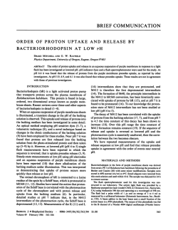
Electronic Supplementary Material (ESI) for RSC Advances. This journal is © The Royal Society of Chemistry 2015 Microbial adaptation to ionic liquids M. S. Álvarez, A. Rodríguez*, M. A. Sanromán, F. J. Deive * Electronic Supplementary Information Materials Ionic liquids C2MIMC2SO4 (>99%) and C2MIMC1SO4 (>99%) were purchased from IoLiTec. C2PyC2SO4 (>99%) was supplied by Merck. P4441C1SO4 (>97%) was kindly donated by CYTEC industries. All of them were subjected to vacuum (P = 2·10−1 Pa) and moderate temperature (T = 343.15 K) for several days to remove possible traces of solvents and moisture, always prior to their use. The ionic liquids were kept in bottles under inert atmosphere until use. Microorganisms and culture medium Pseudomonas stutzeri, Staphylococcus warneri and consortium C26b were grown in a mineral medium composed of: Na2HPO42H2O 8.5 g/L, KH2PO4 3.0 g/L, NaCl 0.5 g/L, NH4Cl 1.0 g/L, MgSO4·7H2O 0.5 g/L, CaCl2 14.7·10-3 g/L, CuSO4 0.4 mg/L, KI 1.0 mg/L, MnSO4·H2O 4.0 mg/L, ZnSO4·7H2O 4.0 mg/L, H3BO3 5.0 mg/L, FeCl3·6H2O 2.0 mg/L. 15 g/L of glucose were also included in the culture medium. 1,2 Trametes versicolor and Shewanella oneidensis were grown in a medium containing glucose (15 g/L), yeast extract (15 g/L), NH4Cl (0.75 g/L), KH2PO4 (2 g/L), MgSO4·7H2O (0.5 g/L), CaCl2·2H2O (0.1 g/L) and KCl (0.5 g/L).3 Halobacterium salinarum and Anoxybacillus flavithermus were cultured in Luria-Bertani medium, composed of 10 g/L trypticase, 5 g/L yeast extract and 10 g/L NaCl. The latter was isolated in a Galician hot spring, as previously reported.4,5 Phanerochaete chrysosporium BKM-F-1767 (ATCC 24725) was grown in Kirk medium, as reported elsewhere.6 Thermus thermophilus HB27 was kindly provided by Dr. J. Berenguer (Universidad Autonoma, Madrid, Spain). This microorganism was grown in a liquid medium containing (g/L, in distilled water) 8 trypticase, 4 yeast extract and 3 NaCl.7 The microorganism inocula were obtained by cultivating the microorganisms in 250 mL-flasks capped with cellulose stoppers, containing the corresponding culture media, and after the stationary phase was reached, the biomass was separated by centrifugation at 5.000xg for 10 min, at 4ºC. The humidity was removed by vacuum drying and the dried cells (pellets) were stored at -20ºC in Eppendorf tubes. The same amount of cells was used for subsequent cultures, since each pellet (with a given cell concentration determined after a calibration absorbance at 600 nm vs. dried cell weight) was resuspended in the corresponding IL-containing culture medium, and it was used to inoculate 100 mL of medium (to yield an initial biomass concentration of 3%). Effect of ionic liquids on microorganisms Two different media were used for investigating the effect of the three selected ionic liquid families: a mineral (MM) and a rich medium (RM) with compositions detailed by Moscoso et al (2012)1 and Deive et al (2010),5 respectively., The pH of each medium was adjusted according to the previously reported, namely: P. chrysosporium pH 4.5, T. versicolor and S. oneidensis pH 7, consortium C26b, St. warneri, A. flavithermus, A. salinarum and T. thermophilus HB27 pH 7.5, and P. stutzeri pH 7.2. All the liquid media were sterilized by autoclaving at 121ºC for 20 min. They were then inoculated with the corresponding microorganisms at 37ºC for the mesophiles, 60ºC for A. flavithermus and 70ºC for T. thermophilus HB27. The cultures were carried out in 96 well plates containing different concentrations of ionic liquids (between 0.005M to 1.5 M). MLC was ascertaining by inoculating plates without the ionic liquid pressure. Microscopy analysis Scanning electron microscopy (SEM) images were taken in a FEI-Quanta 200 environmental scanning electron micro-scope using an accelerating voltage of 15 kV (Electron Microscopy Service, C.A.C.T.I., University of Vigo). Microbial acclimation The stirred tank bioreactor (Biostat B, Sartorius, Germany) consisted of a 3Ljacketed glass column filled with 2 L of mineral medium described above containing 200 M of C2MIMC2SO4. The temperature was maintained at 37 ºC by circulation of thermostatted water, and the pH was adjusted to 8. The bioreactor was inoculated with actively growing cells of P. stutzeri from 24 h flask cultures (3% v/v). Humidified air was supplied continuously at 0.17 vvm and the agitation was set at 100 rpm. Bioreactor was operated for two months. Biopolymer production The adapted P. stutzeri was cultured in 250 mL Erlenmeyer flasks containing 50 mL of mineral medium. After 48 h, maximum biopolymer concentration was detected. Therefore, the culture medium was centrifuged at 14000 rpm and 4ºC to separate the cells. The supernatant was reserved for biopolymer recovery. Afterwards, a mixture containing the supernatant and ethanol in a 1:2 ratio was kept at 4ºC for 2 hours and then centrifuged at 14000 rpm and 4ºC. Biopolymer characterization The biopolymer composition was ascertained after a preliminary acid hydrolysis step, followed by the injection in an HPLC (Agilent 1100) equipped with a RI detector. Composition determination was carried out by direct comparison with standards such as glucose, fructose, sucrose, maltose and rhamnose. Fig. S1 SEM image of P. stutzeri after two months in a stirred tank bioreactor without the presence of the ionic liquid 1.2 0.9 0.9 0.6 0.6 0.3 0.3 0.0 0 2 Staphylococcus 6 8 warneri 4 0 2 0.0 Shewanella oneidensis 6 8 4 0.9 0.9 0.6 0.6 0.3 0.3 Absorbance Absorbance Adapted Pseudomonas stutzeri Pseudomonas stutzeri Absorbance Absorbance 1.2 0.0 Consortium C26b 0.0 2 4 Days 6 8 10 Absorbance 0.9 0.6 0.3 0.0 0 2 4 Days 6 8 10 Fig. S2 Microbial growth in the absence of ionic liquids in rich () and mineral () medium of Pseudomonas stutzeri, adapted Pseudomonas stutzeri, Staphylococus warneri, Shewanella oneidensis and Consortium C26b. 0.9 0.9 0.6 0.6 0.3 0.3 0.0 0 2 4 6 8 Phanerochaete chrysosporium 0 2 6 Trametes versicolor 8 4 0.0 0.9 0.9 0.6 0.6 0.3 0.3 Absorbance Absorbance 1.2 Anoxybacillus flavithermus Thermus thermophilus Absorbance Absorbance 1.2 0.0 0.0 Halobacterium salinarum 2 4 Days 6 8 10 Absorbance 0.9 0.6 0.3 0.0 0 2 4 6 8 10 Days Fig. S3 Microbial growth in the absence of ionic liquids in rich medium of Thermus thermophilus, Anoxybacillus flavithermus, Trametes Phanerochaete chrysosporium and Halobacterium salinarum versicolor, 0.8 0.8 0.6 0.6 0.4 0.4 0.2 0.2 2 Staphylococcus 6 8 warneri 4 0 2 0.0 Shewanella oneidensis 6 8 4 1.0 1.0 0.8 0.8 0.6 0.6 0.4 0.4 0.2 0.2 0.0 0 2 4 6 Consortium C26b 8 Absorbance 1.0 0 Absorbance 1.2 1.0 0.0 0.0 0 2 4 Days 6 8 10 X Data 1.0 Absorbance Adapted Pseudomonas stutzeri Pseudomonas stutzeri Absorbance Absorbance 1.2 0.8 0.6 0.4 0.2 0.0 0 2 4 6 8 10 Days Fig. S4 Microbial growth at MIC concentration of C2MIMC1SO4 in rich () and mineral () medium of Pseudomonas stutzeri, adapted Pseudomonas stutzeri, Staphylococus warneri, Shewanella oneidensis and Consortium C26b. 1.0 0.8 0.8 0.6 0.6 0.4 0.4 0.2 0.2 0 2 4 Phanerochaete chrysosporium 6 8 0 2 4 6 0.0 Trametes 8versicolor 1.0 1.0 0.8 0.8 0.6 0.6 0.4 0.4 0.2 0.2 0.0 0 2 4 Absorbance Anoxybacillus flavithermus 1.0 0.0 Absorbance 1.2 Thermus thermophilus Absorbance Absorbance 1.2 0.0 Halobacterium salinarum 6 8 2 4 6 8 10 Days Absorbance 1.0 0.8 0.6 0.4 0.2 0.0 0 2 4 Days 6 8 10 Fig. S5 Microbial growth at MIC concentration of C2MIMC1SO4 in rich medium of Thermus thermophilus, Anoxybacillus flavithermus, Trametes versicolor, Phanerochaete chrysosporium and Halobacterium salinarum 1.2 Adapted Pseudomonas stutzeri 1.0 1.0 0.8 0.8 0.6 0.6 0.4 0.4 0.2 0.2 0.0 0.0 Absorbance 0Staphylococcus 2 4 warneri 8 0Shewanella 2 4 oneidensis 6 8 1.0 0.8 0.8 0.6 0.6 0.4 0.4 0.2 0.2 0 Consortium 2 C26b 0.0 4 6 8 0 2 4 Days 6 8 10 X Data 1.0 Absorbance 6 1.0 0.0 Absorbance Pseudomonas stutzeri Absorbance Absorbance 1.2 0.8 0.6 0.4 0.2 0.0 0 2 4 6 8 10 Days Fig. S6 Microbial growth at MIC concentration of C2MIMC2SO4 in rich () and mineral () medium of Pseudomonas stutzeri, adapted Pseudomonas stutzeri, Staphylococus warneri, Shewanella oneidensis and Consortium C26b 1.0 0.8 0.8 0.6 0.6 0.4 0.4 0.2 0.2 0Phanerochaete 2 chrysosporium 4 6 8 0Trametes versicolor 2 0.0 4 6 8 1.0 1.0 0.8 0.8 0.6 0.6 0.4 0.4 0.2 0.2 0.0 0 Halobacterium 2 salinarum 4 Absorbance 1.2 Anoxybacillus flavithermus 1.0 0.0 Absorbance Thermus thermophilus Absorbance Absorbance 1.2 0.0 6 8 0 2 4 6 8 10 Days Absorbance 1.0 0.8 0.6 0.4 0.2 0.0 0 2 4 Days 6 8 10 Fig. S7 Microbial growth at MIC concentration of C2MIMC2SO4 in rich medium of Thermus thermophilus, Anoxybacillus flavithermus, Trametes versicolor, Phanerochaete chrysosporium and Halobacterium salinarum 0.8 0.8 0.6 0.6 0.4 0.4 0.2 0.2 2 Staphylococcus 6 8 warneri 4 0 2 0.0 Shewanella oneidensis 6 8 4 1.0 1.0 0.8 0.8 0.6 0.6 0.4 0.4 0.2 0.2 0.0 0 2 4 6 Consortium C26b 8 Absorbance 1.0 0 Absorbance 1.2 1.0 0.0 0.0 0 2 4 Days 6 8 10 X Data 1.0 Absorbance Adapted Pseudomonas stutzeri Pseudomonas stutzeri Absorbance Absorbance 1.2 0.8 0.6 0.4 0.2 0.0 0 2 4 6 8 10 Days Fig. S8 Microbial growth at MIC concentration of C2PyC2SO4 in rich () and mineral () medium of Pseudomonas stutzeri, adapted Pseudomonas stutzeri, Staphylococus warneri, Shewanella oneidensis and Consortium C26b 1.2 1.2 Anoxybacillus flavithermus Thermus thermophilus Absorbance 0.8 0.8 0.6 0.6 Absorbance 1.0 1.0 0.4 0.4 0.2 0.2 0.0 Absorbance 0 2 4 Phanerochaete chrysosporium 6 8 0 2 4 6 Trametes 8versicolor 1.0 1.0 0.8 0.8 0.6 0.6 0.4 0.4 0.2 0.2 0.0 0 2 4 Halobacterium salinarum 6 8 Absorbance 0.0 0.0 0 2 4 6 8 10 Days Absorbance 1.0 0.8 0.6 0.4 0.2 0.0 0 2 4 6 Days 8 10 Fig. S9 Microbial growth at MIC concentration of C2PyC2SO4 in rich medium of Thermus thermophilus, Anoxybacillus flavithermus, Trametes Phanerochaete chrysosporium and Halobacterium salinarum versicolor, 0.6 0.4 0.4 0.2 0.2 0 2 4 Staphylococcus warneri 6 0 2 4 0.0 Shewanella 6 oneidensis 0.6 0.6 0.4 0.4 0.2 0.2 0.0 0 2 4 Consortium C26b 6 Absorbance 0.6 0.0 Absorbance 0.8 Adapted Pseudomonas stutzeri Pseudomonas stutzeri Absorbance Absorbance 0.8 0.0 0 2 4 Days 6 8 X Data Absorbance 0.6 0.4 0.2 0.0 0 2 4 6 8 Fig. S10 Microbial growth at MIC concentration of P4441C1SO4 in rich () and mineral () medium of Pseudomonas stutzeri, adapted Pseudomonas stutzeri, Staphylococus warneri, Shewanella oneidensis and Consortium C26b 0.5 0.4 0.4 0.3 0.3 0.2 0.2 0.1 0.1 0 2 Phanerochaete chrysosporium 4 6 80 2 6 Trametes versicolor 8 4 0.0 0.5 0.5 0.4 0.4 0.3 0.3 0.2 0.2 0.1 0.1 0.0 Absorbance 0.5 0.0 Absorbance 0.6 Anoxybacillus flavithermus Thermus thermophilus Absorbance Absorbance 0.6 0.0 0 2 4 Days 6 0 2 4 Days 6 8 Fig. S11 Microbial growth at MIC concentration of P4441C1SO4 in rich medium of Thermus thermophilus, Anoxybacillus flavithermus, Trametes Phanerochaete chrysosporium and Halobacterium salinarum versicolor, Fig. S12 HPLC chromatogram of the hydrolysed biopolymer (blue) and standard of conventional oligosaccharides (green). References 1. F. Moscoso, F.J. Deive, M.A. Longo and M.A. Sanromán, Bioresource Technol. 2012, 104, 81. 2. F. Moscoso, I. Teijiz, F.J. Deive and M.A. Sanromán, Bioresource Technol. 2012, 119, 270. 3. M.A. Fernández de Dios, A. González del Campo, F.J. Fernández , M. Rodrigo, M. Pazos and M.A. Sanromán, Bioresource Technol., 2013,148, 39. 4. F. Moscoso, M. Sieira, A. Domínguez, F.J. Deive, M.A. Longo and M.A. Sanromán, J. Chem. Technol. Biotechnol. In Press (DOI 10.1002/jctb.4232). 5. F.J. Deive, A. Domínguez, T. Barrio, F. Moscoso, P. Morán, M.A. Longo and M.A. Sanromán, J. Hazard. Mater., 2010, 82, 735. 6. M. Tien, T.K. Kirk, Methods Enzymol., 1988,161, 238. 7. F. J. Deive, E. Carvalho, L. Pastrana, M. L. Rua, M. A. Longo, M. A. Sanroman, Bioresource Technol. 2009, 100, 3630-3637.
© Copyright 2026