
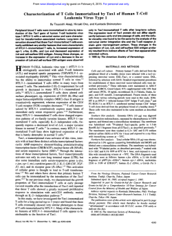
Double-Negative (CD4 - CD8 - ) T Cells From Adult T-cell
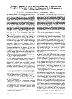
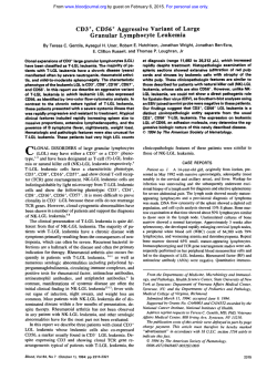
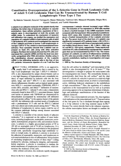
From www.bloodjournal.org by guest on February 6, 2015. For personal use only.
Double-Negative (CD4 - CD8 - ) T Cells From Adult T - c e l l Leukemia Patients
Also Have Poor Expression of the T - c e l l Receptor ap/CD3 Complex
By Hitoshi Suzushima, Norio Asou, Shintaro Nishimura, Kouji Nishikawa, Jian-xiang Wang, Toshiya Okubo,
Makoto Naito, Toshio Hattori, and Kiyoshi Takatsuki
W e present four patients with adult T-cell leukemia (ATL)
derived from a novel T-cell subset (CD4-, CD8- [doublenegative, DN], T-cell receptor [TCR] &). In the ATL cells
of these patients, neither gene nor surface expression of
CD4 and CD8 antigens was detected. Clinical and laboratory data showed no difference between DN-ATL and
CD4+ATL patients. In contrast to typical CD4+ATL cells,
DN-ATL cells were shown to express the protein and messenger RNA (mRNA) for S1 OO@ in immunocytochemical
assay and the reverse-transcription polymerase chain reaction assay. The mean fluorescence intensity of the TCR/
CD3 complex was extremely low in all four DN-ATL patients
as well as in typical CD4+ ATL. All four patients had TCR
@ and y chain gene rearrangements, with deletion of TCR
6 chain gene and mRNA expression for TCR a , @, and CD3
6 but not for TCR y and 6 chain genes. Thus, CD4- CD8TCR a@T cells are also a target for human T-cell lympho-
A
mice23-27 and these lymphocytes have also been detected in
the peripheral blood of patients with lupus nephritis." A human counterpart of the DN TCR a/3' lymphocyte has also
been detected in normal peripheral bloodz9 and skin,30 although the biologic role of this subset has not yet been well
characterized.
We recently reported of a patient with DN-ATL, in whom
leukemic cells proliferated in the gastrointestinal tract epitheli~m.~
In' vitro studies have shown that HTLV-I infects
not only T cells but also B cells and monocyte^.^*-^^ However,
HTLV-I-induced leukemic cells derived from CD4- T cells
have not yet been fully characterized. In this report, we describe the clinical features and the results of TCR gene analysis
in four patients with DN-ATL.
DULT T-cell leukemia (ATL)'.' is etiologically associated with the human T-cell lymphotropic virus type
I (HTLV-I).3.4By its diverse clinical features, ATL has been
classified into acute, chronic, lymphoma, and smoldering
types.' Most of the ATL cells found in any of these subtypes
are derived from CD3+, CD4+, CD8-, T-cell receptor (TCR)
ap' T cells, and they universally express activated T-cell antigens, such as HLA-DR and CD25,6although the expression
level of HLA-DR in acute ATL cells is very low compared
with that in chronic ATL cells.7 Diminished surface expression of the TCR/CD3 complex is also a striking feature of
ATL cells.'-"
TCR a and /3 chain expression by lymphocytes is associated
with the expression of either CD4 or CD8, which are accessory
molecules involved in major histocompatibility complex
(MHC) class 11- or class I-restricted T-cell recognition, re~pectively.'~-'~
Most mature lymphocytes that lack CD4 and
CD8 usually express the TCR yS hai in,'^-'^ which is not
MHC-restricted.16 A few lymphocytes with the CD4-, CD8(double-negative[DN]) TCR ab+ phenotype have been found
in the mouse t h y m ~ s , 'which
~ - ~ ~preferentially express TCR
VpS family gene product^."^^^^^^ Accumulation of DN TCR
ab' lymphocytes has been reported in the peripheral lymphoid organs of MRL/MP-lpr/lpr (Ipr) autoimmune
From the Second Department of Internal Medicine and the Second
Department of Pathology, Kumamoto University Medical School,
Kumamoto; and The Institute for Virus Research, Kyoto University,
Kyoto, Japan.
Submitted May 27, 1992; accepted October 15, 1992.
Supported by a grant-in-aid for ScientiJicResearchfrom the Ministry ofEducation, Science and CultureofJapan, and Science Research
expensesfor Health and WelfareProgramsfrom the Ministry of Health
and Welfare, Japan.
Address reprints request to Hitoshi Suzushima. MD, Second Department of Internal Medicine, Kumamoto UniversityMedical School,
1-1-1Honjo, Kumamoto 860, Japan.
The publication COSIS of this article were defrayed in part by page
charge payment. This article must therefore be hereby marked
"advertisement" in accordance with 18 U.S.C. section I734 solely to
indicate this fact.
0 1993 by The American Society ofHematology.
0006-4971/93/8 I 04-0008$3.00/0
1032
tropic virus type I-induced leukemogenesis. In addition,
expressionof the TCR 4 C D 3 complex on the DN-ATL cells
was further diminished by the addition of anti-CD3 or antiTCR @. monoclonal antibody. These results suggest that
the decreased expression of the TCR a@/CD3complex by
ATL cells plays a key role in the development of ATL. irrespective of CD4 expression.
0 1993 by The American Society of Hematology.
MATERIALS AND METHODS
Patients and cell line. Cells from four patients with DN-ATL
(three acute and one lymphoma type) were extensively studied. The
disease of one of these patients (patient no. 4) was previously reported." The diagnosis of ATL was made by the detection of serum
anti-HTLV-I antibody and the monoclonal integration of HTLV-I
proviral DNA in leukemic cells.35Clinical subtyping was performed
according to the previously described riter ria.^ Lymph node biopsy
was performed after obtaining informed consent according to the
guidelines of the Kumamoto University Medical School Committee
for the Protection of Human Subjects. The human myeloid leukemia
cell line (HL60), human T-cell leukemia cell lines (Jurkat and PEER),
and ATL cell line (SKT-IB)" used in this study were cultured in
RPMI 1640 medium supplemented with 10%heat-inactivated fetal
calf serum (FCS), 2 mmol/L I-glutamine, 100 U/mL penicillin, and
100 pg/mL streptomycin.
Cell preparation and phenotypic analysis. Peripheral blood
mononuclear cells (PBMNC) were obtained from normal volunteers
and the acute ATL by Ficoll-Conray density gradient centrifugation.
Normal T cells were then prepared from PBMNC by rosette formation
with sheep red blood cells (RBCs). Lymph node cells were obtained
from lymphoma type ATL by passing lymph node biopsy specimens
through a steel mesh.
The following murine monoclonal antibodies (MoAbs) were used
for indirect immunofluorescence assays: CD2 (OKTI I), CD3
(OKT3), CD4 (OKT4), and CDX (OKTX) (Ortho Diagnostics, Raritan, NJ); CD3 (Leu4), CD4 (Leu3a), CDX (Leu2a), and TCR a@
(WT3 1) (Beckton Dickinson Monoclonal Center, Mountain View,
CA); HLA-DR (Nula) (Nichirei CO, Ltd, Tokyo, Japan); TCR y6
Blood, Vol81, No 4 (February 15). 1993: pp 1032-1039
From www.bloodjournal.org by guest on February 6, 2015. For personal use only.
1033
DN-ATL CELLS HAVE POOR TCR/CD3 EXPRESSION
Table 1. Clinical Findings of Untreated Patients With DN-ATL
~
Patient
No
1
2
3
4
Organ Involvement
Clinical
Subtype
Age/Sex
Sk
Lu
GI
LN
Lymphoma
Acute
Acute
Acute
65/M
89/M
40/M
72/M
-
+
-
+
+
-
-
-
-
-
+
+
-
+
-
WBC
(XlOS/L)
LDH
(UlL)
Ca
(mg/dL)
Survival
7.2
19.2
41.8
17.1
647
1,949
1,812
4,103
9.2
11.7
11.0
9.5
13<
8
15
16
(mo)
Normal ranges: WBC, 3.5 to 8.5; LDH, 130 to 250; Ca. 8.7 to 10.3.
Abbreviations: Sk, skin; Lu. lung; GI, gastrointestinal tract; LN, lymph node; WBC, white blood cell count; LDH, lactic dehydrogenase.
(TCR 61 and 6TCSI) (T Cell Science Inc, Cambridge, MA); and
CD25 (anti-Tac; provided by Dr T. Uchiyama, Kyoto University).
The cells were further incubated with fluorescein isothiocyanate
(FITC)-conjugated goat F(ab)’, antimouse IgG (Sigma, St Louis, MO),
and analyzed with a FACScan (Becton Dickinson Immunocytometry
Systems). VAK5 (anti-p24 of human immunodeficiency virus type
1) was used as isotype-matched control Ab (IgG,) for WT3 1. PBMNC
from three normal volunteers were simultaneously analyzed with
OKT3 and WT31 as a control and the relative mean fluorescence
intensity (MFI) was calculated as follows:
MFI of the Antigen on Patient Cells
x 100.
Average MFI of the Antigen
on PBMNC From the 3 Normal Volunteers
Two-color analysis was performed using cells from patient no. 2, a
FACStar (Becton Dickinson Immunocytometry Systems), and the
following MoAbs: an FITC-labeled anti-CD4 MoAb (Nu-T,,,; Nichirei CO),an anti-CD25 MoAb (Cosmo Bio CO,Ltd, Tokyo, Japan),
an anti-TCR a@ MoAb (TCR-I), a PE-labeled antLCD8 MoAb
(LeuZa),an anti-CD4 MoAb (Leu3a; Becton Dickinson Monoclonal
Center), and an anti-CD25 MoAb (Immunotech SA, Marseille,
France). Terminal deoxynucleotidyl transferase (TdT) was detected
with indirect immunofluorescenceassay using a Capi kit (Calpis Food
Industries, Tokyo, Japan) according to the manufacturer’s instructions. TCR a@ was also examined with immunocytochemical staining
using @F1(T Cell Science Inc). Peroxidase-antiperoxidase(PAP)staining technique with anti-S100 were performed on methanol-fixed
PBMNC cytospin smears with an S lOO/PAP kit (DAKO Corporation,
Carpinteria, CA).
Southern and Northern blot analysis. DNA was prepared from
the mononuclear cells of each patient by proteinase K digestion followed by phenol-chloroform extraction. The DNA was digested with
restriction enzymes, subjected to electrophoresis on 0.7% agarose
gels, and transferred to nitrocellulose filters (Hybond C extra; Amersham Intemational plc, Buckinghamshire, UK). The filters were hy-
bridized with radiolabeled probes at 42°C for 12 hours, washed, and
then exposed to x-ray film at -80°C.
Total cellular RNA was isolated by ultracentrifugation on a guanidinium isothiocyanate/CsClz gradient. Ten micrograms of total
RNA was subjected to electrophoresis on 1% agarose-formaldehyde
denaturing gel, and was transferred to a nitrocellulose filter (Hybond
C extra). Hybridization was then performed as described above. The
following probes were used for Southern and Northern blotting: a
0.39-kb Hpa 11-Hpa I1 fragment of the Ca gene,36a 1.6-kb EcoRIEcoRI fragment of a Cy probe:’ a 1.5-kb EcoRI-EcoRI fragment of
cDNA containing the C6-specific segment3*(provided by Dr T.W.
Mak), a 3.0-kb HindIII-EcoRI fragment hybridizing to the CO1 gene,
a 4.0-kb EcoRI-EcoRI fragment containing the J/32 gene39(provided
by Dr H. Sakano), a 0.62-kb Pst I-Xho I fragment of the CD3 6 gene4’
(provided by Dr C. Terhorst), a 0.7-kb HindIII-EcoRI fragment hybridizing to the J y l gene4’ (provided by Dr T.H. Rabbitts), and a
1.5-kb Pst I-BamHI fragment of a CD8 probe4’ (provided by Dr P.
Kavathas). The CD4 probe was synthesized from Jurkat cells by the
reverse transcription-polymerase chain reaction (RT-ER). The 0.77kb NCOI-Taq I fragment of a p-actin probe was purchased from
Oncor Inc43(Gaithersburg, MD).
RT-PCR. Ten micrograms of cellular RNA was subjected to reverse transcription with 20 pmol 3’-specific primer and IO U avian
myeloblastosis virus reverse transcriptase (Seikagaku Kogyo CO,Ltd,
Tokyo, Japan) at 37°C for I hour. One tenth of each c-DNA sample
was coamplified using a 5‘- and 3’-SlOOP primer at a final concentration of 0.2 pmol/L in each reaction. Amplification was performed
with 2.5 U Taq polymerase (Thermus aquaticus DNA polymerase;
Bethesda Research Laboratories Life Technologies, Inc, Gaithersburg,
MD) in a Cetus/Perkin-Elmer thermocycler for 30 cycles under the
following conditions: melting at 95°C for 30 seconds, annealing at
55°C for 30 seconds, and extension at 72°C for 1 minute. The 5’sense SlOO@ primer used was 5‘ TGC AGC AAG GAG ACC AGG
AA 3’ (residues 33-52 in the first exon) and the 3’-antisense primer
was 5‘ GAA CTC GTG GCA GGC AGT AG 3’ (residues 335-316
in the third exon)?4 After the reaction, one tenth of each sample was
Table 2. Surface PhenotvDes of DN-ATL Cells
Patient
No.
CD2
( T I 1)
1
98
2
99
3
99
4
85
CD4
(OKT4)
(Leu3a)
3
6
3
3
11
4
1
L
CD8
(OKT8)
(Leu2a)
8
7
2
7
9
2
0
1
CD3 (OKT3)
8
(1 4/700:2)
49
(58/730:8)
98
(104/650: 16)
89
(150/750:20)
TCRag
(WT31)
TCRy6
(TCR61)
(6TCS1)
5
15/145:10)
9
(30/151:20)
81
(39/140:28)
53
(59/155:38)
1
0
1
0
4
0
3
0
HLA-DR
(Nu-la)
CO25
(Tad
TdT
10
95
0
6
36
0
9
9
0
14
92
0
Numbers are actual percentages of positive cells. Values in parentheses are MFl/nMFI:rMFI.
Abbreviations: rMFI, relative MFI; nMFI, average MFI value of the amntigen on PBMNC from the three normal volunteers.
From www.bloodjournal.org by guest on February 6, 2015. For personal use only.
1034
SUZUSHIMA ET AL
tained at diagnosis are shown in Table 1. All patients were
males aged 40 to 89 years. Three had acute ATL and the
other lymphoma ATL. Proliferation of leukemic cells in the
skin, lungs, and/or gastrointestinal tract was observed in three
of the four DN-ATL patients. The morphologies of peripheral
blood cells corresponded to that of typical ATL cells with
convoluted and coarse nuclei. Two of the four DN-ATL patients had hypercalcemia, which is frequently observed in
ATL.5All four patients received intensive chemotherapy and
their survival did not differ from that of patients with CD4+
acute ATL.5
Immunophenotypic analysis. The results of phenotypic
analysis of DN-ATL cells at diagnosis are shown in Table 2.
More than 85% of the cells from all DN-ATL patients were
CD2+and the cells from these patients were also CD4-, CD8-,
and TCR 78- (less than 12%). Lack of CD4 or CD8 antigens
was confirmed using two different MoAbs in each case (CD4,
OKT4 and Leu3a; CD8,OKT8 and Leu2a). Two-color analysis showed that most of the CD25' cells of patient no. 2
were CD4- and CD8- (Fig I). Cells from three DN-ATL
patients had CD25 antigens, whereas the expression of HLADR antigens on cells from any of the four patients was very
low, a similar finding to typical acute CD4+ATL.' Cells from
all three acute DN-ATL patients were CD3+, but the MFI
values were extremely low compared with those for normal
T cells. The lymph node cells from the lymphoma type DNATL patient were CD3-. Cells from two DN-ATL patients
were TCR a@- (less than 10%). However, MFI value for TCR
a@ in patient no. 2 as well as patients no. 3 and 4 was higher
than those for VAKS, isotype-matched negative control Ab.
Similar to the results for CD3 antigen expression, the MFI
values for TCR a@ chain expression by DN-ATL cells were
significantly decreased. These observations were confirmed
by the expression of TCR a@ chain with the other MoAb,
n
.
I
0
0
U
U
c D 2s
c 0 25
Fig 1. Two-cdor immunofluorescence analysis of peripheral
blood " n u c l e a r cells from an acute DN-ATL patient (patient no.
2). Cells were simultaneously analyzed with FITC- or PE-labeled
MoAbs, as indicated beside each dot plot. The percentages of cells
in each population are shown in the boxes.
subjected to Southern blot analysis. The probes used for hybridization
were 20-mer oligonucleotides derived from 3'-primers by S-juxtaposition and labeled by the Sendlabeling method.
RESULTS
ClinicalJieaturesand laboratory data of DN-ATL. 'The
clinical and laboratory data of the patients in this study ob(0
0
(
0
P
a
I
-I
1
2
3
I
4
-10.5
CB1
J71
1
2
3
4
--
18.5
13.5
-4.0
- 11.0
JB2
C6
Y
-
Y-
- 4.0
kb
kb
Fig2. S o u t h e m h y b r i d i
analysis of the TCR B (CB1 or
582)chain g e m in patients with
DN-ATL using EcoRI-digested
DNA. TCRr(J.rl) and 6 (a)
chain
genes were also analyzed using
BemHI-digested DNA. HL60
DNA sewed as a control for the
gennline configuration.The lane
numbers correspond to the patient numbers listed in the tables.
From www.bloodjournal.org by guest on February 6, 2015. For personal use only.
1035
DN-ATL CELLS HAVE POOR TCR/CD3 EXPRESSION
TCR
TCR
-1.6
-1.3
e
pF
rwwwm=~-
rp
- 1.7
5
- 2.2
-
- 1.5
a
-1.3
1.0
kb
E
&
Fig3. Northemhybrid*ation
analysis of total cellular RNA
from DN-ATL cells using constant region probes for the TCR
a,B, 7, and 8 chain genes, the
CD3 ti chain gene, and the CD4
and CD8 antigen genes. RNA
from Jurkat, SKT-1B, and PEER
cells sewed as the positive control. The lane numbers correspond to the patient numbers
listed in the tables.
5
CD3 - ' *
6
/3FI. Over 90% of cells in patients no. 2 and 4 were reacted
weakly with BFI. TdT was negative (less than 15%) in cells
from all DN-ATL patients.
TCR a, 8. y, and 6 chain gene rearrangements. To characterize further the TCR molecules in DN-ATL cells, we first
examined the gene configuration of each chain of the TCR
by Southern blot analysis. In patients no. 1 and 2, whose
cells did not show surface expression of the TCR, the TCR
/3 and y chain genes were rearranged, as they also were in
patients no. 3 and 4 (Fig 2). The TCR 6 chain gene, which
lies between the V a and Ja loci, showed biallelic deletion in
all cases, suggesting that TCR a chain genes were assembled.
The profile of rearrangements of the TCR /3 and y chain
genes were different in each case (Fig 2).
Expression of TCR. CD3 6. CD4, and CD8 genes. To
verify whether the absent or decreased surface expression of
TCR, CD4, and CD8 antigcns on DN-ATL cells was caused
by a lack of gene expression, we examined the mRNA for
these molecules by Northern blot analysis (Fig 3). Mature
transcript.. for the TCR (Y and /3 chain genes were detected
in DN-ATL, but TCR y and 6 chain mRNA could not be
found in any of the DN-ATI, cells. CD3 d mRNA was also
expressed in all cases. These results indicate that abnormal
surface expression of the TCR/CD3 complex by DN-ATL
cells was not caused by defective transcription of this complex.
CD4 and CD8 mRNA were not detectable in any of the DNATL cells (Fig 3), confirming the lack of surface expression
of these antigens.
Effectofanri-CD3MoAb on sut$ace expression ofihe TCR/
CD3 complex. In CD4' ATL cells, expression of the TCR/
CD3 complex on the cell surface decreases following stimulation with anti-CD3 MoAb? To examine whether expres-
1
2
3
4
3.0
0.1
cm
-2.0
- 2.4
CO8
kb
kb
sion of the TCR/CD3 complex by DN-ATL cells would also
be diminished despite their lack of CD4 and CD8 antigens,
cells from patient no. 4 were used. Similar to CD4' ATL
cells, the surface expression of CD3 antigen was diminished
normal T cell
DN-ATL
anti-CD3 (Leu41
Fig4. Representativehistograms of CD3 antigen expression after stimuli with anti-CD3 MoAb. PBMNC from patient no. 4 and
m a l PBMNC were cultured with 1 rg/mL OKT3 MoAb. After 18
hours, the cells were stained with Leu4 and analyzed as described
in Materials and Methods. The MFI values for CD3 antigen expression are shown in the upper right-hand part of each histogram.
From www.bloodjournal.org by guest on February 6, 2015. For personal use only.
SUZUSHIMA ET AL
1036
D
by the addition of I pg/mL OKT3 MoAb for 18 hours to
cells from patient no. 4 or normal PBMNC (Fig 4). We also
obtained a similar result after the addition of 1 pg/mL WT3 I
MoAb (data not shown).
Expression of the gene and protein of SlOOS by DN-ATL
cells. Sl00/3 protein was recently reported to be expressed
by the cells from some patients with aggressive T-cell chronic
lymphoproliferativedisease (T-CLPD),which showed a double-negative phen0type.4~To examine whether DN-ATL cells
also expressed SlOOfi, we analyzed the product and mRNA
for this gene in DN-ATL and CD4' ATL by immunocytochemical assay and the RT-PCR. We examined the cytoplasmic SI00 protein in PBMNC from patients no. I and 2
using the PAP-staining technique. In the majority of the cells
from both patients, SI00 protein was detected with strong
or weak intensity in each cell (Fig 5C and D). In contrast,
this protein was not detected in either the cells from a patient
with CD4'ATL or normal PBMNC (Fig SA and B). A band
corresponding to the amplified SlOO/3gene was clearly shown
in DN-ATL patients no. I through 3, and was barely detectable in CD4' ATL (Fig 6). However, these faint bands
were considered to be caused by contamination with normal
SIOOfi-positivecells, because no band was detected in SKTI B cells derived from the original CD4' ATL cells.
DISCUSSION
Most ATL cells have the CD3+, CD4+, CD8-, and TCR
as+ phenotype, so that ATL has been considered to be a
mature T-cell leukemia! This report is of four patients with
Fig 5. The expression of
S100 protein in the cytoplasm
of PBMNC smear from a normal
volunteer (A), a patient with
CD4+ ATL (B), patient no. 1 (C).
and patient no. 2 (D). PAPstaining technique was used in
this assay.
ATL who express an aberrant phenotype of CD4-, CD8-. It
is unlikely that this double-negative phenotype was caused
by lack of the OKT4 epitope. The lack of CD4 and CD8
mRNA expression in these patients confirmed that their leukemic cells were derived from a double-negativesubset. It is
also unlikely that this double-negative phenotype was derived
from immature T cells because of its expression of the CD3
antigen but not TdT. Most mature lymphocytes having a
double-negative phenotype are known to bear the TCR y6
chain. However, leukemic cells from all the DN-ATL patients
did not react with anti-TCR y6 MoAbs. On the other hand,
cell surface expression of TCR a@ was observed in two patients with acute ATL (patients no. 3 and 4), although the
MFI values were low. The leukemic cells from all DN-ATL
patients had rearrangements of the TCR /3 and y chain genes
associated with allelic deletion of the TCR 6 chain gene. This
genotype is usually observed in CD3+ TCR a@' mature Tcell neoplasia, including ATL.39*46Furthermore, leukemic
cells from all the DN-ATL patients showed expression of
TCR a@and CD3 6 mRNA but not TCR y6 chain mRNA.
These findings indicate that the DN-ATL cells were derived
from a TCR a/3-positivebut TCR 76-negative subset. Some
ATL patients with an aberrant phenotype have been reported
previously, such as CD2-, CD4' CD8' (double-positive),or
CD4- CD8' (CD8 ~ingle-pitive).4'~~
Similar to typical ATL,
the ATL cells of these patients also showed surface expression
of the TCR afi chain, and no ATL cells expressing the TCR
y6 chain have ever been reported to our knowledge. Taken
together with these observations, our DN-ATL patients show
From www.bloodjournal.org by guest on February 6, 2015. For personal use only.
1037
DN-ATL CELLS HAVE POOR TCR/CD3 EXPRESSION
1
2
3
4
5
6
7
SlOO4
- 303
Fig 6. Sl 00,9 gene mRNA expressionin DN-ATL cells. The RTPCR was used for the detection of S1008 mRNA as described in
Materials and Memods. The size of the amplied SlOOB DNA was
303 bp. TCR CU was used as an internal control. Lane 1, SKT-1B;
lane 2, patient no. 1; lane 3, patient no. 2; lane 4, patient no. 3;
lanes 5 to 7, CD4* ATL.
that TCR a@+ T cells with or without the CD4 phenotype
are the sole target of HTLV-I-induced leukemogenesis.
A subset of TCR aj3 lymphocytesthat lacks CD4 and CD8
expression has been detected in normal human peripheral
blood; these cells responded to interleukin-2 (IL-2), IL-3, and
IL-4, and had lytic activity when their TCR complex was
activated.29The DN-ATL cells we examined only responded
to IL-2 and IL-4, similar to typical ATL cells.5oCD4-, CDK,
TCRaF lymphocyteshave also been detected in normal human skin, and they produced IL-2. tumor necrosis factor a
(TNF-a), and interferon gamma (IFN-y) when they were
stimulated with anti-CD3 MoAb and phorbol myristate acetate.." Interestingly, they had the same surface phenotype
as DN-ATL cells (CD2+,CD7-, CD25', except for HLADR') after they were cultured with 1L-2 for 4 weeks, and the
leukemic cells from one of our DN-ATL patients expressed
mRNA of TNF-a and IFN-y without any stimulation (Suzushima et al. unpublished data). However, in this study only
one of the four DN-ATL patients had prominent skin invasion by ATL cells. Therefore, it remains unclear whether
DN-ATL cells originate from DN-T cells residing in the skin.
Several cases of DN-T-cell lymphoma have been reported,
which had many features in common with our DN-ATL
cases." They were widespread at presentation, with frequent
cutaneous, pulmonary, and bone marrow involvement, and
they have a rapid course even with aggressive combined chemotherapy. However, HTLV-I or TCR studies were not performed in those lymphomas. DN-T-CLPD associated with
massive hepatosplenomegaly but without significant lymphadenopathy or cutaneous involvement have been reported
recently, and leukemic cells from those patients expressed
SlOOj3 protein, which is known to be expressed by less than
3% of normal circulating lymphocyte^.^' Although these
SlOOj3+ normal lymphocytes have a suppressor immunophenotype, with expression of CD2, CD8, and CDl I b, their
role in normal T-cell function remains o b s c ~ r e . ~
We
~-~~
showed that cells from the DN-ATL we examined expressed
the product and mRNA for the SlOOBgene. Although HTLVI proviral DNA was not detected in SI OO&positive CLPD?'
it seems that the normal counterpart of the leukemic cells in
DN-ATL is the same as that in SlOOj3-positive CLPD.
In typical CD4' ATL cells, the MFI values of TCR aj3
and CD3 antigens are specifically decxa~ed.~-"
Furthermore,
surface expression of the TCR/CD3 complex by leukemic
cells from lymphoma type ATL is lower than in acute
ATL.'.IO." In addition, the relative proportion of TCR aj3
antigen-positive cells is lower than that of CD3 antigen-positive cells using our antibodies,'.'0*" resulting in a CD3', TCR
aj3- phenotype." Among the four DN-ATL patients, the MFl
values for TCR CUBand CD3 antigens were decreased in two
cases of acute ATL (patients no. 3 and 4). Leukemic cells
from the lymphoma type ATL patient (patient no. 1) had no
surface expression ofeither TCR aj3 or CD3 antigens, whereas
cells from the remaining acute ATL patient (patient no. 2)
reacted with anti-CD3 but not anti-TCR a@ MoAb. Thus,
surface expression of the TCR/CD3 complex was specifically
decreased in DN-ATL as well as in typical ATL. This supports
our hypothesis that diminished expression of this complex
plays a key role in the development of ATL. Because expression ofthe TCR/CD3 complex on DN-ATL cells was further
diminished by the addition of anti-CD3 or anti-TCR a@
MoAbs similar to that on CD4'ATL cells: it seems that the
TCR/CD3 complex is downmodulated by some in vivo
stimuli, although these cells have neither the CD4 nor CD8
antigens that are necessary for assembling MHC-TCR complexes.
In mice, a subset of TCR aj3 lymphocytes without CD4
or CD8 antigen expression has been detected among mature
thymocyte^,'^-^^ which preferentially expresses TCR Vj3 8
gene p r o d u ~ t s . ' ~The
* ' ~DN-ATL
~~~
cells we examined did
not have a common TCR Vj3 family gene product (Suzushima
et al, unpublished data). Expansion of DN-T cells has also
been reported in autoimmune mice homozygous for the /pr/
lpr (lpr) gene23-27
and in peripheral blood lymphocytes from
systemic lupus erythematosis patients.2xThese /pr DN-T cells
had low surface levels of TCR aj3 protein and normal or
increased amounts of TCR a and j3 mRNA.23These similarities between / p i DN-T cells and DN-ATL cells raise a
possibility that both types ofcells respond to unknown stimuli
acting via the TCR aj3 and causing downmodulation of TCR/
CD3 complex expression on the cell surface. The TCR a/?is
considered to recognize antigens presented by MHC molecules acting with CD4 or CD8, and it is not known whether
DN-T cells that bear TCR a@ are able to recognize antigens.
However, Ipr DN-T cells are reported to be involved in the
generation of autoantibodies by interaction with surface Igs
on Lyl+ B cells through the TCR
The proliferation of
DN TCR as" cells was also reported in a patient with combined immunodeficiency.s6These DN-T cells recognized cu-
From www.bloodjournal.org by guest on February 6, 2015. For personal use only.
SUZUSHIMA ET AL
1038
taneous tissue and were responsible for the reactions resembling those of graft-versus-host disease. Further analysis of
TCR a@ligands is considered to provide some insight into
the mechanism of the possible downmodulation of surface
expression of the TCR/CD3 complex in DN-ATL.
ACKNOWLEDGMENT
We thank Drs T.M. Mak, H. Sakano, T.H. Rabbitts, C. Terhorst,
and P. Kavasath for providing DNA probes; Dr T. Uchiyama for
providing Tac monoclonal antibody; and Drs T. Watanabe, H. Natori,
and R. Nakano for providing the samples in this study.
REFERENCES
I . Takatsuki K, Uchiyama T, Sagawa K, Yodoi J: Adult T cell
leukemia in Japan, in Seno S, Takaku F, Irino S (eds): Topics in
Hematology. Amsterdam, The Netherlands, Excepta Medica, 1977,
P 73
2. Uchiyama T, Yodoi J, Sagawa K, Takatsuki K, Uchino H:
Adult T-cell leukemia: Clinical and hematologic features of 16 cases.
Blood 5048 I , 1977
3. Poiesz BJ, Ruscetti FW,Gazdar AF, Bunn PA, Minna JD,
Gallo R C Detection and isolation of type-C retrovirus particles from
fresh and cultured lymphocytes of a patient with cutaneous T-cell
lymphoma. Proc Natl Acad Sci USA 77:7415, 1980
4. Hinuma Y, Nagata K, Hanaoka M, Nakai M, Matsumoto T,
Kinoshita K, Shirakawa S, Miyoshi I: Adult T cell leukemia: Antigen
in an ATL cell line and detection of antibodies to the antigen in
human sera. Proc Natl Acad Sci USA 78:6476, I980
5. Kawano F, Yamaguchi K, Nishimura H, Tsuda H, Takatsuki
K: Variation in the clinical courses of adult T-cell leukemia. Cancer
55:851, 1985
6. Hattori T, Uchiyama T, Tobinai T, Takatsuki K, Uchino H:
Surface phenotype of Japanese adult T-cell leukemia cells characterized by monoclonal antibodies. Blood 58:645, 1981
7. Shirono K, Hattori T, Hata H, Nishimura H, Takatsuki K
Profiles of expression of activated cell antigen on peripheral blood
and lymph node cells from different clinical stages of adult T-cell
leukemia. Blood 73: 1664, 1989
8. Tsuda H, Takatsuki K: Specific decrease in T3 antigen density
in adult T-cell leukemia cells: I. Flow microfluorometric analysis. Br
J Cancer 50:843, 1984
9. Matsuoka M, Hattori T, Chosa T, Tsuda H, Kuwata S, Yoshida
M, Uchiyama T, Takatsuki K: T3 surface molecules on adult T cell
leukemia cells are modulated in vivo. Blood 67:1070, 1986
IO. Shirono K, Hattori T, Matsuoka M, Asou N, Takatsuki K:
Adult T cell leukemia cell lines that originated from primary leukemic
clones also had a defect of expression of CD3-T cell receptor complex.
Leukemia 2:728, 1988
11. Suzushima H, Hatton T, Asou N, Wang J, Nishikawa K,
Okubo T, Anderson P, Takatsuki K Discordant gene and surface
expression of the T-cell receptorlCD3 complex in adult T-cell leukemia cells. Cancer Res 5 1 :6084, 1991
12. Gay D, Maddon P, Sekaly R, Talle MA, Godfrey M, Long E,
Goldstein G, Chess L, Axel R, Kappler J, Marrack P: Functional
interaction between human T-cell protein CD4 and the major histocompatibility complex HLA-DR antigen. Nature 328:626, I987
13. Gabert J, Langlet C, Zamoyska R, Pames JR, Schmitt-Verbulst
AM, Malissen B Reconstitution of MHC class I specificity by transfer
of the T cell receptor and Lyt-2 genes. Cell 50545, 1987
14. Brenner MB, McLean J, Dialynas DP, Strominger JL, Smith
JA, Owen F‘L,Seidman JG, Ip S, Rosen F, Krangel MS: Identification
of a putative second T-cell receptor. Nature 322:145, 1986
15. Fenini S, Bottino C, Biassoni R, Poggi A, Sekaly RP, Moretta
L, Moretta A: Characterization of CD3+, CD4-, CD8- clones ex-
pressing the putative T cell receptor y gene product. J Exp Med 166:
277, 1987
16. Lanier LL, Ruitenberg JJ, Phillips JH: Human CD3+ T lymphocytes that express neither CD4 nor CD8 antigens. J Exp Med
164:339, 1986
17. Budd RC, Miescher GC, Howe RC, Lees RK, Bron C,
MacDonald HR: Developmentally regulated expression of T cell receptor p chain variable domains in immature thymocytes. J Exp
Med 166577, 1987
18. Fowlkes BJ, Kruisbeek M, Ton-That H, Weston MA, Coligan
JE, Schwartz RH, Pardoll DM: A novel population of T-cell receptor
CY@
bearing thymocytes which predominantly expresses a single Vp
gene family. Nature 329:25 I , I987
19. Crispe IN, Moore NW, Husmann LA, Smith L, Bevan MJ,
Shimonkevitz R P Differentiation potential of subsets of CD4-CD8thymocytes. Nature 329:336, 1987
20. Ceredig R, Lynch F, Newman P: Phenotypic properties, interleukin 2 production, and developmental origin of a “mature” subpopulation of Lyt-2- L3T4- mouse thymocytes. Proc Natl Acad Sci
USA 84:8578, 1987
21. Wilson A, Ewing T, Owens T, Scollay R, Shortman K: T cell
antigen receptor expression by subsets of Ly-2-L3T4- (CD4-CD8-)
thymocytes. J Immunol 1401470, 1988
22. Miescher GC, Howe RC, Lees RK, MacDonald H R CD3associated alp and y/6 heterodimeric receptors are expressed by distinct populations of CD4-CD8- thymocytes. J Immunol 140 1779,
1988
23. Miescher GC, Budd RC, Lees RK, MacDonald H R Abnormal
expression of T cell receptor genes in Lyt2-L3T4- lymphocytes of
/pr mice: Comparison with normal immature thymocytes. J Immunol
138:1959, 1987
24. Hashimoto Y, Yui K, Littman D, Greene M: T-cell receptor
genes in autoimmune mice: T-cell subsets have unexpected T-cell
receptor gene programs. Proc Natl Acad Sci USA 84583, 1987
25. Budd RC, Schreyer M, Miescher GC, MacDonald HR: T cell
lineages in the thymus of Ipr/lpr mice. J Immunol 139:2200, 1987
26. Marcos MA, Toribio ML, de la Hera A, Marquez C, Gaspar
ML, Martinez AC: Mutual cell interactions and the selection of immune repertories. Immunol Today 9:204, 1988
27. Davignon JL, Cohen PL, Eisenberg RA: Rapid T cell receptor
modulation accompanies lack of in vitro mitogenic responsiveness
of double negative T cells to anti-CD3 monoclonal antibody in MRL/
Mp-lpr//pr mice. J Immunol 141:1848, 1988
28. Shivakumar S, Tsokos GC, Datta SK: T cell receptor alp expressing double negative (CD4-/CD8-) and CD4’ T helper cells in
human augment the production of pathogenic anti-DNA autoantibodies associated with lupus nephritis. J Immunol 143:103, 1989
29. Londei M, Verhoef A, Berardinis PD, Kissonerghis M, Grubech-Loebenstein BG, Feldmann M: Definition of a population of
CD4-8-T cells that express the ap T-cell receptor and respond to
interleukin 2, 3, and 4. Proc Natl Acad Sci USA 86:8502, 1989
30. Groh V, Fabbi M, Hochstenbach F, Maziarz RT, Strominger
JL: Double-negative (CD4-CDK) lymphocytes bearing T-cell receptor CY and p chains in normal human skin. Proc Natl Acad Sci USA
865059, 1989
31. Hattori T, Asou N, Suzushima H, Takatsuki K, Tanaka K,
Naito K, Natori H, Oizumi K Leukemia of novel gastrointestinal
T-lymphocyte population infected with HTLV-I. Lancet 337:76, 1991
32. Yamamoto N, Matsumoto Y, Koyanagi Y, Tanaka Y, Hinuma
Y: Unique cell lines harbouring both Epstein-Barr virus and adult
T-cell leukemia virus, established from leukemia patients. Nature
299:367, 1982
33. Longo DL, Gelmann EP, Cossman J, Young RA, Gallo RC:
Isolation of HTLV-I-transformed B-lymphocyte clone from a patient
with HTLV-associated adult T-cell leukemia. Nature 3 10:505, 1984
From www.bloodjournal.org by guest on February 6, 2015. For personal use only.
DN-ATL CELLS HAVE POOR TCR/CD3 EXPRESSION
34. Dhawan S, Streicher HZ, Wahl LM, Miller N, Louie AT,
Goldfarb IS, Jackson WL, Casali P, Notkins AL: Model for studying
virus attachment: 11. Binding of biotinylated human T cell leukemia
virus type I to human blood mononuclear cells potential targets for
human T cell leukemia virus type I infection. J Immunol 147:102,
1991
35. Yoshida M, Seiki M, Yamaguchi K, Takatsuki K Monoclonal
integration of human T-cell leukemia provirus in all primary tumors
of adult T-cell leukemia suggests causative role of human T-cell leukemia virus in the disease. Proc Natl Acad Sci USA 81:2534, 1984
36. Tanagi Y, Chan A, Chin B, Minden M, Mak T W Analysis
of cDNA clones specific for human T cells and the (Y and 0 chains
of the T-cell receptor heterodimer from a human T-cell line. Proc
Natl Acad Sci USA 82:3430, 1985
37. Yoshikai Y, Toyonaga B, Koga Y, Kimura N, Griesser H,
Mak T W Repertorie of the human T cell gamma genes: High frequency of nonfunctional transcripts in thymus and mature T cells.
Eur J Immunol 17:119, 1987
38. Takihara Y, Champagne E, Griesser H, Kimura N, Tkachuk
D, Reimann J, Okada A, Alt FW,Chess L, Minden M, Mak T W
Sequence and organization of the human T cell 6 chain gene. Eur J
Immunol 18:283, 1988
39. Matsuoka M, Hagiya M, Hattori T, Asou N, Maeda S, Shimada
K, Tsai SC, Sakano H, Takatsuki K Gene rearrangements of T cell
receptor /3 and y chains in HTLV-I infected primary neoplastic T
cells. Leukemia 2234, 1988
40. van den Elsen P, Shepley B-A, Borst J, Coligan JE, Markham
AF, Orkin S, Terhorst C Isolation of cDNA clones encoding the
20K T3 glycoprotein of human T-cell receptor complex. Nature 3 12:
413, 1984
4 1. Lefranc M-P, Rabbitts TH: Two tandemly organized human
genes encoding the T-cell y constant-region sequence show multiple
rearrangement in different T-cell types. Nature 3 16:464, 1985
42. Kavasath P, Sukhatme VP, Herzenberg LA, Parnes J P Isolation of the gene encoding the human T-lymphocyte differentiation
antigen Leu-2 (T8) by gene transfer and cDNA subtraction. Proc
Natl Acad Sci USA 81:7688, 1984
43. Cleveland DW, Lopata MA, MacDonald RJ, Cowan NJ, Rutter WJ, Kirschner MW: Number and evolutionary conservation of
a-and /3-tubulin and cytoplasmic 0- and y-actin genes using specific
cloned cDNA probes. Cell 20:95, 1980
44. Allore RJ, Friend WC, OHanlon D, Neilson KM, Baumal
R, Dunn RJ, Marks A Cloning and expression of the human S IOOP
gene. J Biol Chem 265:15537, 1990
45. Hanson CA, Bockenstedt PL, Schnitzer B, Fox DA, Kueck
B, Braun DK: S 100-positive,T-cell chronic lymphoproliferative dis-
1039
ease: An aggressive disorder of an uncommon T-cell subset. Blood
78:1803, 1991
46. Asou N, Hattori T, Matsuoka M, Kawano F, Takatsuki K
Rearrangements of T-cell antigen receptor 6 chain gene in hematologic
neoplasms. Blood 74:2707, 1989
47. Yamada Y, Kamihira S, Amagasaki T, Kinoshita K, Kusano
M, Ikeda S, Toriya K, Suzuyama J, Ichimaru M: Changes of adult
T cell leukemia cell surface antigens at relapse or at exacerbation
phase after chemotherapy defined by use of monoclonal antibodies.
Blood 64:440, 1984
48. Matsumoto T, Matsumoto M, Kikuchi H, Yunoki K: Immunologic characterization of lymphoid tissue tumor cells from adult
T-cell leukemia (ATL) and peripheral T-cell lymphoma (PTCL) in
an ATL-endemic area. Jpn J Clin Hematol 27:693, 1986
49. Kawano F, Tsukamoto A, Satoh M, Sanada I, Shido T, Suzushima H, Asou N, Takatsuki K Poorly expressed CD2 antigen on
the leukemic cells of adult T-cell leukemia implicate more aggressive
clinical course? Leuk Res 14: 107 1, 1990
50. Hattori T, Sakai K, Suzushima H, Asou N, Takatsuki K, Matsuzaki Y, Nakauchi H, Natori H, Oizumi K Double negative (CD4-,
CD8-) TCR aP' adult T cell leukemia cells proliferated vigorously
in the presence of IL-2 or IL-4. Myelodysplastic syndrome and cytokines. Excepta Medica, 1990, p 137
5 I . Grogan TM, Fielder K, Range1 C, Jolley U, Wirt DP, Hicks
MJ, Miller TP, Brooks R, Greenberg B, Jones S Peripheral T-cell
lymphoma: Aggressive disease with heterogeneous immunotypes. Am
J Clin Pathol 83:279, 1985
52. Takahashi K, Yoshino T, Sonobe H, Kayashi K: Immunocytochemical characterization of S- 1OOP-positive human T-lymphocytes by a double immunostaining method. Virchows Arch [B] 53:
375, 1987
53. Sansoni P, Rowden G, Manara GC,Ferrari C, De Panfilis G:
S- 100-positive T-cells are largely restricted to a CD8-positive, 9.3negative subset. Virchows Arch [B] 53:301, 1987
54. Ferrari C, Sansoni P, Rowden G, Manara GC, Torresani C,
De Panfilis G: One half of the CD 1 1b+ human peripheral blood Tlymphocytes coexpresses the S-100 protein. Clin Exp Immunol 72:
357, 1988
55. De Panfilis G, Rowden G, Manara GC,Ferrari C, Torresani
C, Sansoni P: The S-IOOP protein in normal human peripheral blood
is uniquely present within a discrete suppressor-T-cell compartment.
Cell Immunol 144:398, 1988
56. Wirt DP, Brooks EG, Vaidya S, Klimpel GR, Waldmann TA,
Goldblum RM: Novel T-lymphocyte population in combined immunodeficiency with features of graft-versus-host disease. N Engl J
Med 321:370, 1989
From www.bloodjournal.org by guest on February 6, 2015. For personal use only.
1993 81: 1032-1039
Double-negative (CD4- CD8-) T cells from adult T-cell leukemia
patients also have poor expression of the T-cell receptor alpha
beta/CD3 complex
H Suzushima, N Asou, S Nishimura, K Nishikawa, JX Wang, T Okubo, M Naito, T Hattori and K
Takatsuki
Updated information and services can be found at:
http://www.bloodjournal.org/content/81/4/1032.full.html
Articles on similar topics can be found in the following Blood collections
Information about reproducing this article in parts or in its entirety may be found online at:
http://www.bloodjournal.org/site/misc/rights.xhtml#repub_requests
Information about ordering reprints may be found online at:
http://www.bloodjournal.org/site/misc/rights.xhtml#reprints
Information about subscriptions and ASH membership may be found online at:
http://www.bloodjournal.org/site/subscriptions/index.xhtml
Blood (print ISSN 0006-4971, online ISSN 1528-0020), is published weekly by the American
Society of Hematology, 2021 L St, NW, Suite 900, Washington DC 20036.
Copyright 2011 by The American Society of Hematology; all rights reserved.


© Copyright 2026