
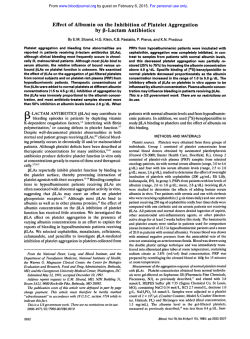
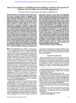
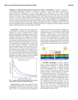
Platelet Glycoprotein IIb-III, (ffIIbp3 Integrin) Confers Fibrinogen
From www.bloodjournal.org by guest on February 6, 2015. For personal use only.
Platelet Glycoprotein IIb-III, ( f f I I b p 3 Integrin) Confers Fibrinogen- and
Ac tiva tion-Dependen t Aggregation on Heterologous Cells
By Mony M. Frojmovic, Timothy E. O‘Toole, Edward F. Plow, Joseph C. Loftus, and Mark H. Ginsberg
To analyze molecular mechanisms of platelet aggregation,
w e have studied the aggregation of Chinese hamster ovary
(CHO) cells expressing between 1 and 4 x lo5 recombinant
human glycoprotein (GP) 11,-111, molecules per cell (A5 cells).
These cells aggregated as measured by the disappearance of
single cells during rotary agitation. Aggregation was dependent on the presence of extracellular fibrinogen ( ~ 5 0 0
nmol/L) and divalent cations, and required prior activation of
the GPII,-Ill,. A synthetic peptide (GRGDSP) and monoclonal
anti-GPII,-Ill, antibody (2G12) that block platelet aggregation also blocked aggregation of these cells. Parent CHO cells
or those expressing recombinant GPII,-Ill, containing a point
mutation that causes variant thrombasthenia both failed to
aggregate when stimulated in the presence of fibrinogen.
These data show that GPII,-Ill, is the only unique platelet
surface component required for aggregation.
o 1991 by The American Society of Hematology.
P
KL).These two Abs, respectively, report on the activation dependent allbP3receptor for Fg” and on the resting heterodimer
c~mplex.’~
After incubation at room temperature for 30 to 60
minutes, samples were diluted to 0.5 mL with Tyrodes immediately
preceding FCM ana lyse^.'^
HYSIOLOGIC platelet aggregation is essential for
normal hemostasis. Such aggregation requires the
presence of glycoprotein (GP) IIh-IIIa’ in the “activated
~tate,”’.~
ie, in a state competent to bind fibrinogen (Fg) or
other adhesive macromolecules with high affinity. Although
the aggregation response requires adhesive protein binding
centered on Fg,’,2,4,7
I’ it is clear that additional “postoccupancy” event^'.^^^^" Is are also required for normal
aggregation. In particular, contributions from other unique
platelet components from the surface or internal
to
the aggregation response have been difficult to evaluate.
We therefore analyzed the capacity of recombinant GPI1,111, to support aggregation of heterologous cells.
In the present report, we have used two novel tools to
“isolate” the role of GPI1,-111, in supporting cellular
aggregation: (1) recombinant GPI1,-111, inserted into a
non-platelet cellular environment (Chinese hamster ovary
[CHO] cells); and ( 2 ) activating antibodies directed against
GPIII, previously shown to directly transform GPI1,-111,
into a high-affinity Fg receptor.I6 The aggregation of CHO
cells required functional GPII,,-IIIa,activation, and divalent
cation-dependent Fg binding. Thus, GPI1,-111, is the only
unique platelet component required for Fg-mediated platelet aggregation.
MATERIALS AND METHODS
Generation and Analysis of Stable Cell Lines
CHO cells were cotransfected with equal amounts of CD3a and
CD2b,” and a CDM8 vector containing the neomycin resistance
gene (CDNeo) in a 3 0 1 ratio as described.” Cloned cell lines were
established by single cell sorting in a FACStar (Becton Dickinson,
San Jose, CA) and maintained in media without G418 (Geniticin;
GIBCO, Grand Island, NY). Cell lines were established using
normal human platelet allbP3
(A5),and for allbP3
containing a point
mutation in p3abrogating Fg binding (BCAM). These two cell lines
were readily distinguishable by the presence (A5) and absence
(BCAM) of upregulation of binding of the anti-LIBS1 monoclonal
antibody (MoAb) following incubation of the cells with RGDcontaining peptides.’,
Fluorescence Measurements by Flow Cytometry (FCM)
Cells prepared in suspension (see below) at 2.5 x lo5 in 25 FL
Tyrodes buffer (NaC1 140 mmol/L, KC12.7 mmol/L, NaHCO, 0.4
mmollL, containing albumin [ l mgimL], Ca” [2 mmol/L], Mg” [2
mmol/L], glucose [l mgimL], all adjusted to pH 7.4; hereafter
referred to as Tyrodes), were incubated with fluoresceinated
(F1TC)-PAC1 (10 pg/mL)I8 or FITC-4F10I9 in the presence or
absence of additional agonists andlor inhibitors (final volume, 50
Blood, Vol78, No 2 (July 15). 1991: pp 369-376
Activating MoAbs
The principal MoAb used was one previously raised against
purified al,,Ps,Ab6220;we used both the IgG and Fab fragments of
this Ab (IgG62 and Fab62). In a few initial experiments we also
used the Fab fragment of the IgG, P41, which was raised against
intact platelets for its capacity to stimulate PAC1 (and Fg)
binding.l6 For one study (Table l),we also used the Fab fragments
of Mab33 IgG (Fab33), which bind to GPIII,, as previously
reported.*’ The IgGs were purified from ascitic fluid on protein
A-sepharose (BioRad, Richmond, CA); the Fab fragments were
prepared by digestion of the IgGs with papain (2001 wtiwt of IgG
to papain) for 6 hours at 37T, and finally purified with protein A
sepharose (sodium dodecyl sulfate-polyacrylamide gel electrophoresis [SDS-PAGE] showed the presence of less than 8% undigested
heavy chain).
Immunoprecipitation: Analysis of GPII, and 111,
Stable allbP3transfectants and wild-type CHO cells were surface
labeled by the lactoperoxidase-glucose oxidase method and octylglucoside extracts prepared. Immunoprecipitations with specific
antibody were preformed as previously described” and analyzed by
reducing and nonreducing SDS-PAGE, followed by autoradiography. The recombinant GPI1,-111, is antigenically and functionally
similar to platelet GPII,-III,.16 To compare the molecular size of
these two proteins, recombinant GPI1,-111, was immunoprecipitated from surface labeled A5 cells and its mobility on SDS-PAGE
From the Department of Physiology, McGill University, Montreal,
Quebec, Canada.
Submitted October 22, 1990; accepted March 12, 1991.
Supported in part by the Medical Research Council of Canada
(M.M.F.),and by National Institutes of Health Grant Nos. HL-28235
and HL-31950.
Presented in part at the 63rd Scientific Sessions of the American
Heart Association, Dallas, TX, November 1990.
Address reprint requests to Mony M. Frojmovic, PhD, Department
of Physiology, McGill University, 3655 Drummond, #1102, Montreal,
Quebec, Canada H3G lY6.
The publication costs of this article were defrayed in part by page
charge payment. This article must therefore be hereby marked
“advertisement” in accordance with 18 U.S.C. section 1734 solely to
indicate this fact.
0 1991 by The American Society of Hematology.
0006-4971I9117802-0012$3.00/0
369
From www.bloodjournal.org by guest on February 6, 2015. For personal use only.
FROJMOVIC ET AL
370
Table 1. Comparison of Fg-Dependent Aggregation of A5 Cells
Activated With Antibodies Against GPIII,
%PAmax*
Antibody
Mean 2 SD
Range
Fab62
IGg62
Fab (P41)
65 f 13 (8)t
61 2 15 (8)
50 (1)
40-80
38-84
-
'PAmax values for a given preparation were obtained by comparing
particle counts for A5 cells activated with and without Fg (500 nmol/L);
each of these samples was preactivated with Ab (6 pmol/L in all cases)
for 30 minutes and then rotated for 15 to 20 minutes with subsequent
fixation: PAmax values are the average for PA, and PA, (see Materials
and Methods).
tThe number of different A5 cell suspensions evaluated is shown in
parentheses; four common preparations were used for Fab62 and
lgG62, yielding 57 f 13and 55 f 7 %PAmax, respectively.
was compared with purified platelet GPI1,-111, electrophoresed in
the adjacent lane. Recombinant GPIII, exactly comigrated with
platelet GPIII,, but the recombinant GPII, had a slightly greater
mobility (apparent Mr = 125,000) than the platelet protein (apparent Mr = 133,000), consistent with altered glycosylation in the
CHO cells.
Preparation of Cell Suspensions
Monolayers of cells in culture were harvested at room temperature by one wash with phosphate-buffered saline (PBS), pH 7.4;
incubated with 3.5 mmol/L EDTA (5 minutes); and washed via
pelleting (2X) with Tyrodes to give suspensions containing = 5 x
lo7particledml. Such suspensions usually contained unacceptably
high levels of aggregated cells, with greater than 75% of cells in
aggregates of greater than 4 cells per aggregate; moreover, the
preparations varied from day to day, as reported by others?'
Attempts to grow and maintain single cells directly in suspension
culture, as described by Harper and Juliana,= proved unsuccessful.
Therefore, we determined the minimal final concentration of
TPCK-Trypsin needed to yield largely single cells with negligible
higher-order aggregates. We used 0.01% (final concentration)
TPCK-Trypsin, added directly for a further 5 minutes of incubation
J
to the 3.5 mmol/L EDTA above. A solution of 10% fetal calf serum
and soybean trypsin inhibitor (1 mg/mL) in Tyrodes was then
added (1 vol), followed by pelleting and two washes with final
resuspension in Tyrodes at = 10' cells/mL, to optimize neutralization of trypsin. These trypsin-resuspended cells typically contained
74% f 10% singlets, 18% f 7% doublets, 4% f 3% triplets,
negligible higher-order aggregates for A5 (n = 5 ) , CHO, or BCAM
cells; 96% f 2% of all cells were viable (Trypan blue exclusion
test). The cell suspensions were incubated and gently mixed at
room temperature for 1/2 hour before activatiodaggregation
assays, and were normally used within 3 hours.
Aggregation of A5 Cells in Wells Rotated At 75 rpm
A modification of the procedure recently described for studies of
aggregation of L cell transfectants was sed.'^ Typically, 124 FL of
unactivated A5 cells (2 x lO'/mL) was added to one side of each
well of a 24-well tissue culture plate plus 77 FL of buffer; 24 pL of
Ab62 was added to a second side of each well and all mixed by
gentle swirling at 0 times; the mixture was incubated for greater
than 30 minutes at room temperature; and then 75 p L of buffer or
Fg solution was pipetted in, mixed with rapid hand-swirling ( < 3
seconds), and then rotated at 75 rpm on a American Rotator V
(American Dade) gyrotory shaker at R T for greater than 20
minutes. To arrest aggregation, 400 FL of 1% glutaraldehyde (GA)
(or 2% formaldehyde [FA] on two occasions yielding the same
results) was added directly into the rotating suspension and
allowed to rest at room temperature for greater than 15 minutes
before analysis. In a few experiments, Fg was added at the same
time as activating Ab.
Parameters of Aggregation
Three parameters were typically used to describe cell aggregation in this study: the percent of particles aggregated (%PA),
derived from the changes in singlet counts ( % P A ) or from the
changes in total particle counts (%PA,), and aggregate size. The
general equation for %PA due to changes in particle counts is:
%PA = (1 - N,/N,) x 100 (equation 1); where No and N,are the
number of particles per unit volume at time 0 (control sample) and
t (time for aggregation). Thus, %PA can be determined by
microscopy from changes in singlet counts (%PAJ, as well as in
100
h
8
Y
PAS
PAT
Aggregate size
40
20
0
A 5*
CHO*
BCAM*
Fig 1. Extent of aggregation of A5 venus"control"
CHO cells incubated with Ab62 and Fg. The A5*,
CHO*, and BCAW are t h e same cells preactivated
with Fab62 as described in t h e Legend t o Fig 7.
Sub-aliquots of these suspensions were taken from
t h e activated cells with and without Fg addition, and
the particle distribution and size of aggregates determined by microscopic counting as in Materials and
Methods. The actual percent of aggregation due to
decreases in singlets (%PA,) or total particles (%P&)
is shown, while the mean sizes of aggregates determined from the average number of cells per aggregate have been normalized to that seen for the A5*
cells = 3 f 1 cells per aggregate seen for CHO* or
BCAM" cells versus 250 cells per aggregate seen for
A5* cells.
From www.bloodjournal.org by guest on February 6, 2015. For personal use only.
a&
AND CELL AGGREGATION
37 1
120
BUnu
ae
Y
-m
C
0
60-
0)
aLI
0
0
a
40-
0
PAS
PAT
Aggregate
size
Fig 2. Aggregation of Ab62 (IgG)-actbated A5 cells is Fg-dependent. The aggregation and size of aggmgates formed is presented as % P A ,
%PA,, and relative aggregate size (see Materials and Methods). Cell counts and aggregate size were determined for A5 cells; A5 cells plus Fg (500
nmol/L); A5 cells plus lgG62 (6.1 pmol/L); or A5 cells plus lgG62 (30 minutes) followed by addition of Fg (SO0 nmollL). Each of these suspensions
(300 pL) was incubated for 30 minutes without Fg in the microtiter wells, followed by addition of buffer or in one case, Fg (500 nmol/L); gyrotated
at 75 rpm for 15 minutes; fixed with glutaraldehyde and analyzed (see Materials and Methods). Cell counts before gyrotation were 7 x l b / m L for
all suspensions. The %PA, values are mean values determined both by microscopy and FCM. Aggregate size was determined as the number of
cells per aggregate and normalized t o 50 cells/aggregate as 100%. The bars represent one standard deviation for three t o four values determined
for any one parameter (see Materials and Methods). Similar results were obtained with a second A5 preparation.
total particle counts (%PAT), and by FCM for %PA, (see below).
All three methods generally gave very similar results, but FCM was
a much faster method. Cross-checkingof methods was conducted
in a number of experiments 10 avoid artifacts, including possible
disaggregation due to flow in the FCM. Microwopic and FCM
estimates of %PA gave, respectively. less than 10% and F / r
variations in duplicates for the same suspension. Unless othenvise
specified, %PA, in the Results is an average for duplicate samples
determined
and FCM and
refers an
average for all three methods. The data is presented graphically as
average values when only duplicate measurements have heen made
for a given parameter (Fig I ) , or as mean 2 standard deviation
(SD)when three or more values have been pooled (Figs 2 and 3).
Aggregation Determined by Particle Counting
purfide Co"n'inR
M ; , - ~ , ~ c oCell
~ . suspensions were viewed on a hemocytometer
with a phase-contrast &iss microwope ( 2 0 magnification);
~
singlets and multiplets (2 or more cells per aggregate) were
counted as individual particles ( 21 0 particles per sample) for
determinations of the total singlet count (N,) and total particle
count (NT) present per
Of cell suspension*
as PreviouslY
reported in platelet studies." For the evaluation of size distribution
of particles, the frequency distribution of singlets and of doublets
to higher-order aggregates was recorded ( L I
0 particles per
"1
100
Fig 3. Fg-dependent aggregation of A5 cells requires calcium and actbated GPllb-llla. The %PA,
%PA,, and relative aggregate size are shown for (1)
A5 cells maximally aggregated with lgG62 (6.1
pmollL) and Fg (500 nmol/L) as in Fig 2, and (2) for
inhibitory added to the At cells immediately before
Ab62 addition: EDTA (5 mmol/L), RGDSP (1 mmollL),
2G12, and a non-blocking Ab15 (60 pglmL). The
experimental and assay conditions are otherwise
identical t o those described in Fig 2.
PAS
PAT
J
T
1
Aggregate
9120
From www.bloodjournal.org by guest on February 6, 2015. For personal use only.
FROJMOVIC ET AL
372
sample): phase-contrast optics ( 2 0 ~was
) found more useful than
bright field for counting cells within aggregates, with an uncertainty
of 2 10% to 20% for aggregates containing greater than =50 cells
per aggregate. Aggregate size was obtained by averaging the
number of particles containing greater than 2 cells per aggregate
( 2 2 0 particles counted), and then generally expressed as a
percentage of maximal aggregate size, normally 250 cells per
aggregate.
FCM. For large experiments, we used the more efficient FCM
to determine the particle count per unit volume (N,) of the A5 cell
suspensions, as recently reported for studies of neutrophils.26
Briefly, the Becton Dickinson FACScan was used by determining
the flow time (FT) required to count a fixed number of A5 particles
(typically 3,000). The glutaraldehyde-fixed A5 suspensions in the
above aggregation mixture were generally diluted a further threefold with particle-free Hematall (N, = 5 x 10'/mL); 500 FL was
added to Becton Dickinson Falcon tubes (#2052), and taken up
into the FACScan at the high flow rate (FR) setting. Cells were
discriminated from noise by gating on forward scatter. Using
FACScan Research software; the FT to measure 3,000 particles
was recorded for the different A5 cell preparations. Because the
FACScan draws a sample at a constant FR, the number of particles
per unit volume (N) = 3,0004FR x FT). It follows that N,/N, =
FTJFT,. Substituting into equation 1 yields: %PA, = (1 - FTJ
FT,) x 100 (equation 2); yielding %PA, directly from a comparison
of FTs for control (FTJ and aggregating (FT,)
samples.
Other Reagents
Monoclonals IgG 4F10 and 2G12, specific for the a,,,,P,complex,
were generous gifts from Virgil Woods (Univ. of California, San
Diego). Other monoclonals were produced or obtained as described."' The peptide GRGDSP was prepared as previously
described." These proteins and antibodies were stored at -70°C.
thawed at 37°C just before use, and diluted with Tyrodes-albumin;
they were spun in a microfuge (Eppendorf; Brinkman, Westbury,
NY) at room temperature for 5 minutes to remove any large
aggregates. TPCK-Trypsin (230 f 5 U/mg protein; Worthington,
NJ) was made up in Tyrodes (free of albumin) just before use. All
other reagents or chemicals obtained were of the highest available
quality.
RESULTS
Characterization of Recombinant GPII,-Ill, and its
Activation in CHO Cells
We found that the treatment of A5 cells with low doses of
trypsin was required to dissociate small aggregates in
EDTA suspensions. This treatment had little effect on the
quantity (Fig 4A) or structure of GPI1,-111, in A5 cells (Fig
4B). A5 cells treated in this manner could also be stimulated to express binding sites for the PAC1lRantibody (Fig
5) or Fg (Fig 6). Fab fragments of IgG 62 produced maximal
PAC1 binding at = 6 pmol/L, Fab and 4 pmol/L IgG.
Ab62-stimulated specific Fg binding reached steady state
by 60 minutes, whether activated by IgG62 or by Fab62
fragments lacking the Fc portion (not shown), with 2 75%
of maximal binding within 30 minutes (Fig 6). The time
course for Fg binding was similar when the antibody and Fg
were added simultaneously or the cells were preactivated
with Fab62 for 30 minutes before Fg addition. In succeeding experiments we routinely preactivated with Ab62 for 30
to 60 minutes before both Fg binding and A5 aggregation
studies to ensure optimal Fg-receptor expression.
Fig 4. (A) Fluorescence histograms showing GPllb-llla on A5 cells
resuspended with and without trypsin. A5 cells were resuspended
from the same preparation of cells in culture using EDTA (-) and
EDTA plus trypsin (---); incubated with buffer (A) or with FITC-AMF10
(recognizing the q,,p3 complex) (9) for 60 minutes, and analyzed with
the FACStar FCM (see Materials and Methods). (9) Analysis of
GPllb-llla subunits from A5 cells resuspended with and without
trypsin. Detergent lysate was prepared from surface-iodinated A5
cells resuspendedwith EDTA without (0) and with trypsin (TRYP).The
lysate was immunoprecipitatedwith an anti-GPII,-Ill, antibody (454)
(lanes 1 through 4), or treated with control serum (NRS) (lanes 5
through 6). The resulting products were analyzed by SDS-PAGE for
nonreducing (-pME) and reducing (+pME) 7% gels, with subsequent
autoradiography.
From www.bloodjournal.org by guest on February 6, 2015. For personal use only.
u , ,AND
~ ~CELL
~ AGGREGATION
373
60
at 75 rpm, large visible aggregates formed within = 5 to 12
minutes of gyrotation. The actual percent of single cells or
particles maximally recruited into aggregates, normally
evaluated after 15 to 30 minutes gyrotation, was identical
for Ab62, whether evaluated for the Fab or IgG form
(Table 1). This was the case even with the large variations in
the absolute %PA max values, which varied by up to
twofold (see range in Table 1).Similar aggregation was also
observed with activation induced by Fab fragments of an
additional anti-GPIII, antibody (P41 in Table 1).
Although trypsinization of cells grown in culture is a
widely used technique to obtain a monodispersed cell
suspension, causing minor (Fig 4) or negligible (Fig 5)
changes in the ciIIbp3receptor on the A5 cells, we examined
the possible effects on aggregation due to this isolation
procedure. We found similar results as shown in Table 1
when we evaluated A5 cells resuspended only with EDTA
and no trypsin: %PA (average of %PA, and %PA,) was
50% 2 5% for preactivation with 6 kmol/L Fab62 (conditions as in Table 1). It must be emphasized that such studies
without trypsin were not usually feasible due to variations
in the degree of aggregates present before activation, but in
the above example greater than 70% of particles contained
5 3 cells per aggregate, with the balance containing from 4
to greater than 10 cells per aggregate.
As observed for the Fg binding time course (Fig 6),
similar maximal aggregation was obtained for A5 cells
whether the Fab62 was added together with 500 nmol/L Fg
(60% 2 5%, 230 minutes rotation) or cells from the same
A5 preparation were first preactivated for 30 minutes
before Fg addition (71% at 30 minutes rotation). Almost
identical behavior was also found with a distinct Fab (P41)
previously reported16to yield similar activation and expression of Fg-binding sites on A5 cells as for Fab62: 50% -C 2%
and 58% aggregation, respectively, for joint or preaddition
of the Fab (P41) to A5 cells and Fg.
40
Functional GPII,-Ill, Is Required for Cell Aggregation
300
I
I
1o1
1oo
1o2
103
104
Fluorescence Intensity
Fig 5. Expression of PAC-1 binding sites on A5 cells activated with
lgG62. Trypsinized A5 cell suspensions were incubated with (A)
buffer, (B) FITC-PAC1, and (C through E) FITC-PAC1 plus 1.2,4.1, and
6.1 pmol/L of lgG62 for 35 minutes and analyzed with a FACStar FCM
(see Materials and Methods).
Aggregation of CHO Cells Bearing Recombinant
GPII,-I Ila
When A5 cells were preactivated with Ab62, followed by
Fg addition (500 nmol/L) and gyrotation in the plastic wells
100
1
.... ........... -TI
/.
1.
I
1
E
m
LL
80
20
0
P
,
,
,
0
20
40
60
80 100 120 140
Time (min)
Fig 6. Time course for Fg binding t o A5 cells activated with lgG62.
The specific binding of '"I Fg t o A5 cells is plotted as a percentage of
maximal binding attained at 60 t o 120 minutes versus the time of
incubation of the A5 cells with 500 nmol/L Fg at room temperature,
conducted as previously described." This was determined for 7.1 t o
as
7.2 pmol/L Fab 62 (--; open symbols) and 6.1 pmol/L lgG62 (-;
closed symbols) as activators. For one cell preparation, Fab 62 was
and
added together with the '9 Fg at 0 time (0);for a second (0,O)
third (A, A)preparation, the Ab62 was added t o the A5 cells for 30 t o
45 minutes before addition of the '"I Fg. The 100% values for
'"I-Fg-specific binding corresponded t o mean values of 185,000 and
133,000 Fg molecules per A5 cell, respectively, for activation with
lgG62 and Fab62. Nonspecific binding of Fg, determined by incubating
the A5 cells with GRGDSP (1 mmol/L) and Fg, was linear over time and
typically 25% t o 30% of total (uncorrected) Fg binding found at 15
minutes of incubation.
To determine if aggregation required functional GPI1,III,, the aggregation of wild-type CHO cells, CHO cells
expressing transfected normal aIIbp3
(A5),and CHO cells
expressing transfected aImps(BCAM) incapable of binding
Fg, were c~mpared.'~
The A5 cells used in these studies
contained between 1 to 4 x 10' recombinant GPI1,-111,
molecules per cell as assayed from the binding of antibody
2Gl2.I' In addition, we confirmed by FCM analysis, using
FITC-labeled antibodies directed at the GPI1,-111, complex, that the A5 and BCAM cells compared in our
aggregation studies had similar surface-expressed quantities of GPIIb-IIIa,as previously r e p ~ r t e d . ' ~
Addition of 500 nmol/L Fg to antibody-activated cells
caused large visible aggregates of A5 cells to form within
= 10 minutes of gyrotation (Fig 7C and D). The large visible
aggregates formed with the A5 cells + Fg persisted with the
fixation used for the photomicroscopy, with many aggregates containing greater than 50 cells per aggregate. The
micrograph in Fig 7C may, in fact, underrepresent the very
large aggregates seen in Fig 7D, because the mixing and
From www.bloodjournal.org by guest on February 6, 2015. For personal use only.
FROJMOVIC ET AL
374
Fig 7. CHO cells expressing normal recombinant
GPII,-III, can aggregate in the presence of Fg and
Ab62 (Fab). Photomicrographs of cells fixed after 60
minutes of preactivation at room temperature with
7.2 pmol/L Fab62 and placed in gyrotation for 20
minutes at room temperature with 500 nmol/L Fg
were obtained for (A) CHO cells, (B) BCAM cells, and
(C and 0 ) A5 cells observed in t w o different fields of
the same preparation. Total particle counts before
gyrotation and Fg addition were 1 t o 3 & 0.1 x
lO'lmL for all cell suspensions. Micrographs of activated cells without Fg, or of unactivated cells -c Fg
were indistinguishable from (A) and (B) for all three
cell types. Pictures were taken using a bright field
lox objective and 1.6~optovar (Zeiss) for 50 pL of
fixed cell suspensions placed on similar sized wells
on glass slides prepared with vacuum grease, with
cover glass sealing this well. The data for particle size
and aggregation is given in Table 1.
fixation appeared to diminish the size of unusually large
aggregates. In contrast, neither the CHO nor BCAM cells
showed aggregate formation (Fig 7A and B). Thus, aggregation requires functional GPII,-III,. This concept is further
supported by the quantitative data for platelet aggregation
measured from changes in singlets (%p&) Or in total
Particle count (%PAT), and from relative m ~ aggregate
n
size, obtained for CHO, A5,or BCAM cells (Fig 1).
Aggregation of A5 Cells Requires Both
Activation and Fg
We next demonstrated that negligible aggregation occurs
for A5 cells in the presence of only Fg (500 nmol/L) or only
IgG62 (6.1 kmol/L). However, the combination of preactivation with IgG62, and subsequent addition of Fg to the A5
cells undergoing collisions with gyrotation of the suspension at 75 rpm, gave similar extent and size of aggregate
formation (Fig 2), as reported for Fab62-induced aggregation (Fig 1). This was again the case whether evaluating
%PA, %PA,, or relative aggregate size (Fig 2).
Aggregation of Ab62-Activated A5 Cells Requires
Divalent Cations and Fg Binding Sites
To determine if Fg binding to activated recombinant
GPII,-III, was required for aggregation, we examined the
effects of inhibition of Fg binding on this reaction. We
found that EDTA (5 mmol/L), GRGDSP (1 mmol/L), and
monoclonal 2G12 (60 kg/mL), which all inhibit Fg binding
to GPIIh-IIIa,'4~R
all effectively blocked the aggregation of
A5 cells induced by IgG62 and Fg (Fig 3). A control Ab15
(60 p,g/mL), known to bind to GPIII, without affecting Fg
binding,'" did not affect the A5 cell aggregation (Fig 3). The
elevated aggregation seen with GRGDSP inhibition is not
significantly different from that seen for unactivated A5
cells treated with GRGDSP, which gave %PA, %PAT,and
relative size of 31%, 25% ? 6%, and 26% ? 2%, respectively.
DISCUSSION
Human recombinant GPI1,-111, can mediate cell aggregation in the membrane environment of CHO cells if the
following conditions are satisfied: (1) the GPI1,-111, is
From www.bloodjournal.org by guest on February 6, 2015. For personal use only.
q e P B AND CELL AGGREGATION
375
creted from a-granules), thought to form a TSP-mediated
capable of binding Fg; (2) this integrin is activated to form a
complex with Fg that stabilizes macroaggregates; (3) GP1,high-affinity receptor for Fg, previously shown to have kd =
11,, a receptor for collagen, which may modulate GPIIb110 & 18 pmol/L16; (3) divalent cations and Fg are both
IIIa3’; and (4) the PADGEM protein or GMP-140, whose
present; and (4) the Fg binding site on the activated
appearance on the platelet surface parallels TSP secretion
GPI1,-111, is accessible. Based on these requirements,
from ~u-granules.”~~’
In addition, the platelet contains intertransfected GPI1,-111, confers an aggregation response on
CHO cells that mimics aspects of human platelet aggreganal membrane pool^,"^^^ and secretable adhesive proteins,
including TSP, vWf, and additional Fg, all of which may
tion.
Physiologic platelet aggregation triggered by agonists
confer unique and complex contributions to the functional
such as thrombin require internal signalling.z8The antibodexpression of GPI1,-111,-Fg in supporting platelet aggregaies used here to activate GPI1,-111, do so without signal
tion. It has also been shown that the membrane lipid
transduction.I6 Thus, cellular aggregation appears to reenvironment may modulate the functional activity of intequire GPI1,-111, activation and may not depend on the
grim3‘
multitude of other cellular responses to agonist. Further,
Our results demonstrating aggregation of A5 cells in the
this approach now makes possible the systematic evaluation
absence of many of the above platelet components suggest
of the structural elements of GPI1,-111, required for arlbP3- that these platelet factors serve in the expression and
mediated aggregation. In addition, the relative contriburegulation of the Fg receptor, but are not directly required
tions to aggregation of intracellular signalling, and of
for the minimal aggregation reported here. Nevertheless, in
surface-changes in platelets can now be dissected from the
stirred suspensions, as normally used to study platelet
activation of GPI1,-111, by comparing platelets with the
aggregation,” no significant aggregation of A5 cells was
GPI1,-111, transfected cells. The binding of Fg or another
observed. Thus, the more complex platelet may have
GPI1,-111, ligand is required for platelet aggregati~n.’,’,~,’.’~ properties not present in A5 cells needed to support high
Events following Fg binding are needed to allow its funcshear aggregation. Future studies comparing platelet and
tional expression in eliciting platelet-platelet aggregation,
A5 cell efficiencies of aggregation under well-controlled
likely involving (1) further conformational changes in the
conditions33 may help to further define these ancillary
occupied
( 2 ) interactions with cytoskelet~n,’~~~~
elements.
(3) clustering of the receptors in activated cells before
cell-cell on tact,''^^^^^^ and (4) localization of Fg occupied
ACKNOWLEDGMENT
receptors on surface projection^.^^'^^^^ The platelet also
contains many other membrane glycoproteins,’,’ including:
We gratefully acknowledge the technical help of Jane Forsythe,
(1) about 25,000 GPIb molecules in the plasma membrane
Alison Glass, and Tim Halloran, and the use of the FACScan
that can bind von Willebrand factor (vWf); ( 2 ) GPIV
generously provided by Dr L. Sklar (Research Institute of Scripps
Clinic, La Jolla, CA).
believed to be a receptor for thrombospondin (TSP; seREFERENCES
1. Plow EF, Ginsberg MH: Cellular adhesion: GPI1,-111, as a
prototypic adhesion receptor. Prog Hemost Thromb 9:117, 1989
2. Nurden AT: Platelet membrane glycoproteins and their
clinical aspects, in Verstraete M, Vermylen J, Lijnen R, Arnout J
(eds): Thrombosis and Haemostasis. Leuven, Belgium, Leuven
University, 1987, p 93
3. Phillips DR, Charo JF, Parise LV, Fitzgerald L A The
platelet membrane glycoprotein 11,-111, complex. Blood 71331,
1988
4. Peerschke EIB: The platelet fibrinogen receptor. Semin
Hematol22:241, 1985
5. Coller BS: A new murine monoclonal antibody reports an
activation-dependent change in the conformation and/or microenvironment of the platelet glycoprotein 11,-111, complex. J Clin
Invest 76:101,1985
6. Shattil SJ, Hoxie JA, Cunningham M, Brass L F Changes in
the platelet membrane glycoprotein 11,-111, complex during platelet activation. J Biol Chem 258:10240, 1985
7. Asch AS, Leung LLK, Polley MJ, Nachman RL: Platelet
membrane topography: Colocalization of thrombospondin and
fibrinogen with the glycoprotein 11,-111, complex. Blood 66:926,
1985
8. Gartner TK, Power JW, Beachey EH, Bennett JS, Shattil SJ:
The tetrapeptide analogue of the alpha chain and decapeptide
analogue of the gamma chain of fibrinogen bind to different sites
on the platelet fibrinogen receptor. Blood 66:3050,1985
9. Shattil SJ, Budzynski A, Scrutton MC: Epinephrine induces
platelet fibrinogen receptor expression, fibrinogen binding, and
aggregation in whole blood in the absence of other excitatory
agonists. Blood 73:150,1989
10. Loftus JC, Albrecht RM: Redistribution of the fibrinogen
receptor of human platelets after activation. J Cell Biol 995322,
1984
11. Isenberg WM, McEver RP, Phillips DR, Shuman MA,
Bainton DF: The platelet fibrinogen receptor: An immunogold
surface replica study of agonist-induced ligand binding and receptor clustering. J Cell Biol104:1655, 1987
12. Peerschke EIB: Decreased accessibility of platelet-bound
fibrinogen to antibody and enzyme probes. Blood 74582,1989
13. Frelinger AL 111, Lam SC-T, Plow EF, Smith MA, Loftus
JC, Ginsberg MH: Occupancy of an adhesive glycoprotein receptor
modulates expression of an antigenic site involved in cell adhesion.
J Biol Chem 263:12397,1988
14. Kakaiya RM, Kiraly TL, Cable RG: Concanavalin A induces
patchingkapping of the platelet membrane glycoprotein IIJIII,
complex. Thromb Haemost 59:281,1988
15. Santoso S, Zimmerman U, Neppert J, Mueller-Eckhardt C:
Receptor patching and capping of platelet membranes induced by
monoclonal antibodies. Blood 67:343,1986
16. O’Toole TE, Loftus JC, Du X, Glass AA, Ruggeri ZM,
Shattil SJ, Plow EW, Ginsberg MH: Affinity modulation of the
From www.bloodjournal.org by guest on February 6, 2015. For personal use only.
376
allbP3
integrin (platelet GPIIb-IIIa)is an intrinsic property of the
receptor. Cell Regulation 1:883, 1990
17. O’Toole TE, Loftus JC, Plow EF, Glass AA, Harper JR,
Ginsberg MH: Efficient surface expression of platelet GPI1,-111,
requires both subunits. Blood 74:14,1989
18. Taub R, Gould RJ, Garsky VM, Ciccarone TM, Hoxie J,
Friedman PA, Shattil SJ: A monoclonal antibody against the
platelet fibrinogen receptor contains a sequence that mimics a
receptor recognition domain in fibrinogen. J Biol Chem 264:259,
1989
19. Woods VL, Wolff LE, Keller DM: Resting platelets contain
a substantially centrally located pool of glycoprotein 11,-111, complex which may be accessible to some but not other extracellular
proteins. J Biol Chem 261:15242,1986
20. Frelinger AL 111, Cohen I, Plow EF, Smith MA, Loftus JC,
Ginsberg MH: Selective inhibition of integrin function by antibodies specific for ligand-occupied receptor conformers. J Biol Chem
265:6346,1990
21. Plow EF, Loftus J, Levin E, Fair D, Dixon D, Forsyth J,
Ginsberg MH: Immunologic relationship between platelet membrane glycoprotein GPII,JII, and cell surface molecules expressed
by a variety of cells. Proc Natl Acad Sci USA 83:6002,1986
22. Kaesberg PR, Ershler WB, Esko JD, Mosher DF: Chinese
hamster ovary cell adhesion to human platelet thrombospondin is
dependent on cell surface heparan sulfate proteoglycan. J Clin
Invest 83:994,1989
23, Harper PA, Juliano RL: Isolation and characterization of
Chinese hamster ovary cell variants defective in adhesion to
fibronectin-coated collagen. J Cell Biol87:755,1980
24. St John T, Myer J, Idzerada R, Gallatin WM: Expression of
CD44 confers a new adhesive phenotype on transfected cells. Cell
60:45,1990
25. Frojmovic MM, Milton JG, Gear AL: Platelet aggregation
FROJMOVIC ET AL
measured in vitro by microscopic and electronic particle counting.
Methods Enzymol169:134,1989
26. Rochon YP, Frojmovic MM: Dynamics of human neutrophil
aggregation dynamics evaluated by flow cytometry. J Leuk Biol (in
press, 1991)
27. Loftus JC, O’Toole TE, Plow EF, Glass A, Frelinger AL 111,
Ginsberg MH: A P3integrin mutation abolishes ligand binding and
alters divalent cation-dependent conformation. Science 249:915,
1990
28. Haslam RJ: Signal transduction in platelet activation, in
Verstraete M, Vermylen J, Lijnen R, Arnout J (eds): Thrombosis
and Haemostasis. Leuven, Belgium, Leuven University Press, 1987,
p 147
29. Hensler ME, Frojmovic MM, Taylor RG, Hantgan RR,
Lewis JC: Platelet morphological changes and fibrinogen receptor
localization: Initial responses in ADP-activated human platelets.
Am J Pathol (submitted)
30. Coller BS, Beer JH, Scudder LE, Steinberg MH: Collagenplatelet interactions: Evidence for a direct interaction of collagen
with platelet GPIa/IIa and an indirect interaction with platelet
GPIIbiIIIa mediated by adhesive proteins. Blood 74:182, 1989
31. George JN, Pickett EB, Saucerman S, McEver RP, Kunicki
TJ, Keiffer N, Newman PJ: Platelet surface glycoproteins. Studies
on resting and activated platelets and platelet membrane microparticles in normal subjects, and observations in patients during adult
respiratory distress syndrome and cardiac surgery. J Clin Invest
78:340, 1986
32. Conforti G, Zanetti A, Pasquali-Ronchetti I, Quaglino D Jr,
Neyroz P, Dejana E: Modulation of vitronectin receptor binding by
membrane lipid composition. J Biol Chem 265:4011,1990
33. Bongrand P: Physical Basis of Cell-Cell Adhesion. Boca
Raton, FL, CRC, 1988
From www.bloodjournal.org by guest on February 6, 2015. For personal use only.
1991 78: 369-376
Platelet glycoprotein IIb-IIIa (alpha IIb beta 3 integrin) confers
fibrinogen- and activation-dependent aggregation on heterologous
cells
MM Frojmovic, TE O'Toole, EF Plow, JC Loftus and MH Ginsberg
Updated information and services can be found at:
http://www.bloodjournal.org/content/78/2/369.full.html
Articles on similar topics can be found in the following Blood collections
Information about reproducing this article in parts or in its entirety may be found online at:
http://www.bloodjournal.org/site/misc/rights.xhtml#repub_requests
Information about ordering reprints may be found online at:
http://www.bloodjournal.org/site/misc/rights.xhtml#reprints
Information about subscriptions and ASH membership may be found online at:
http://www.bloodjournal.org/site/subscriptions/index.xhtml
Blood (print ISSN 0006-4971, online ISSN 1528-0020), is published weekly by the American
Society of Hematology, 2021 L St, NW, Suite 900, Washington DC 20036.
Copyright 2011 by The American Society of Hematology; all rights reserved.


© Copyright 2026