
Dr. John B. Davies
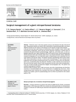
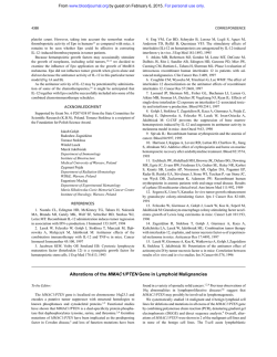
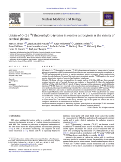
1/28/2015
OBJECTIVES
• Provide a brief review of cancer epidemiology
• Discuss the clinical evaluation of ocular melanocytic
VRS RETINAL UPDATE:
tumors
CHOROIDAL MELANOMA
• Highlight new developments in gene expression profiling
f
for choroidal melanoma
• Class 1 - lower risk of metastasis
• Class 2 - higher risk of metastasis
John B. Davies, MD
January 31, 2015
• Discuss case examples
NUMBERS OF SURVIVORS FOR THE MOST
COMMON CANCERS IN THE US AS OF JANUARY
2014
MALES
FEMALES
PROSTATE
2,975,970
BREAST
3,131,440
COLON / RECTUM
621,430
UTERINE
624,890
MELANOMA
516,570
COLON / RECTUM
624,340
BLADDER
455,520
MELANOMA
528,860
ALL SITES
6,876,600
ALL SITES
7,607,230
Cancer Treatment & Survivorship Facts and Figures 2014-2015, American Cancer Society
American Cancer Society
American Cancer Society
American Cancer Society
1
1/28/2015
CANCER STATISTICS
MALES
RISK OF
DEVELOPING (%)
RISK OF DYING
FROM (%)
PROSTATE
15.0
2.7
7.4
LUNG
1 IN 13
4.8
COLON
1 IN 20
BLADDER
ALL SITES
American Cancer Society
1 IN 7
3.8
1 IN 26
43.3
6.5
2.0
0.9
22.8
US National Cancer Institute’s Surveillance Epidemiology and End Results (SEER) Database, 2009-2011
CANCER STATISTICS
FEMALES
RISK OF
DEVELOPING (%)
RISK OF DYING
FROM (%)
BREAST
12.3
2.7
1 IN 8
6.2
LUNG
4.7
1 IN 16
4.5
COLON
1.9
1 IN 22
UTERINE
ALL SITES
2.7
0.6
1 IN 37
37.8
OUR FOCUS TODAY:
CHOROIDAL MELANOMA
19.3
US National Cancer Institute’s Surveillance Epidemiology and End Results (SEER) Database, 2009-2011
CHOROIDAL MELANOMA
• Most common primary intraocular tumor in adults
• Incidence in the US: 6 to 7 cases per million
• Affects 1,200 - 1,500 people in the US each year
• Usually diagnosed in the sixth decade of life
• Most cases are sporadic without a positive family
history of the disease
CHALLENGES
• Despite many advances in the diagnosis and
treatment, there has not been a corresponding
improvement in survival rates
• Early micro-metastasis have already occurred before
treatment of the primary tumor
• Prolonged latency before the metastatic disease
manifests itself
2
1/28/2015
CHOROIDAL MELANOMA: RISK
FACTORS
OCULAR MELANOCYTOSIS
• Caucasian race
• Light iris color (blue or gray)
• Cutaneous
C t
nevi,i atypical
t i l cutaneous
t
nevi,i ffreckles,
kl
iiris
i nevii
• Other melanocytic conditions
• Ocular melanocytosis
• Oculodermal melanocytosis (nevus of Ota)
• ? sunlight exposure, welding
OCULODERMAL
MELANOCYTOSIS (NEVUS OF
OTA)
SITES OF OCULAR
INVOLVEMENT
• Uveal tract: 95%
• Choroid - most common
• Ciliary body
• Iris
• Other sites (conjunctiva): 5%
PATHOLOGY
PATHOLOGY
3
1/28/2015
CELL TYPES
PRESENTATION
• Location and size determine presenting symptoms
• Peripheral lesions may go undetected for a
• Spindle A
long time
• Spindle B
• Posterior lesions may lead to visual changes
early on
• Epithelioid
• Ciliary body tumors may displace the iris or lead to
• Mixed
sectoral cataracts
• May erode through the iris or sclera
CLINICAL FEATURES
CLINICAL FEATURES
• Brown, elevated, dome shaped mass
• Variable degree of pigmentation
• Orange lipofuscin pigment
• Subretinal fluid
• May break through Bruch’s membrane leading to a
collar-button or mushroom-shaped configuration
ULTRASONOGRAPHY
Anterior
CHOROIDAL NEVI
Posterior
• Benign melanocytic uveal lesions
• Typically found on routine dilated
B Scan
fundus exam
• Usually asymptomatic
• In the United States, prevalence
A Scan
Low internal
reflectivity
ranges from 4.6 to 7.9% in
Caucasians
4
1/28/2015
DISTINGUISHING FEATURES
NEVUS
•
Clearly defined
margins
•
Flat or slightly elevated
•
Associated drusen
•
Remains stable in size
CHOROIDAL NEVI: NATURAL
HISTORY
MELANOMA
•
Indiscrete margins
• Risk of malignant transformation: 1 in 8,845
•
More elevated
• Increased risk with age:
•
Subretinal fluid
•
Orange pigment
•
Growth
• By age 80, risk of transformation is 0.78%
Singh, Ophthalmology 2005
Kivela, Ophthalmology 2006
“TO FIND SMALL OCULAR MELANOMA
USING HELPFUL HINTS DAILY”
RISK FACTORS FOR
MALIGNANCY
• Thickness > 2 mm
• Fluid
• Symptoms
• Orange pigment
• Margin within 3 mm of optic disc
“To Find Small
Ocular Melanoma
Using Helpful
Hints Daily”
RISK FACTORS
RISK OF GROWTH
AT 5 YEARS (%)
MANAGEMENT
0
3
OBSERVE
1
38
OBSERVE Q 4-6
MONTHS
3 OR MORE
>50
CONSIDER
TREATMENT
• Ultrasonographic Hollowness
• Absence of Halo
• Absence of Drusen
MANAGEMENT OF INTERMEDIATE
RISK LESIONS
TREATMENT
• Radiation
• Plaque brachytherapy
• External beam radiation therapy
• Enucleation
• Photographic and ultrasonographic / OCT
documentation
• Transpupillary Thermal Therapy (TTT)
• Periodic reassessment for signs of growth
5
1/28/2015
PLAQUE BRACHYTHERAPY
COLLABORATIVE OCULAR
MELANOMA STUDY (COMS)
• Small tumor trial - observational
• Medium tumor trial - enucleation vs. brachytherapy
• Large tumor trial - enucleation vs. enucleation +
external beam radiation
COMS: SMALL TUMOR STUDY
OBSERVATION
204 PATIENTS
5 YEAR ALL CAUSE
MORTALITY (%)
5 YEAR
MORTALITY W ITH
METASTATIC
MELANOMA (%)
6
1
• Features predictive of tumor growth:
• Increased tumor thickness and diameter, orange
pigment, absence of drusen, absence of adjacent
RPE changes, pinpoint hyperfluorescence on
angiography
COMS: MEDIUM TUMOR STUDY
ENUCLEATION
660 PATIENTS
IODINE-125
BRACHYTHERAPY
5 YEAR ALL CAUSE
MORTALITY (%)
5 YEAR
MORTALITY W ITH
METASTATIC
MELANOMA (%)
19
11
18
9
COMS: MEDIUM TUMOR STUDY
657 PATIENTS
6
1/28/2015
COMS: LARGE TUMOR STUDY
MOLECULAR CLASSIFICATION
OF UVEAL MELANOMA
• Similar survival regardless if patients were treated with
external beam radiation or not prior to enucleation
• Gene expression profiling can identify patients at high
risk for metastasis
5 YEAR ALL CAUSE
MORTALITY (%)
ENUCLEATION
506 PATIENTS
EXTERNAL RADIATION +
ENUCLEATION
• Commercially available test: DecisionDx-UM, an
expression profile of 15 genes
43
• Tumor sample obtained from small-gauge needle
38
biopsy
497 PATIENTS
MOLECULAR CLASSIFICATION
OF UVEAL MELANOMA
MOLECULAR CLASSIFICATION
OF UVEAL MELANOMA
• Class 1 - lower risk of metastasis
• Offer individualized management
• Genes expressed
p
are more similar to normal uveal
melanocytes
• Class 2 - higher risk of metastasis
• Genes expressed are similar to primitive,
undifferentiated cells
PROGNOSTIC POWER OF GENE
EXPRESSION PROFILING
• Prognosis
• Metastatic surveillance
• Clinical trial opportunities
• Ultimate goal: improve patient outcomes
DRIVER MUTATIONS IN UVEAL
MELANOMA
CLASS 1
CLASS 2
GNAQ / GNA11
BAP1
SF3B1
EIF1AX
7
1/28/2015
GNAQ / GNA11
BAP1
• Tumor suppressor gene, chromosome 3p21
• Oncogenes: disable GTPase activity leading to
constitutive activation of cell signaling pathways
• Found in premalignant nevi and uveal melanoma of all
stages
• Thought to be an early initiating event in tumorigenesis
but not necessarily sufficient for the development of a
metastatic tumor
SF3B1
• Removes ubiquitin from substrates
• Roles in gene expression, cell cycle regulation, cellular
identity
• Found in the majority of Class 2 tumors, rarely in Class 1
• In most metastasizing Class 2 tumors, one copy of BAP1 is
mutated and the other is absent through loss of the entire
chromosome (strong relationship between Class 2 tumors
and monosomy 3)
EIF1AX
• Splicing Factor 3B Subunit 1
• Eukaryotic Translation Initiation Factor 1A,
1A X-linked
• Alters mRNA splicing
• Associated with Class 1 tumors
• Associated with Class 1 tumors
8
1/28/2015
71 YO ♀ WITH A CHOROIDAL TUMOR
• Worsening vision OD x 1 year
• Choroidal lesion not previously noted on past diabetic
eye exams
• 20/30 OD, 20/25 OS
B SCAN
IMPRESSION / PLAN
66 YO ♂ WITH A CHOROIDAL TUMOR
• Choroidal melanoma
• Exudative retinal detachment
• Enucleation vs. plaque brachytherapy vs. external
beam radiation
• First told of a nevus in the right eye 6 years ago
• No vision changes
• 20/30 OD, 20/20 OS
• Class 1
9
1/28/2015
B SCAN
79 YO ♀: AMD AND WORSENING VA OD
IMPRESSION / PLAN
• 2008: VA OD 20/25
• Intermediate risk choroidal melanocytic tumor
• Observation
• 2011: VA OD 20/100
• History of colon cancer in 2006 s/p surgery and
chemotherapy
20/100
20/20
10
1/28/2015
• Angiographic features consistent with occult choroidal
neovascularization
• Begin treating with Avastin
• After first injection, VA improved to 20/30
• Variable SRF over subsequent months, 4 total Avastin
injections
• Macular lesion continues to enlarge
• Concern for choroidal mass: metastatic colon cancer
vs. amelanotic melanoma?
2011
2011
2013
2013
11
1/28/2015
• Systemic work-up unremarkable
• PET scan: negative for metastasis
• Diagnosis: subfoveal amelanotic choroidal melanoma
• June 2013: brachytherapy with 16 mm plaque
• Exudative retinal detachment
CONCLUSIONS
CONCLUSIONS
• Risk factors growth of choroidal nevi and malignant transformation: “To
• Prostate and breast cancer account for the most new
cases of cancer in the US in men and women,
respectively
• Lung cancer accounts for the most cancer-related
deaths in both men and women
• Choroidal melanoma is the most common primary
intraocular malignancy in adults
Find Small Ocular Melanoma Using Helpful Hints Daily”
• Thickness > 2 mm, Fluid, Symptoms, Orange pigment, Margin within
3 mm of optic disc, Ultrasonographic Hollowness, absence of Halo,
absence of Drusen
• Gene expression profiling predicts the risk of metastasis
• Class 1 - lower risk
• Class 2 - higher risk
• Our understanding of the mutations that drive the development of ocular
melanoma may lead to targeted therapy in the future and improved
patient outcomes.
12


© Copyright 2026