Surgical management of a giant retroperitoneal teratoma - revista
Rev Mex Urol 2014;74(4):234-237
ÓRGANO OFICIAL DE DIFUSIÓN DE LA SOCIEDAD MEXICANA DE UROLOGÍA,
COLEGIO DE PROFESIONISTAS, A.C.
www.elsevier.es/uromx
Clinical case
Surgical management of a giant retroperitoneal teratoma
F. R. Zamora-Varelaa,*, A. Castro-Alfarob, J. C. Navarro-Vargasb, A. Scavuzzob, Z. A.
Santana-Ríosb, P. F. Martínez-Cerverab and M. A. Jiménez-Ríosb
a
Urology Speciality Residency, Hospital Regional “Dr. Valentín Gómez Farías”, ISSSTE, Guadalajara, Jal., Mexico
b
Urology Division, Instituto Nacional de Cancerología, Mexico City, Mexico
KEYWORDS
Testis;
Nonseminomatous;
Giant teratoma;
Mexico
Abstract Testicular cancer is the most common cancer in men between the ages of 20 and 35
Palabras clave
Testículo; No
seminoma; Teratoma
gigante; México.
Manejo quirúrgico de un teratoma retroperitoneal gigante
years. Teratomatous elements are found in 50% to almost 80% of nonseminomatous germ cell tumors
(NSGCTs) and there are few reports in the literature on the resection of large-scale teratomas.
A 28-year-old man had a past history of orchiectomy, which revealed a tumor with an 80%
teratomatous composition, and he did not receive adjuvant treatment; 6 months later he
presented with gastrointestinal symptomatology, weight loss, and lumbar pain, as well as a
palpable abdominal mass. Alpha-fetoprotein (AFP) and β-subunit human chorionic gonadotropin
(β-hCG) hormone levels were elevated. Plain chest x-ray showed metastatic lesions. Abdominal
computed tomography (CT) scan identified a giant retroperitoneal tumor extending from the
renal vessels to the left iliac vessels. The patient was given chemotherapy and his tumor marker
(TM) values descended to normal. A control post-chemotherapy CT scan showed tumor
progression and so the giant retroperitoneal tumor was resected. The iliac artery was infiltrated,
and in turn, no dissection plane was observed. Therefore, en bloc resection was carried out at
the level of the iliac vessels; a Dacron® graft was placed on the iliac artery with no complications.
Flow was verified through intraoperative Doppler ultrasonography (US). The patient was
released in good general condition.
Advanced stage teratomas have a high recurrence rate after chemotherapy and therefore
resection is recommendable due to the compression of neighboring structures. Giant
retroperitoneal tumor resection has a mortality rate of up to 6%. Residual tumor resection was
offered to our patient, given his symptomatology and in an effort to provide him with a better
quality of life.
Resumen El cáncer testicular es el cáncer más común en edades de 20 a 35 años, el teratoma
abarca desde un 50% hasta casi el 80% de los tumores de células germinales no seminomatosos
(TCGNS); la resección de TCGNS de gran dimensión está bajamente reportada en la literatura
médica.
* Corresponding author at: Paseo de las Brisas N° 4128-302, Colonia Lomas Altas, C.P. 45120, Zapopan, Jal., México. Telephone: 01 (33)
3749 3944. Email: [email protected] (F. R. Zamora-Varela).
Surgical management of a giant retroperitoneal teratoma 235
Se presenta caso de masculino de 28 años de edad, con antecedente de orquiectomía de estirpe
teratoma 80%, sin recibir tratamiento adyuvante; 6 meses después inicia con sintomatología
gastrointestinal, pérdida de peso y dolor lumbar, así como masa abdominal palpable; con la alfa-fetoproteína (AFP) y la hormona gonadotrofina coriónica fracción-β (hCG-β) elevadas. La radiografía simple de tórax mostró lesiones metastásicas. La tomografía computarizada (TC) de
abdomen evidencia tumor retroperitoneal gigante, desde los vasos renales hasta vasos ilíacos
izquierdos. Recibió quimioterapia logrando descender los marcadores tumorales (MT) hasta valores normales. En la TC de control posquimioterapia se observa progresión tumoral, por lo que
se realiza resección del tumor retroperitoneal gigante. Durante el procedimiento, a nivel de los
vasos ilíacos se resecó en bloque debido a no tener plano de disección por infiltración a la arteria ilíaca, allí se colocó un injerto de Dacrón®, sin complicaciones. Se verificó su flujo con ultrasonido (US) Doppler transquirúrgico. El paciente se dio de alta en buenas condiciones generales.
Los teratomas en etapas avanzadas tienen alta tasa de recaída después de la quimioterapia, por
lo que es recomendable la resección debido a la compresión de estructuras vecinas. La resección de tumores retroperitoneales gigantes tiene una mortalidad de hasta el 6%. Debido a la
sintomatología del paciente y con el fin de ofrecerle una mejor calidad de vida, se optó por
brindarle la posibilidad de la resección del tumor residual.
0185-4542 © 2014. Revista Mexicana de Urología. Publicado por Elsevier México. Todos los derechos
reservados.
Introduction
Testicular cancer is the most common cancer in men
between the ages of 20 and 35 years. It makes up 1% of all
neoplasias in men. Teratoma elements are found in 55% to
79% of the nonseminomatous germ cell tumors (NSGCTs) and
metastasis has been reported in 27% to 46%. Approximately
30% to 40% of men with NSGCTs have signs of metastatic
disease at tumor presentation onset. The curative rate for
advanced disease varies between 80% and 90% with a
multidisciplinary approach that includes platin-based
chemotherapy followed by retroperitoneal lymph node
dissection (RPLND) of the residual disease.1-3 Elevated tumor
markers (TMs) despite primary chemotherapy are indicative
of cancer persistence. RPLND is one of the standard
managements of patients with NSGCTs at all stages. It is used
as first-line treatment in patients with non-metastatic
disease as an alternative to primary chemotherapy or
surveillance and is used as first or second-line therapy in
patients with regional retroperitoneal lymph node metastasis.
And finally, RPLND is used for treating residual disease after
chemotherapy in patients with metastatic NSGCT.2,4
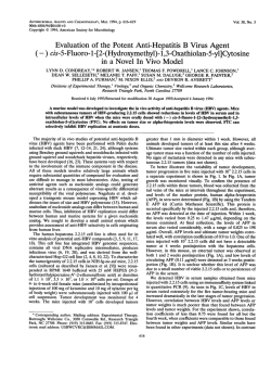
the left iliac vessels (fig. 1). He received 4 cycles of
chemotherapy with bleomycin, etoposide, and cisplatin
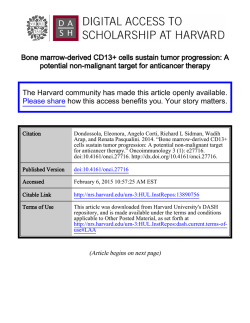
(BEP) and the TMs descended to normal values. The postchemotherapy control CT scan revealed tumor progression
(fig. 2) and so the decision was made to resect the giant
retroperitoneal tumor that measured approximately 30 x 22
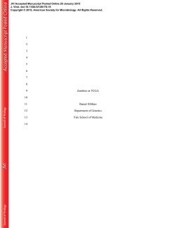
x 6 cm and weighed 1,500 g. The tumor surrounded the
aorta, the vena cava, the superior and inferior mesenteric
artery and the left common iliac vessels (fig. 3). En bloc
resection was necessary because the tumor was found to be
invading the iliac artery and, as a result, no dissection plane
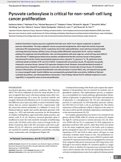
was observed. A Dacron® graft was placed on the artery with
no complications (fig. 4) and flow was verified through an
intraoperative Doppler ultrasound (US).
Case presentation
A 28-year-old man had a past history of orchiectomy that
revealed a tumor with an 80% teratomatous composition,
and he did not receive chemotherapy. Six months later he
presented with gastrointestinal symptomatology, weight
loss of 5 Kg in 2 months, and lumbar pain. Abdominal
examination revealed a fixed, painful, palpable mass. TMs
were: lactate dehydrogenase (LDH) 216, alpha-fetoprotein
(AFP) 2,372, and β-subunit human chorionic gonadotropin
(β-hCG) 395. A plain chest film showed lesions consistent
with tumor activity that was then confirmed by computed
tomography (CT), which also identified a giant
retroperitoneal tumor extending from the renal vessels to
Figure 1 Retroperitoneal tumor extending from the renal vessels; infiltration towards the left iliac vessels is observed.
236
Figure 2 Post-chemotherapy control tomography scan showing
giant tumor progression after 4 cycles of bleomycin, etoposide,
and cisplatin (BEP).
F. R. Zamora-Varela et al
Figure 3 Retroperitoneal resection of the giant tumor; aorta
and vena cava are observed. En bloc tumor resection.
There was a 5L blood loss and the total surgery duration
was 7.30 hours. The patient had no postoperative
complications and was released from the hospital in good
general condition. He presented with diuresis of 0.7 mL/
Kg/h, warm extremities, adequate coloring, and distal
pulses.
Discussion
The advent of chemotherapy considerably changed
testicular tumor treatment and today it has high curative
percentages. However, the presence of teratoma in the
primary tumor has been a predictor for incomplete response
to chemotherapy in cases of advanced disease. 2 Studies
report that patients with NSGCTs in stages II and III with
teratoma in the primary tumor, compared with patients
without teratoma, have little radiologic response (defined
as a residual mass of 15 mm or less) when treated with
cisplatin.5,6
Advanced disease management is more difficult due to
the aggressiveness of the chemotherapy. The appearance of
residual disease obliges us to be more careful in relation to
treatment because retroperitoneal resection of large
masses is a complex procedure that is not exempt from
complications, with a surgical mortality rate of 6% in
advanced disease.2 Notwithstanding the fact that lymph
node dissection continues to be standard treatment in
NSGCTs, it is indicated in cases of failed chemotherapy
when there is a giant teratomatous residual mass
compressing neighboring structures.7,8 Furthermore, a high
proportion of patients already presenting with advanced
disease have shown a considerable risk for recurrence,
despite complete resection of the residual mass.5
Conclusions
The resection of giant retroperitoneal tumors has a
mortality rate of up to 6%. Residual tumor resection was
Figure 4 Dacrón® arterial graft placement.
offered to our patient given his symptomatology and in an
effort to provide him with a better quality of life. These
giant retroperitoneal tumors should be treated in specialized
centers and by highly experienced surgeons, given the
formidable complexity of the procedure.
Conflict of interest
The authors declare that there is no conflict of interest.
Financial disclosure
No financial support was received in relation to this article.
Surgical management of a giant retroperitoneal teratoma References
1. Masterson TA, Shayegan B. Clinical Impact of Residual
Extraperitoneal Masses in Patients with Advanced
Nonseminomatous Germ Cell Testicular Cancer. Urology
2012;79(1):156-159.
2. Capitanio U, Jeldres C, Perrotte P, et al. Population-based
study of perioperative mortality after retroperitoneal
lymphadenectomy for nonseminomatous testicular germ cell
tumors. Urology 2009;74(2):373-377.
3. Carver BS, Cronin AM. The total number of retroperitoneal
lymph nodes resected impacts clinical outcome after
chemotherapy for metastatic testicular cancer. Urology
2010;75(6):1431-1335.
237
4. Coogan CL, Foster RS. Postchemotherapy retroperitoneal lymph
node dissection is effective therapy in selected patients with
elevated tumor markers after primary chemotherapy alone.
Urology 1997;50(6):957-962.
5. Rabbani F, Farivar-Mohseni H. Clinical outcome after
retroperitoneal lymphadenectomy of patients with pure
testicular teratoma. Urology 2003;62(6):1092-1096.
6. Karam JA, Raj GV. Growing Teratoma Syndrome. Urology
2009;74(4):783-784.
7. Ozen H, Ekici S. Resection of Residual Masses alone: An
alternative in surgical therapy of metastatic testicular germ
cell tumors after chemotherapy. Urology 2001;57 (2):323-327.
8. Giménez Bachs JM, Salinas Sánchez AS. Cirugía de gran masa
residual tras quimioterapia en tumor germinal testicular
avanzado. Actas Urol Esp 2004;28(3):230-233.
© Copyright 2026
