
IL-18 is produced by prostate cancer cells and sec
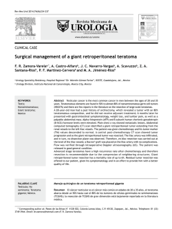
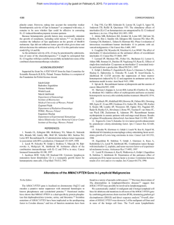
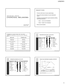
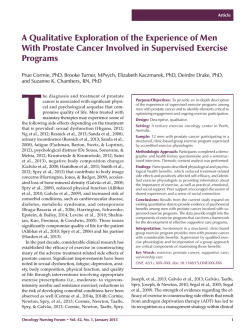
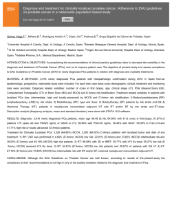
Publication of the International Union Against Cancer Int. J. Cancer: 106, 827– 835 (2003) © 2003 Wiley-Liss, Inc. IL-18 IS PRODUCED BY PROSTATE CANCER CELLS AND SECRETED IN RESPONSE TO INTERFERONS Sophie LEBEL-BINAY1,2, Nicolas THIOUNN3, Gonzague DE PINIEUX4, Annick VIEILLEFOND4, Bernard DEBRE´ 3, Jean-Yves BONNEFOY2, Wolf-Herman FRIDMAN1,5 and Franck PAGE` S1,5* 1 INSERM U 255, Centre de Recherches Biome´dicales des Cordeliers, Paris, France 2 Centre d’Immunologie Pierre Fabre, Saint-Julien en Genevois Cedex, France 3 Service d’Urologie, Hoˆpital Cochin, AP-HP, Paris, France 4 Service d’Anatomie Pathologique, Hoˆpital Cochin, AP-HP, Paris, France 5 Service d’Immunologie Biologique, Hoˆpital Europe´en Georges Pompidou, AP-HP, Paris, France Murine models have shown that IL-18 has antiangiogenic and antitumor effects, but little is known about IL-18 production in human tumors. We investigated IL-18 expression in clinically localized prostate cancers by immunohistochemistry and showed that 75% of the prostate cancers studied (27/36 cases) presented with tumor cells producing IL-18. Prostate tumor cell lines PC-3, DU 145 and LNCaP synthesized the immature form of IL-18 (p24). IFN-␥ produced in prostate cancers induced caspase-1 mRNA and IL-18 secretion of tumor cell lines, which was inhibited by the cellpermeable Tyr-Val-Ala-Asp-aldehyde caspase-1 inhibitor (YVAD-CHO). Interestingly, IFN-␣ also induced IL-18 secretion of the poorly differentiated cell line PC-3. PC-3 and DU 145, but not the well-differentiated cell line LNCaP, expressed IL-18R␣ (IL-1Rrp) protein and transcripts for IL18R (AcPL). Exogenous IL-18 increased mitochondrial activity of both cell lines evaluated by the tetrazolium (MTT) assay but did not influence their proliferation. This indicated that prostate tumor cells could secrete IL-18 in response to IFN-␥ in the tumor microenvironment and that IL-18 could act as a autocrine/paracrine factor for the tumor. In the cohort of patients studied, IL-18 expression in prostate cancers (with up to 10% of tumor cells stained) was associated with a favorable outcome and equally predictive as pathologic stage on multivariate analysis (log rank test, p ؍0.02). Tumor IL-18 production is a novel physiopathologic feature of prostate cancer and appears to be a favorable event in the course of the disease. Modulation of IL-18 production by interferons could have a beneficial clinical effect, which deserves further investigation. © 2003 Wiley-Liss, Inc. Key words: IL-18; caspase-1; prostate cancer; prognosis; IFN-␥; IFN-␣ IL-18 was first identified as an IGIF based on its ability to induce high levels of IFN-␥ secretion by both NK cells and Th1 clones.1,2 IL-18 belongs to the IL-1 family,3 lacks a signal sequence2 and is processed into an 18 kDa mature form by caspase1.4 It is mainly produced by macrophages and dendritic cells. However, we and others have shown that IL-18 is also synthesized by nonimmune cells.5 IL-18 potentiates IL-12-induced Th1 development6,7 and plays an important role in T-cell proliferation.2 In addition, it enhances FasL-mediated cytotoxicity of NK cells and Th1 cells8,9 and has proinflammatory properties by inducing chemotactic molecules for macrophages, polymorphonuclear neutrophils and inflammatory cytokines such as TNF-␣ or IL-1.10 –12 Murine models have demonstrated the antitumor activity of IL-18, either by systemic administration13,14 or in tumors genetically modified to constitutively express IL-18.15 Antitumor effects are mediated by IL-18-induced IFN-␥, NK cells and T CD4ϩ Fas-dependent cytotoxicity.13–16 In vitro, IL-18 added to cultures of tumor cells, NK cells, dendritic cells and T cells induces tumor cell death by enhancing NK cell cytotoxicity, which activates dendritic cells to promote a specific CTL response.17 This could position IL-18 as an important bridge between innate and adaptive antitumor immune responses. Some antitumor effects of IL-18 may also be mediated via nonimmunologic mechanisms as IL-18 has antiangiogenic properties in vitro and immunohistochemistry studies have revealed hypovascularization of IL-18-treated tumors.15,18 Little is known about the in vivo presence of IL-18 in human tumors. In a previous study, we observed that IL-18 protein was expressed at various levels in colon cancers and that loss of transcripts at the tumor site of the 2 downstream targets of IL-18, i.e., IFN-␥ and FasL, was associated with the concomitant presence of distant metastases.19 These results suggested a role of IL-18 in the control of tumor spreading in humans, in line with the antitumor properties of IL-18 observed in murine models. Research in prostate cancer has focused on the role of IFN-␥ in tumor progression. In vitro, IFN-␥ causes cycle arrest of prostate tumor cell lines, induces the cyclin-dependent kinase inhibitor p21WAF1, downregulates neu/HER-220 and decreases the metastatic potential of some prostate cancer cell lines.21 IFN-␥ is produced by stromal and epithelial cells in normal prostate and prostate cancers,22 and analysis of the transcriptional profile of prostate cancers by Affymetrix (Santa Clara, CA) Genechip technology has shown that about 30% of malignant tumors present downmodulation of IFN-␥-inducible molecules.23 This may indicate modulation of IFN-␥ in tumors, but no information is available about the existence of the IFN-␥-inducing cytokine IL-18 in normal prostate and prostate cancers. We therefore investigated the IL-18 status of prostate cancers and its association with clinical outcome. Abbreviations: CI, confidence interval; CTL, cytotoxic T lymphocyte; DAB, diaminobenzidine; ECL, enhanced chemiluminescence; HRP, horseradish peroxidase; IGIF, IFN-␥-inducing factor; IL-18R, IL-18 receptor; MAb, monoclonal antibody; NK, natural killer; PSA, prostate-specific antigen; RR, relative risk; STAT, signal transducer and activator of transcription; TBS, TRIS-buffered saline; Th1, T-helper 1; TNF, tumor necrosis factor. Grant sponsor: Centre d’Immunologie Pierre Fabre; Grant sponsor: Association pour la Recherche sur le Cancer: Pˆole d’Etude du Microenvironnement Tumoral. The first 2 authors contributed equally to this work. *Correspondence to: Service d’Immunologie Biologique, Hˆopital Europ´een Georges Pompidou, 20-40 rue Leblanc, 75 908 Paris Cedex 15, France. Fax: ϩ33-1-56-09-20-80. E-mail: [email protected] Received 9 August 2002; Revised 2 January, 24 February 2003; Accepted 3 March 2003 DOI 10.1002/ijc.11285 828 LEBEL-BINAY ET AL. MATERIAL AND METHODS Patients and clinical features A total of 43 patients with localized prostate carcinoma treated by radical prostatectomy at Cochin Hospital, Paris, were included.24 Gleason score and pathologic TNM stage were reevaluated in each case by 2 pathologists and a urologist. Patients had newly diagnosed tumors and did not receive any adjuvant treatment after surgery. Bone metastases were assessed by bone X-ray and bone scan. Extraosseous metastases were assessed by surgical biopsy. Recurrence was defined as a significant elevation of PSA and/or new symptoms due to local tumor recurrence. Of the 36 patients selected (7 cases excluded for insufficient quality of IL-18 staining), 6 died from prostatic disease, 14 had a favorable clinical outcome with no recurrence or metastases and 22 relapsed, with bone metastases in 11 cases. Median follow-up was 95 months (range 75–115). Immunohistochemistry Archival formalin-fixed, paraffin-embedded tissues were available for all 43 lesions. Ten specimens of normal prostate and 15 specimens of typical benign prostatic hyperplasia were also selected. A single morphologically representative block was selected per case. Sections (5 m) were air-dried, deparaffinized and heated 3 times for 5 min in a microwave oven in citrate buffer (pH 6.0). Sections were first incubated for 5 min with 3% hydrogen peroxide aqueous solution to quench endogenous peroxidase activity. Goat polyclonal IgG anti-IL-18 was used for IL-18 detection (R&D Systems, Abingdon, UK). This antibody recognized the inactive precursor and mature form of IL-18. Sections were incubated for 60 min with the antibodies at a dilution of 1:50. Biotinylated conjugate and streptavidin peroxidase were applied for 15 min, and DAB chromogen was used as a peroxidase substrate complex (all from the LSABϩ kit; Dako, Copenhagen, Denmark). Incubations were performed at room temperature. Tissue sections were then counterstained with Harris hematoxylin and mounted with aqueous mounting medium (Dako). Intrinsic positive controls for immunoreactivity in each section were IL-18-stained cells in the stroma (macrophage-like cells). A control using IgG from nonimmunized goats (R&D Systems) at the same final concentration as that of anti-IL-18 IgG was carried out for each case, to exclude nonspecific binding. Immunohistochemical staining was evaluated by 2 pathologists (A.V. and G.deP.) blinded to patient outcome, in each case. The percentage of IL-18ϩ tumor cells was calculated as the mean value obtained from 3 different fields randomly selected (at ϫ400) within the same section. Results were expressed as 0%, Յ10%, 11–33%, 34 – 66% and Ն67%. Staining cut-off for statistical evaluation was 10%. Cells and cell cultures Prostate cancer cell lines LNCaP, PC-3 and DU 145 (all from the ATCC, Manassas, VA) were maintained in DMEM supplemented with 10% heat-inactivated FCS, 100 U/ml of penicillin, 50 g/ml of streptomycin (all from GIBCO, Grand Island, NY) and 50 g/ml of Plasmocyn (Cayla, Toulouse, France). RT-PCR RNA was isolated from cells by the RNeasy Kit procedure and treated with RNAse-free DNase (all from Qiagen, Valencia, CA). RT-PCR was performed using 1 g of total cellular RNA incubated with AMV reverse transcriptase, primer oligo(dT), dNTP and RNase inhibitor for 1 hr at 42°C (cDNA synthesis kit; Boehringer-Mannheim, Indianapolis, IN). PCR amplification was performed as previously described.19 The sequences of the 5Ј and 3Ј oligonucleotide primers and the sizes of their products were as follows: IL-18, 5Ј-GCT TGA ATC TAA ATT ATC AGT C-3Ј, 5Ј-GAA GAT TCA AAT TGC ATC TTA T-3Ј, 342 bp; caspase-1, 5Ј-GGT CCT GAA GGA GAA GAG AA-3Ј, 5Ј-AGG CCT GGA TGA TGA TCA CC-3Ј, 842 bp; IL-1Rrp, 5Ј-TTG GAG TGA TGA CAG GAA CAC-3Ј, 5Ј-CAT CAG ATA GGT CGT TAC TAC TAC C-3Ј, 223 bp; AcPL, 5Ј-GGT TAT TAC TCC TGC GTG C-3Ј, 5Ј-CCA TTT TCT TCC CCG AAC ATC C-3Ј, 273 bp; HPRT, 5Ј-TTC AAA TCC AAC AAA GTC TG-3Ј, 5Ј-AGC ACT GAA TAG AAA TAG TGA TAG A-3Ј, 278 bp. The authenticity of PCR products was verified by diagnostic restriction digests. Amplification was conducted as follows: for caspase-1: step 1 was 1 cycle at 94°C for 5 min, step 2 was 40 cycles at 96°C for 2 min, 55°C for 1.5 min and 72°C for 2 min and step 3 was 1 cycle at 72°C for 10 min; for IL-18, IL-1Rrp, AcPL and HPRT: step 1 was 1 cycle at 94°C for 5 min, step 2 was 40 cycles at 94°C for 30 sec, 57°C for 30 sec and 72°C for 30 sec and step 3 was 1 cycle at 72°C for 10 min. Half of the PCR products were electrophoresed on 1.5% agarose gel, stained with ethidium bromide and photographed under UV light. Detection of IL-18 and IL-18R␣ by flow cytometry Cells were detached from the bottom of dishes (Falcon, Becton Dickinson Labware, Franklin Lakes, NJ) with cell dissociation solution (Sigma, St. Louis, MO), using the procedure recommended by the manufacturer. IL-18 was detected with the 2-step fixation and permeabilization Intrastain kit (Dako), according to the manufacturer’s instructions. Briefly, following fixation and permeabilization, 5 ϫ 105 cells were incubated with a mouse antihuman proIL-18 MAb (R&D Systems) or with the isotypematched MAb (mouse IgG1, R&D Systems; final concentration 20 g/ml) for 20 min at room temperature. After washing, cells were incubated for 20 min with antimouse IgG-FITC conjugate (1:50, Sigma). Cells were then washed and analyzed using Cell Quest software on a FACSCalibur (Becton Dickinson, San Jose, CA). To analyze cell-surface IL-18R␣ expression, 5 ϫ 105 cells were incubated with 20 g/ml goat anti-IL-18R␣ polyclonal IgG (R&D Systems) at 4°C for 30 min. As control, staining was performed with nonspecific isotype-matched polyclonal antibody (R&D Systems). Cells were then washed twice and incubated for 30 min on ice with biotinylated rabbit antigoat IgG (1:50; Vector, Burlingame, CA). After washing, cells were incubated at 4°C for 30 min with streptavidin-FITC (1:50; Pharmingen, San Diego, CA) and then analyzed using Cell Quest software on a FACSCalibur. Immunoblot analysis Cell pellets of the human prostate cancer cell lines were lysed in 1% Triton buffer containing a cocktail of protease inhibitors (Boehringer-Mannheim). For each cell line, 100 g of protein, estimated by the DC protein assay (Bio-Rad, Hercules, CA), were solubilized with Laemmli sample buffer and subjected to SDSPAGE (15%) using reduction conditions. Proteins were transferred by a semidry transblot system; the blot was blocked for 2 hr with 5% w/v nonfat dry milk and 0.05% Tween-20 in TBS and incubated with 1 g/ml of anti-hIL-18 MAb (R&D Systems) for 6 hr at room temperature. Blots were washed and then incubated for 45 min with HRP-conjugated antimouse IgG at 1:6,000 (Bio-Rad). After washing, IL-18 was detected using the ECL detection kit (Amersham, Arlington Heights, IL). The Mr of the proteins was estimated by comparison with the position of a standard (Kaleidoscope 39-3 kDa, Bio-Rad), using recombinant IL-18 (Chemicon, Temecula, CA) as control. Cell treatment for caspase-1 induction and IL-18 production Caspase-1 induction. Cell lines (1.5 ϫ 106 cells) were seeded in 20 ml of culture medium in a 75 cm2 flask (Falcon, Becton Dickinson). After incubation for 1 day at 37°C, medium was replaced and IFN-␥ (Abcys; Valbiotech, Paris, France) added to the culture at a final concentration of 1,000 U/ml. After 24 hr of incubation at 37°C, cells were collected by trypsinization and submitted to RNA extraction and RT-PCR for caspase-1, as described above. IL-18 secretion induced by interferons. Cell lines were seeded at 1.5 ϫ 106 cells (for collection at 24, 48 and 72 hr) and at 1 ϫ 106 cells (for collection at 96 hr) in 20 ml of culture medium in a 75 cm2 flask. After 1 day of incubation at 37°C, medium was replaced and IFN-␥ (1,000 U/ml Abcys, Valbiotech) or IFN-␣ (1,000 and IL-18 PRODUCTION BY PROSTATE CANCER 5,000 U/ml Roferon-A; Roche, Mannheim, Germany) was added to the culture. After incubation for 24, 48, 72 or 96 hr, supernatants were collected, centrifuged at 4,000g for 15 min to remove cells and debris and stored at –20°C until analysis. Specimens were then thawed at room temperature and concentrated by Centricon-Plus 20 (Millipore, Bedford, MA) at 4,000g for 30 min. The IL-18 level was then recorded with a human IL-18 ELISA kit for determination of active IL-18 (Medical & Biological Laboratories, Nagoya, Japan).25 Values were weighted by the concentration factors obtained and normalized for the total number of tumor cells initially seeded to allow a valid comparison between all conditions. Experiments were performed 3 times. Inhibition of caspase-1. DU 145 cells were seeded in 75 cm2 flasks and preincubated with 10 M of the cell-permeable TyrVal-Ala-Asp-aldehyde caspase-1 inhibitor (YVAD-CHO; Calbiochem, Darmstadt, Germany) for 1 hr at 37°C. Cells were then stimulated with 1,000 U/ml of IFN-␥. Caspase-1 inhibitor was added to the culture every 24 hr. Supernatants were collected at 48 and 72 hr, centrifuged and frozen until IL-18 determination. Experiments were performed 3 times. Tetrazolium (MTT) assay Cells were seeded on a flat-bottomed 96-well plate at 5 ϫ 104 cells/well in RPMI-1640 with 10% FCS. Twenty-four hours later, cultures were downshifted to serum-free medium, to which recombinant IL-18 (0 –100 ng/ml, Chemicon) was added. After 4 days of culture, mitochondrial activity was assessed by adding 50 g of the vital dye MTT (Sigma) to culture. The blue dye taken up by the cells after 4 hr of incubation was dissolved in 0.04 N HClisopropanol (100 l/well), and absorbance at 550 nm was read on an automated microplate reader. Data are means of 3 separate experiments. Proliferation assay Cells were seeded at 5,000 cells/well in 96-well flat-bottomed microtiter plates, and IL-18 (Chemicon) at various concentrations was added at the time of plating. Plates were incubated at 37°C in 5% CO2 for 38 hr and then labeled for 10 hr with 1 Ci/well 3 H-thymidine. Cells were harvested onto glass fiber filter paper, and the incorporated radioactivity was measured by liquid scintillation counting. All samples were measured in triplicate in at least 2 independent experiments. Statistical analysis The association between IL-18 staining and clinicopathologic variables was tested using the 2 test. The primary outcome was disease-specific relapse, which was determined from the date of radical prostatectomy. Data were evaluated for disease relapse using univariate and multivariate analyses in a Cox proportional hazards model for IL-18 staining and other clinical and pathologic predictors of outcome. The statistical significance of differences between means of IL-18 secreted was evaluated by Student’s unpaired t-test. p Ͻ 0.05 was required for significance. RESULTS IL-18 is expressed in normal prostate and prostate cancers Prostate cancers and distant normal prostate tissue of 36 patients with newly diagnosed localized prostate carcinoma treated by radical prostatectomy24 were subjected to IL-18 immunostaining. In normal prostate tissue, the IL-18 signal was consistently observed in basal cells and some scattered cells in the stroma with the shape and size of macrophages, whereas epithelial cells did not express IL-18 (Fig. 1a). Figure 1b shows a characteristic IL-18 immunoreactivity in areas of basal cell hyperplasia. No IL-18 staining was observed in epithelial cells of typical adenomatous hyperplasia. Positive tumor immunostaining for IL-18 was detected in 27 of the 36 prostate cancers studied. The IL-18 signal was located in the cytoplasm of tumor cells, and some stromal cells also presented IL-18 reactivity (Fig. 1c,d). The pattern of tumor 829 IL-18 expression was heterogeneous. Foci of tumor cells situated away from zones of IL-18 staining (Fig. 1e) did not express IL-18 (Fig. 1f). Percentages of IL-18ϩ tumor cells in each tumor were as follows: 0% (9 cases), Յ10% (8 cases), 11–33% (8 cases), 34 – 66% (8 cases) and Ն67% (3 cases). Overall, 75% of the tumors analyzed (27/36 cases) presented Ͼ10% of tumor cells producing IL-18. IL-18 is produced by prostate tumor cell lines To confirm and extend this finding of the capacity of prostate tumor cells to produce IL-18, we evaluated IL-18 expression of 2 poorly differentiated malignant prostate cell lines (PC-3 and DU 145) and the well-differentiated LNCaP cell line. IL-18 amplification products were detected in all cell lines (Fig. 2a). Intracellular staining for pro-IL-18 by flow cytometry was detected in the representative experiment shown in Figure 2b for the 3 cell lines, PC-3, DU 145 and LNCaP. The only form of IL-18 detected in cell lines by Western blot analysis was the 24 kDa inactive precursor form. LNCaP always displayed the less intense signal for IL-18. IFN-␥ modulates caspase-1 mRNA and IL-18 secretion of prostate tumor cell lines Only small amounts of IL-18 (about 20 pg/ml for 106 cells) were inconsistently detected by ELISA for PC-3, DU 145 and LNCaP (5 separate experiments, data not shown). As IL-18 secretion is preceded by activation of caspase-1, we looked for caspase-1 transcripts in the cell lines. Caspase-1 PCR signals were inconsistently observed in PC-3 and DU 145 despite 40 amplification cycles, while LNCaP did not express transcripts for caspase-1 (Fig. 3a). The caspase-1 mRNA profile of the cell lines could therefore account for the weak and inconsistent IL-18 secretion. IFN-␥ produced in prostate cancers22 has been shown to induce caspase-1 in tumor cell lines derived from other cancers.26 –28 We therefore wondered whether IFN-␥ could modulate caspase-1 in prostate tumor cell lines. Incubation with 1,000 U/ml of IFN-␥ for 24 hr markedly increased caspase-1 mRNA in PC-3 and induced caspase-1 mRNA in DU 145 and LNCaP (Fig. 3a). In addition, 1,000 U/ml of IFN-␥ added to cultures induced IL-18 secretion in a dose- (not shown) and time-dependent manner (Fig. 3b). PC-3 secreted about 4-fold more IL-18 than DU 145, while only very small amounts of IL-18 were detected in the supernatant of LNCaP cells. The effect of IFN-␥ on cell viability was monitored in parallel and did not reveal any increase in cell death as detected by Trypan blue exclusion (data not shown). Evidence of the role of caspase-1 in IFN-␥-induced IL-18 secretion was strengthened by the capacity of a caspase-1 peptide inhibitor, YVAD-CHO, to inhibit about 70% of the IL-18 secreted by DU 145 when stimulated for 72 hr with 1,000 U/ml of IFN-␥ (Fig. 4). This indicates that prostate tumor cells synthesize pro-IL-18 and can secrete the cytokine in response to IFN-␥ in the tumor microenvironment. Prostate tumor cell lines express IL-18R␣ IL-18 can be secreted by prostate tumor cell lines in response to IFN-␥. We looked for expression on tumor cell lines of the 2 chains of IL-18R, i.e., IL-18R␣ the ligand binding chain (IL1Rrp), and IL-18R (AcPL), the receptor signaling chain. The well-differentiated tumor cell line LNCaP did not express transcripts for the 2 chains of IL-18R (Fig. 5a). In contrast, PC-3 and DU 145 presented with IL-1Rrp transcripts, and a faint but consistent RT-PCR signal for AcPL (Fig. 5a). Membrane staining for IL-18R␣ was detected by flow cytometry for PC-3 and DU 145 (Fig. 5b). Tumor cell line reactivity to IL-18 was evaluated for PC-3 and DU 145 under serum-free conditions using a tetrazolium (MTT) assay. PC-3 showed an increase of cellular mitochondrial activity under IL-18 stimulation, with a maximum obtained for a final concentration of 1 ng/ml of IL-18 (Fig. 5c), as evaluated after 4 days of culture. IL-18 stimulated slightly but reproductively DU 145, with a less pronounced effect than that observed for PC-3 (Fig. 5c). Data were in accordance with a functional IL-18R expressed on tumor cells and indicated that IL-18 could act in an 830 LEBEL-BINAY ET AL. FIGURE 1 – Photomicrographs represent IL-18 immunoreactivity patterns in (a) normal prostate, with IL-18 immunostaining detected in basal cells and some stromal cells (arrows) of normal glands; (b) positive staining in areas of basal cell hyperplasia; (c,d) 2 prostate cancers with tumor cells stained for IL-18; (e,f) heterogeneity of IL-18 tumor staining in 2 different areas of the same prostate cancer. Control using IgG from nonimmunized goat at the same final concentration as that of antiIL-18 IgG was carried out for each case and excluded nonspecific binding [magnification ϫ120 for (a,c,d), ϫ50 for (b) and ϫ100 for (e,f)]. autocrine/paracrine way. We evaluated whether IL-18 influenced the growth of the cell lines and did not observe proliferative activity of the cytokine by cell counts (data not shown) and proliferation assay using 3H-thymidine incorporation (Fig. 6). Proliferation assay was also performed under serum-free conditions with similar results (data not shown). Incubation of tumor cell lines PC-3 and DU 145 with IL-18 neutralizing antibody or with antiIL-18R␣ antibody, selected for its ability neutralize human IL18R-mediated biologic activity, did not influence tumor growth (data not shown). IL-18 synthesis by prostate tumor cells and clinical outcome Since IL-18 is known to have antitumor activity, we asked whether it might influence the clinical outcome of patients with prostate cancer. In the cohort of 36 patients studied, no correlation was observed between IL-18 (staining cut-off 10%) and Gleason score or pathologic stage (Table I) even after reanalysis of the data using different cut-off values for tumor IL-18 positivity at 33% and 66%. Univariate analysis of disease relapse in patients stratified on the basis of the level of IL-18 expression (staining cut-off 10%) showed a better outcome of patients who had tumors with high IL-18 expression (RR ϭ 0.4, 95% CI 0.2–1; p ϭ 0.049). This result was confirmed in multivariate analysis, where the p value even increased after adjustment for Gleason score and pathologic stage (RR ϭ 0.3, 95% CI 0.1– 0.8; p ϭ 0.02) (Table II). This supports the notion that tumor staining for IL-18 might be prog- nostic for better outcome, independently of clinicopathologic features. Effect of IFN-␣ on IL-18 secretion by prostate tumor cell lines To look for other immune molecules that could modulate IL-18 in prostate cancers, we evaluated the effect of IFN-␣, which shares some biologic activities with IFN-␥ on prostate tumor cells. Interestingly, 5,000 U/ml of IFN-␣-induced time-dependent IL-18 secretion by the poorly differentiated cell line PC-3 (Fig. 7). This phenomenon was also present at 1,000 U/ml with about 300 ng of IL-18 /106 cells detected at 96 hr (data not shown). In contrast, IL-18 secretion was weak for LNCaP and DU 145 (Fig. 7). These results indicate that IFN-␣ is also capable of modulating IL-18 secretion of prostate tumor cell lines, with a probably less pronounced effect than that observed for IFN-␥. DISCUSSION In our study, we demonstrated that prostate cancer cells produce IL-18 and that IL-18 secretion was modulated by interferons in prostate tumor cell lines. Production of IL-18 by the tumor was associated with a better clinical outcome. Little is known about the capacity of tumor cells to produce IL-18. Pancreatic tumor cells and Burkitt lymphoma cell lines produce IL-18 (F. Page`s et al., unpublished data), and malignant skin tumor cell lines and ovarian carcinomas also secrete IL- IL-18 PRODUCTION BY PROSTATE CANCER 831 18,29,30 suggesting that IL-18 may be produced by a wide range of tumors. In the prostate, although normal epithelial cells do not produce IL-18, about 75% of prostate cancers studied (27/36 cases) presented tumor cells (derived from normal epithelial cells) producing IL-18. IL-18 tumor staining was not associated with Gleason grade or pathologic stage and was heterogeneous, ranging from 10% to 66% of tumor cells producing IL-18. Prostate cancers often present areas with different Gleason grades, indicating varying degrees of differentiation of tumor cells and deteriorating cancer cell architecture. This intratumoral heterogeneity may account for the heterogeneous tumor IL-18 expression. Anti-IL-18 antibodies used in the immunohistochemical study do not discriminate between the immature and active forms of the cytokine, which raises the question of IL-18 secretion by the tumor. At the very least, the staining pattern of IL-18 in prostate FIGURE 2 – (a) RT-PCR analysis of mRNA expression of IL-18 transcripts in PC-3, DU 145 and LNCaP cell lines. HPRT control PCR was performed to monitor RT-PCR amplification efficiency. The myelomonocytic KG-1 cell line is shown as positive control. (b) Intracytoplasmic expression of pro-IL-18 on the 3 prostate tumor cell lines detected by flow cytometry. Shaded and open histograms represent staining with anti-proIL-18 antibody and isotype-matched irrelevant MAb, respectively. (c) Western blot analysis of IL-18 protein expression. AntiIL-18 antibody detected a 24 kDa protein in all prostate cell lines analyzed. Recombinant human IL-18 was used as positive control. FIGURE 3 – (a) RT-PCR analysis of mRNA expression of caspase-1 transcripts in PC-3, DU 145 and LNCaP nonstimulated or stimulated cell lines with 1,000 U/ml IFN-␥ for 24 hr. The myelomonocytic KG-1 cell line is shown as positive control. (b) Means Ϯ SD of IL-18 detected by ELISA in culture supernatants of PC-3, DU 145 and LNCaP cell lines after stimulation with 1,000 U/ml of IFN-␥ for 0, 24, 48, 72 and 96 hr. FIGURE 4 – Means Ϯ SD of IL-18 measured by ELISA of culture supernatant of DU 145 stimulated with IFN-␥ (1,000 U/ml) in the absence (solid bar) or presence (hatched bar) of caspase-1 inhibitor YVAD-CHO for 48 and 72 hr. *Student’s t-test. 832 LEBEL-BINAY ET AL. FIGURE 6 – Effect of recombinant IL-18 (0 –250 ng/ml) on prostate tumor cell lines. Plates were incubated for 38 hr and then labeled for 10 hr with 3H-thymidine. Each point is the mean Ϯ SD of triplicate experiments. TABLE I – DISTRIBUTION OF IL-18 STAINING AS FUNCTION OF GLEASON SCORE AND PATHOLOGIC STAGE IL-18 immunoreactivity FIGURE 5 – (a) RT-PCR analysis of mRNA expression of IL-18R␣ (IL-1Rrp) and IL-18R (AcPL) transcripts in PC-3, DU 145 and LNCaP. (b) Reactivity of anti-IL-18R␣ (IL1-Rrp) antibody with prostate tumor cell lines was analyzed by flow cytometry. Shaded and open histograms represent staining with anti-IL-18R␣ antibody and isotypematched irrelevant antibody, respectively. (c) Effect of recombinant IL-18 on mitochondrial activity of prostate carcinoma cells. Cells were grown in DMEM with 10% FCS. Twenty-four hours later, cells were downshifted to serum-free conditions, and recombinant IL-18 was added at the concentrations indicated on the abscissa. Four days later, mitochondrial activity was assessed by MTT assay. Results are expressed as relative ratios to the IL-18-free controls. Data are means Ϯ SD of 3 separate experiments. cancers indicates that large amounts of mature IL-18 could be rapidly processed and secreted by the tumor in response to tumor cell stimulation. Certain arguments suggest secretion of IL-18 by at least a subset of tumor cells: (i) caspase-1, required for cleavage of pro-IL-18 into an active form, is detected in about 75% of prostate tumors from patients with no endocrine therapy prior to surgery,31 with a heterogeneous pattern of expression within tumor cells,32 and patients evaluated in our series had newly diagnosed pathologic stage TXN0M0 tumors and were untreated before surgery; (ii) using an ELISA kit that specifically detects the mature form of IL-18,25 we detected low concentrations of IL-18 in the culture supernatants of prostate tumor cell lines; (iii) the IFN-␥ produced in prostate tumors22 was able to increase or induce Gleason score 3–6 7–9 Pathologic stage T1–T2 T3–T4 n (%) Յ10% n (%) Ͼ10% n (%) 13 (36.1) 23 (63.9) 4 (23.5) 13 (76.5) 9 (47.4) 10 (52.6) 19 (52.8) 17 (47.2) 9 (52.9) 8 (47.1) 10 (52.6) 9 (47.4) p* 0.14 0.99 *p values were determined by the 2 test. p Ͻ 0.05 was required for significance. caspase-1 mRNA of prostate tumor cell lines, with a subsequent increase of IL-18 secretion in the medium. Taken together, these results suggest that at least a subset of tumor cells within prostate cancers secrete active IL-18. The induction or increase of transcripts for caspase-1 by IFN-␥ has also been observed in cell lines derived from ovarian,26 hepatic27 and colon28 carcinomas. IFN-␥ induced cell apoptosis via activation of caspase-126,27 or sensitized the cells to killing by numerous proapoptotic stimuli.28 We did not detect an increase of cell death following IFN-␥ stimulation in prostate tumor cell lines, as previously published.33,34 Interestingly, the hormone-sensitive cell line LNCaP displayed a different pattern of IL-18 production under IFN-␥ stimulation and IL-18R expression compared to the hormone-independent cell lines PC-3 and DU 145. Upon stimulation by interferons, LNCaP cells reacted with a strong induction of caspase-1 mRNA but nevertheless reproducibly secreted the lowest amounts of IL-18 833 IL-18 PRODUCTION BY PROSTATE CANCER TABLE II – UNIVARIATE AND MULTIVARIATE ANALYSES FOR IL-18 STAINING AND CLINICOPATHOLOGIC VARIABLES WITH RELAPSE-FREE SURVIVAL Variable n IL-18 staining Ͻ10% Ն10% Gleason score 3–6 7–9 Pathologic stage T1–T2 T3–T4 Univariate analysis RR1 95% CI Multivariate analysis p2 RR 95% CI 0.049 17 19 1 0.4 0.02 1 0.3 0.2–1.0 0.1–0.8 0.049 13 23 1 2.5 0.7 1 1.2 1.0–6.4 0.4–4.0 0.01 19 17 1 3.5 1.4–8.8 p 0.02 1 4.1 1.3–12.7 Risk ratio.– Performed using a Cox proportional hazards model. P Ͻ 0.05 was required for significance and is presented in bold. 1 2 FIGURE 7 – Means Ϯ SD of IL-18 measured by ELISA in culture supernatant of PC-3, DU 145 and LNCaP cells stimulated with 5,000 U/ml of IFN-␥ for 0, 24, 48, 72 and 96 hr. with the slowest kinetics among the 3 cell lines analyzed. While induction of caspase-1 is known to precede IL-18 secretion, these data indicate that it is not sufficient and that additional regulatory steps are involved. Caspase-1 is synthesized as an inactive proenzyme that is selectively cleaved after an aspartate residue to produce the active enzyme.35 The apparent kinetic discrepancies between the cell lines may be attributable to different states of caspase-1 activation. LNCaP cells were cultured without androgen. This induces a strong induction of the proto-oncogene Bcl2,36 which may have antagonistic effects on caspase-1 activation.37 In addition, previous reports have demonstrated the absence of reactivity to IFN-␥ of LNCaP in terms of proliferation, induction of STAT-138 and MHC class I modulation.39 Our data show that LNCaP cells also differ from the hormone-independent cell lines for the IFN-␥-inducing cytokine IL-18. Expression of the IL-18 binding moiety of the receptor and the presence of transcripts for the  chain required for optimal transduction signal in PC-3 and DU 145 suggested possible autocrine/ paracrine activity of IL-18 on tumor cells. This was strengthened by the observation of a reproducible increase in mitochondrial activity of the cell lines following IL-18 stimulation. The effect of IL-18 on tumor cells will have to be carefully and extensively evaluated as the IL-18 produced by BF16F10, a murine melanoma cell line expressing IL-18R, is a survival factor promoting FasL expression and decreases the susceptibility of tumor cells for killing by NK cells.40 In a first attempt, we evaluated the proliferation of tumor cells under IL-18 stimulation and did not detect any influence of IL-18 on tumor growth. Furthermore, IL-18R expression by prostate tumor cells may lead to underestimation of the capacity of tumor cells to secrete IL-18, as evaluated by ELISA on culture supernatants. Free IL-18 detected by ELISA could only represent the amount of cytokine not bound to IL-18R. This could also explain why IL-18 in supernatants was only detected 48 hr following IFN-␥ stimulation, whereas caspase-1 transcripts were observed at 24 hr. Apart from our study in colon cancer,19 no publication has addressed the clinical significance of IL-18 expression in tumors. In the present study, prostate cancer patients with little or no tumor IL-18 staining (Ͻ10% of tumor cells stained) were identified to be a population with a high risk of recurrence. Statistical significance was observed despite the small sample size, but the p value was close to the limit required for significance. Reanalysis of the data using a multivariate model adjusted for Gleason score and pathologic stage increased the degree of significance, suggesting a robust difference and indicating that IL-18 staining could be an independent prognostic factor, just as predictive as pathologic stage. This prognostic value now needs to be confirmed and extended in a larger patient cohort. Identification of the mechanisms underlying the beneficial action of IL-18 in terms of prognosis was beyond the scope of our study but needs to be investigated. Several studies have provided evidence that a Th1-type tumor microenvironment, in accordance with the in situ presence of activated cytotoxic T cells or NK cells, favors the control of tumor spreading.41,42 IL-18, a Th1-promoting cytokine, may promote cytotoxic responses in prostate cancers but has also been shown to induce Th2 cytokines in the absence of IL-12.43 This is, however, improbable in prostate cancers as, apart from the locally produced IL-6,44 2 Th1 cytokines potentially induced by IL-18, IL-245 and IFN-␥,22 are increased at the tumor site compared to normal prostate or benign prostatic hyperplasia. An alternative explanation for our data would be that IL-18 could interfere with angiogenesis or the proliferative or metastatic capacity of tumor cells. However, we have to keep in mind that IL-18 has also proinflammatory properties and that proinflammatory cytokines, including IL-146 and TNF-␣,47 promote cancer cell adhesion and metastasis. IL-18 increases expression of VCAM-1 by the hepatic sinusoidal endothelium, which favors adherence of melanoma cells and a role of the cytokine has been demonstrated in the development of hepatic metastases of B16M in vivo.48,49 This could in part explain the detrimental role of high serum concentration of IL-18 in patients with gastric50 or breast51 carcinomas. IL-18 could then have a beneficial effect when produced at the tumor site and be detrimental when its concentration increases in the serum. In conclusion, this study shows that IL-18 is produced by prostate tumor cells and its secretion is increased by the interferons. In patients, production of IL-18 by the tumor could be associated with a better outcome. Data also support the notion that immune molecules such as interferons could influence the local environment in a way that would be beneficial for the patients. ACKNOWLEDGEMENTS We thank Drs. T. Flam and M. Zerbib for providing clinical data, M. Carton for help with statistical analysis and Ms. O. Carrie`re for helpful technical assistance. 834 LEBEL-BINAY ET AL. REFERENCES 1. 2. 3. 4. 5. 6. 7. 8. 9. 10. 11. 12. 13. 14. 15. 16. 17. 18. 19. Okamura H, Nagata K, Komatsu T, Tanimoto T, Nukata Y, Tanabe F, Akita K, Torigoe K, Okura T, Fukuda S, Kurimoto M. A novel costimulatory factor for gamma interferon induction found in the livers of mice causes endotoxic shock. Infect Immun 1995;63:3966 – 72. Okamura H, Tsutsi H, Komatsu T, Yutsudo M, Hakura A, Tanimoto T, Torigoe K, Okura T, Nukada Y, Hattori K, Kurimoto M. Cloning of a new cytokine that induces IFN-gamma production by T cells. Nature 1995;378:88 –91. Ushio S, Namba M, Okura T, Hattori K, Nukada Y, Akita K, Tanabe F, Konishi K, Micallef M, Fujii M, Torigoe K, Tanimoto T, et al. Cloning of the cDNA for human IFN-gamma-inducing factor, expression in Escherichia coli, and studies on the biologic activities of the protein. J Immunol 1996;156:4274 –9. Gu Y, Kuida K, Tsutsui H, Ku G, Hsiao K, Fleming MA, Hayashi N, Higashino K, Okamura H, Nakanishi K, Kurimoto M, Tanimoto T, et al. Activation of interferon-gamma inducing factor mediated by interleukin-1beta converting enzyme. Science 1997;275:206 –9. Pages F, Lazar V, Berger A, Danel C, Lebel-Binay S, Zinzindohoue F, Desreumaux P, Cellier C, Thiounn N, Bellet D, Cugnenc PH, Fridman WH. Analysis of interleukin-18, interleukin-1 converting enzyme (ICE) and interleukin-18-related cytokines in Crohn’s disease lesions. Eur Cytokine Netw 2001;12:97–104. Robinson D, Shibuya K, Mui A, Zonin F, Murphy E, Sana T, Hartley SB, Menon S, Kastelein R, Bazan F, O’Garra A. IGIF does not drive Th1 development but synergizes with IL-12 for interferon-gamma production and activates IRAK and NFkappaB. Immunity 1997;7: 571– 81. Adachi O, Kawai T, Takeda K, Matsumoto M, Tsutsui H, Sakagami M, Nakanishi K, Akira S. Targeted disruption of the MyD88 gene results in loss of IL-1- and IL-18-mediated function. Immunity 1998; 9:143–50. Dao T, Ohashi K, Kayano T, Kurimoto M, Okamura H. Interferongamma-inducing factor, a novel cytokine, enhances Fas ligand-mediated cytotoxicity of murine T helper 1 cells. Cell Immunol 1996;173: 230 –5. Tsutsui H, Matsui K, Kawada N, Hyodo Y, Hayashi N, Okamura H, Higashino K, Nakanishi K. IL-18 accounts for both TNF-alpha- and Fas ligand-mediated hepatotoxic pathways in endotoxin-induced liver injury in mice. J Immunol 1997;159:3961–7. Puren AJ, Razeghi P, Fantuzzi G, Dinarello CA. Interleukin-18 enhances lipopolysaccharide-induced interferon-gamma production in human whole blood cultures. J Infect Dis 1998;178:1830 – 4. Puren AJ, Fantuzzi G, Gu Y, Su MS, Dinarello CA. Interleukin-18 (IFNgamma-inducing factor) induces IL-8 and IL-1beta via TNF alpha production from non-CD14ϩ human blood mononuclear cells. J Clin Invest 1998;101:711–21. Fehniger TA, Shah MH, Turner MJ, VanDeusen JB, Whitman SP, Cooper MA, Suzuki K, Wechser M, Goodsaid F, Caligiuri MA. Differential cytokine and chemokine gene expression by human NK cells following activation with IL-18 or IL-15 in combination with IL-12: implications for the innate immune response. J Immunol 1999; 162:4511–20. Micallef MJ, Yoshida K, Kawai S, Hanaya T, Kohno K, Arai S, Tanimoto T, Torigoe K, Fujii M, Ikeda M, Kurimoto M. In vivo antitumor effects of murine interferon-gamma-inducing factor/interleukin-18 in mice bearing syngeneic Meth A sarcoma malignant ascites. Cancer Immunol Immunother 1997;43:361–7. Osaki T, Peron JM, Cai Q, Okamura H, Robbins PD, Kurimoto M, Lotze MT, Tahara H. IFN-gamma-inducing factor/IL-18 administration mediates IFN-gamma- and IL-12-independent antitumor effects. J Immunol 1998;160:1742–9. Tasaki K, Yoshida Y, Maeda T, Miyauchi M, Kawamura K, Takenaga K, Yamamoto H, Kouzu T, Asano T, Ochiai T, Sakiyama S, Tagawa M. Protective immunity is induced in murine colon carcinoma cells by the expression of interleukin-12 or interleukin-18, which activate type 1 helper T cells. Cancer Gene Ther 2000;7:247–54. Hashimoto W, Osaki T, Okamura H, Robbins PD, Kurimoto M, Nagata S, Lotze MT, Tahara H. Differential antitumor effects of administration of recombinant IL-18 or recombinant IL-12 are mediated primarily by Fas-Fas ligand- and perforin-induced tumor apoptosis, respectively. J Immunol 1999;163:583–9. Tanaka F, Hashimoto W, Okamura H, Robbins PD, Lotze MT, Tahara H. Rapid generation of potent and tumor-specific cytotoxic T lymphocytes by interleukin 18 using dendritic cells and natural killer cells. Cancer Res 2000;60:4838 – 44. Coughlin C, Salhany KE, Wysocka M, Aruga E, Kurzawa H, Chang AE, Hunter CA, Fox, JC, Trinchieri G, Lee WM. Interleukin-12 and interleukin-18 synergistically induce murine tumor regression which involves inhibition of angiogenesis. J Clin Invest 1998;101:1441–52. Pages F, Berger A, Henglein B, Piqueras B, Danel C, Zinzindohoue F, 20. 21. 22. 23. 24. 25. 26. 27. 28. 29. 30. 31. 32. 33. 34. 35. 36. 37. 38. 39. 40. Thiounn N, Cugnenc PH, Fridman WH. Modulation of interleukin-18 expression in human colon carcinoma: consequences for tumor immune surveillance. Int J Cancer 1999;84:326 –30. Kominsky SL, Hobeika AC, Lake FA, Torres BA, Johnson HM. Down-regulation of neu/HER-2 by interferon-gamma in prostate cancer cells. Cancer Res 2000;60:3904 – 8. Street SE, Cretney E, Smyth MJ. Perforin and interferon-gamma activities independently control tumor initiation, growth, and metastasis. Blood 2001;97:192–7. Royuela M, de Miguel MP, Ruiz A, Fraile B, Arenas MI, Romo E, Paniagua R. Interferon-gamma and its functional receptors overexpression in benign prostatic hyperplasia and prostatic carcinoma: parallelism with c-myc and p53 expression. Eur Cytokine Netw 2000; 11:119 –27. Shou J, Soriano R., Hayward SW, Cunha GR, Williams PM, Gao WQ. Expression profiling of a human cell line model of prostatic cancer reveals a direct involvement of interferon signaling in prostate tumor progression. Proc Natl Acad Sci USA 2002;99:2830 –5. De Pinieux G, Flam T, Zerbib M, Taupin P, Bellahcene A, Waltregny D, Vieillefond A, Poupon MF. Bone sialoprotein, bone morphogenetic protein 6 and thymidine phosphorylase expression in localized human prostatic adenocarcinoma as predictors of clinical outcome: a clinicopathological and immunohistochemical study of 43 cases. J Urol 2001;166:1924 –30. Kikkawa S, Shida K, Okamura H, Begum NA, Matsumoto M, Tsuji S, Nomura M, Suzuki Y, Toyoshima K, Seya T. A comparative analysis of the antigenic, structural, and functional properties of three different preparations of recombinant human interleukin-18. J Interferon Cytokine Res 2000;20:179 – 85. Kim EJ, Lee JM, Namkoong SE, Um SJ, Park JS. Interferon regulatory factor-1 mediates interferon-gamma-induced apoptosis in ovarian carcinoma cells. J Cell Biochem 2002;85:369 – 80. Shin EC, Ahn JM, Kim CH, Choi Y, Ahn YS, Kim H, Kim SJ, Park JH. IFN-gamma induces cell death in human hepatoma cells through a TRAIL/death receptor-mediated apoptotic pathway. Int J Cancer 2001;93:262– 8. Ossina NK, Cannas A, Powers VC, Fitzpatrick PA, Knight JD, Gilbert JR, Shekhtman EM, Tomei LD, Umansky SR, Kiefer MC. Interferongamma modulates a p53-independent apoptotic pathway and apoptosis-related gene expression. J Biol Chem 1997;272:16351–7. Park H, Byun D, Kim TS, Kim YI, Kang JS, Hahm ES, Kim SH, Lee WJ, Song HK, Yoon DY, Kang CJ, Lee C, et al. Enhanced IL-18 expression in common skin tumors. Immunol Lett 2001;79:215–9. Wang ZY, Gaggero A, Rubartelli A, Rosso O, Miotti S, Mezzanzanica D, Canevari S, Ferrini S. Expression of interleukin-18 in human ovarian carcinoma and normal ovarian epithelium: evidence for defective processing in tumor cells. Int J Cancer 2002;98:873– 8. Sasaki Y, Ahmed H, Takeuchi T, Moriyama N, Kawabe K. Immunohistochemical study of Fas, Fas ligand and interleukin-1beta converting enzyme expression in human prostatic cancer. BJU Int 1998; 81:852–5. Winter RN, Kramer A, Borkowski A, Kyprianou N. Loss of caspase-1 and caspase-3 protein expression in human prostate cancer. Cancer Res 2001;61:1227–32. Hobeika AC, Etienne W, Cruz PE, Subramaniam PS, Johnson HM. IFN-gamma induction of p21WAF1 in prostate cancer cells: role in cell cycle, alteration of phenotype and invasive potential. Int J Cancer 1998;77:138 – 45. Nakajima Y, DelliPizzi A, Mallouh C, Ferreri NR. Effect of tumor necrosis factor-alpha and interferon-gamma on the growth of human prostate cancer cell lines. Urol Res 1995;23:205–10. Cohen GM. Caspases: the executioners of apoptosis. Biochem J 1997;326:1–16. Gleave M, Tolcher A, Miyake H, Nelson C, Brown B, Beraldi E, Goldie J. Progression to androgen independence is delayed by adjuvant treatment with antisense Bcl-2 oligodeoxynucleotides after castration in the LNCaP prostate tumor model. Clin Cancer Res 1999;5: 2891– 8. Bruckheimer EM, Kyprianou N. Bcl-2 antagonizes the combined apoptotic effect of transforming growth factor-beta and dihydrotestosterone in prostate cancer cells. Prostate 2002;53:133– 42. Kominsky SL, Hobeika AC, Lake FA, Torres BA, Johnson HM. Down-regulation of neu/HER-2 by interferon-gamma in prostate cancer cells. Cancer Res 2000;60:3904 – 8. Bander NH, Yao D, Liu H, Chen YT, Steiner M, Zuccaro W, Moy P. MHC class I and II expression in prostate carcinoma and modulation by interferon-alpha and -gamma. Prostate 1997;33:233–9. Cho D, Song H, Kim YM, Houh D, Hur DY, Park H, Yoon D, Pyun KH, Lee WJ, Kurimoto M, Kim YB, Kim YS, et al. Endogenous interleukin-18 modulates immune escape of murine melanoma cells IL-18 PRODUCTION BY PROSTATE CANCER 41. 42. 43. 44. 45. 46. by regulating the expression of Fas ligand and reactive oxygen intermediates. Cancer Res 2000;60:2703–9. Pages F, Vives V, Sautes-Fridman C, Fossiez F, Berger A, Cugnenc PH, Tartour E, Fridman WH. Control of tumor development by intratumoral cytokines. Immunol Lett 1999;68:135–9. Tartour E, Gey A, Sastre-Garau X, Lombard Surin I, Mosseri V, Fridman WH. Prognostic value of intratumoral interferon gamma messenger RNA expression in invasive cervical carcinomas. J Natl Cancer Inst 1998;90:287–94. Hoshino T, Wiltrout RH, Young HA. IL-18 is a potent coinducer of IL-13 in NK and T cells: a new potential role for IL-18 in modulating the immune response. J Immunol 1999;162:5070 –7. Hobisch A, Rogatsch H, Hittmair A, Fuchs D, Bartsch G Jr, Klocker H, Bartsch G, Culig Z. Immunohistochemical localization of interleukin-6 and its receptor in benign, premalignant and malignant prostate tissue. J Pathol 2000;191:239 – 44. Royuela M, De Miguel MP, Bethencourt FR, Fraile B, Arenas MI, Paniagua R. IL-2, its receptors, and bcl-2 and bax genes in normal, hyperplastic and carcinomatous human prostates: immunohistochemical comparative analysis. Growth Factors 2000;18:135– 46. Lauri D, Bertomeu MC, Orr FW, Bastida E, Sauder D, Buchanan MR. Interleukin-1 increases tumor cell adhesion to endothelial cells 47. 48. 49. 50. 51. 835 through an RGD dependent mechanism: in vitro and in vivo studies. Clin Exp Metastasis 1990;8:27–32. Orosz P, Echtenacher B, Falk W, Ruschoff J, Weber D, Mannel DN. Enhancement of experimental metastasis by tumor necrosis factor. J Exp Med 1993;177:1391– 8. Vidal-Vanaclocha F, Fantuzzi G, Mendoza L, Fuentes AM, Anasagasti MJ, Martin J, Carrascal T, Walsh P, Reznikov LL, Kim SH, Novick D, Rubinstein M, et al. IL-18 regulates IL-1beta-dependent hepatic melanoma metastasis via vascular cell adhesion molecule-1. Proc Natl Acad Sci USA 2000;97:734 –9. Mendoza L, Carrascal T, De Luca M, Fuentes AM, Salado C, Blanco J, Vidal-Vanaclocha F. Hydrogen peroxide mediates vascular cell adhesion molecule-1 expression from interleukin-18-activated hepatic sinusoidal endothelium: implications for circulating cancer cell arrest in the murine liver. Hepatology 2001;34:298 –310. Kawabata T, Ichikura T, Majima T, Seki S, Chochi K, Takayama E, Hiraide H, Mochizuki H. Preoperative serum interleukin-18 level as a postoperative prognostic marker in patients with gastric carcinoma. Cancer 2001;92:2050 –5. Gunel N, Coskun U, Sancak B, Gunel U, Hasdemir O, Bozkurt S. Clinical importance of serum interleukin-18 and nitric oxide activities in breast carcinoma patients. Cancer 2002;95:663–7.
© Copyright 2026