
Retroviral Vector Design for Long-Term Expression in
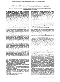
From www.bloodjournal.org by guest on February 6, 2015. For personal use only.
Retroviral Vector Design for Long-Term Expression in Murine
Hematopoietic Cells In Vivo
By Pamela H. Correll, Susan Colilla, and Stefan Karlsson
A series of retroviral vectors containing the human glucocerebrosidase (GC) cDNA driven by various promoters have
been constructed in an attempt t o discover which vector
design can most efficiently transduce murine hematopoietic
stem cells (HSCs) and drive expression of the transferred
gene in hematopoietic cells of mice reconstituted with the
transduced stem cells. The simplest vector, LG, in which the
GC gene is driven by the viral LTR, was the most efficient
vector at infecting HSCs, with an average viral copy number
in hematopoietic tissues of 3 copies/cell in recipient mice.
In general, the viral vectors that contained any additional
promoters or enhancers t o drive expression of either the GC
gene or a selectable marker gene (NeoR)had lower titers
and/or transduced HSCs at a lower efficiency. This was seen
most markedly when the human phosphoglycerate (PGK)
promoter was used t o drive the human GC cDNA. Despite
repeated attempts t o obtain a high titerproducer clone, this
virus consistently produced low titersand subsequently resulted in the lowest proviral copy numbers in long-term reconstituted mice. Only the viral LTR and the PGK promoter
were capable of drivingsignificant levels of human GC RNA
in hematopoietic cells of long-term reconstituted mice, with
a much lower level of RNA generated by an internal herpes
TK or SV40 immediate early promoter. Insertionof the internal transcription unit in the opposite orientation relative t o
the viral LTRs had a detrimental effect on gene expression.
The levels of RNA generated by a hybrid LTR containing the
myeloproliferative sarcoma virus enhancer were higher in
bone marrow-derived macrophages than in nonadherent
cells of the bone marrow when compared with the LG vector. The presence of an internal promotert o drive expression
of the human GC cDNA did notseem t o have a detrimental
effect on expression levels from theviral LTR. In fact, in the
presence of an internal TK or PGK promoter expression from
the LTR was increased despite the presence of lower proviral
copy numbers. Insertion of a second gene (NeoR)into the
vector had a negative impact on long-term expression in
hematopoietic cells in vivo; however, this seems t o be due
solely t o the lower transduction efficiency of this vector.
Overall, the highest levels of GC activity in macrophages
of long-term reconstituted mice were generated by the LG
vector; however, these levels were variable. Vectors containing an internal SV40,TK, or PGK promoter produced
consistent levels of GC activity in these cells, but because
of the lowertransduction efficiency obtained in the presence
of these promoters, simple vectors containing a single gene
driven bythe viral LTR currently remainthe mostpromising
viruses for gene therapy of human hematopoieticdisorders.
This is a US government work. There are no restrictions on
its use.
R
platelets, all mature hematopoietic cells derived from these
pluripotent stem cells, are replaced continuously throughout
life. Transfer of the appropriate gene into HSCs could be
used for treatment of any disorder that affects one or more
of these cell lineages.
Retroviral vectors have been used successfully to transfer
a number of genes into murine HSCs; however, persistent,
long-term expression in the progeny of these cells in vivo
has been historically difficult to obtain.' Vectors that produce
high levels of expression in immortalized fibroblasts or hematopoietic cell lines do not necessarily result in expression
in differentiated progeny of infected HSCs in v ~ v o .It~has
.~
beenunclearwhether the low levels of expression in the
majority of these studies were due to insufficient transcription or low transduction of HSCs. In addition, it is impossible
to directly compare the results from these studies because
of differences in methods of infection, transduction efficiency, and vector design (genes being transferred, regulatory elements present, direction of transcription, and vector
backbone used).
Inan attempt to determine what vector design would be
mostusefulto
drive expression of a transferred gene in
hematopoietic cells, a series of retroviral vectors containing
the human glucocerebrosidase (GC) cDNA under the transcriptional regulation of a variety of promoters and enhancers has been constructed. GC deficiency, commonly
known as Gaucher's d i ~ e a s eis, ~a leading candidate for HSC
gene therapy. It is an autosomal recessive lysosomal storage
disorder characterized by an accumulation of GC in macrophages of the BM, spleen, liver, lung, and brain.6 We previously transferred the human GC gene into murine hemato-
ETROVIRAL VECTORS have been used for the last
decade to transfer genes into mammalian cells for the
purpose of developing gene therapy for inherited disorders,
cancer, and acquired immunodeficiency syndrome (AIDS).
The majority of research to date has focused on bone marrow
(BM) as a target tissue for retroviral gene therapy.' Hematopoietic stem cells (HSCs) are able to persist throughout life
by undergoing proliferation to produce daughter stem cells
(self-renewal) and are also able to differentiate to form all
cells of the hematopoietic system. Erythrocytes, lymphocytes (B and T), granulocytes, monocytes/macrophages, and
From the Molecular and Medical Genetics Section, Developmental and Metabolic Neurology Branch, National lnstitute of Neurological Disease and Stroke, National Institutes of Health,
Bethesda, MD.
Submitted October 25, 1993: accepted May 17, 1994.
Supported in part bythe National Gaucher Foundation by a grant
to S.K.
Performed in partial fuljillment of the requirements for the PhD
degree in genetics at the George Washington University, Washington, DC, by P.H.C.
Address reprint requests to Stefan Karlsson, MD, PhD, Molecular
and Medical Genetics Section, Developmental and Metabolic Neurology Branch, National lnstitute of Neurological Disease and
Stroke, National Institutes of Health, Bldg 10, Room 3004, Bethesda,
MD 20892.
The publication costsof this article were defrayedin part by page
charge payment. This article must therefore be hereby marked
"advertisement" in accordance with 18 U.S.C. section I734 solely to
indicate this fact.
This is a US govemment work There are no restrictions on its use.
OOO6-4971/94/8406-0033$0.00/0
1812
Blood, Vol84, No 6 (September 15). 1994: pp 1812-1822
From www.bloodjournal.org by guest on February 6, 2015. For personal use only.
1813
VECTOR DESIGN FOR LONG-TERMEXPRESSION
poietic progenitor cells’ and stem cells,8 and expression of
potentially therapeutic levels of human GC in the majority
of macrophages from long-term reconstituted mice has been
shown?.“
In this report, we directly compare the transduction efficiency of several retroviral vectors in murine HSCs and
the ability of these viruses to stably express human GC
in macrophages derived from the transduced stem cells. In
comparing these vectors, we are asking several fundamental
questions about retroviral vector design and its effect on
transduction efficiency and expression. How does the inclusion of an internal promoter to the viral construct effect its
ability to infect HSCs and drive expression of the transferred
gene, and how do different promoters compare with respect
to these issues? What happens when the internal transcription
unit is placed in the opposite orientation with respect to the
viral LTRs? Does the addition of a selectable marker gene
affect the ability of the virus to infect HSCs or the ability
of the viral LTR to direct expression of the transferred gene
in hematopoietic cells? Finally, does the choice of retroviral
enhancer have an impact on the ability of the virus to transduce HSCs and/or direct expression of the transferred gene
in the differentiated progeny of these cells? The answers to
these questions have universal importance when designing
retroviral vectors for gene therapy of any disorder involving
the hematopoietic system.
MATERIALS AND METHODS
Retroviral Vectors
All vectors described in this study are based on the LN series of
retroviral vectors.” Construction of the LG9and LGSN” vectors
has been described elsewhere. All other vectors were constructed
from a plasmid (Gl) in which the NeoRgene from LN was removed
and replaced with a multiple cloning site (Genetic Therapy Inc,
Gaithersburg, MD). A 2.3-kb fragment of the human GC cDNA”
was subcloned into the EcoRI site of the polylinker in bluescript
sk(-) (Stratagene, LaJolla, CA), with the 5’ end of the gene towards
the Sac1 site and the 3’ end of the gene towards the Kpn I site
(SKGCS). A polyadenylation signal was added to the 3’ end of the
GC cDNA by ligation of the Nae IIHindIII fragment of PMCINeoPolA into the EcoRV site in SKGCS (SKGCNPolA).
All intenal promoters used were subcloned into the polylinker of
bluescript ks(-) with the 3’ end of the promoter oriented towards
the Sac I site. The 520-bp EcoRYBamHI fragment of PGK-pucl9
(provided by Dr S.H. Orkin, Children’s Hospital, Boston, MA) containing the human phosphoglycerate (PGK) promoter was cloned
into the EcoRI to BamHI sites of bluescript (KSPGK). The 334-bp
Pvu IIIHindIII fragment of pCHllO (Stratagene) containing the
SV40 promoter was blunted and cloned into the EcoRV site of
bluescript and oriented toward the Sac I site of the polylinker
(KSSV). The 280-bp Xho UPsr I fragment of PMCINeo containing
the herpes thymidine kinase promoter and mutant polyoma enhancerI4was cloned into the Xho I to Pst I sites of bluescript (KSTK).
The myeloproliferative sarcoma virus (MPSV) enhancer was cloned
into the 3‘ Moloney’s murine leukemia virus (MoMLV) LTR in G1
by replacing the Sac I-Cla I fragment of the MoMLV LTR with the
600-bp Sac I-Cla I fragment from the MPSV LTR (provided by Dr
D. Bodine, NHLBI, NIH, Bethesda, MD) (GIMP).
Expression cassettes containing the internal promoters in front of
the human GC cDNA were constructed in bluescript by digesting
the plasmids KSSV, KSTK, and KSPGK with EcoRI, S m I, and
Xba I, respectively, all of which cut at the 3’ end of the respective
promoters. A second digest was performed with Xmn I, which cuts
in the ampicillin-resistance gene of the bluescript plasmid. ‘Ihe 1.35-,
1.3-, and 1.5-kb fragments containing the respective promoters were
isolated. SKGCS was cut with a partial EcoRI digest, S m I, or Xba
I, which all cut at the 5’ end of the GC cDNA, and Xmn I in the
ampRgene. The 4.2-bp fragments containing the human GC cDNA
were isolated and ligated to the respective promoter fragments.
The MG vector was constructed by ligating the Nor UXho I fragment of SKGCS into the Not VXho I fragment of GlMP. The SG,
TG, and PG vectors were created by ligation of the Xho I fragments
of SVGC, TKGC, and PGKGC into the Xho I site of G1. Both
orientations of the cassettes were isolated relative to the viral LTR.
A polyadenylation site was added to the vectors in which the GC
gene was in the backwards orientation by removal ofthe Sal I
fragment containing the last 828-bp of the GC cDNA and replacing
this with the Sal I fragment of SKGCNPolA that contains the last
828 bp of the GC cDNA plus the inserted polyadenylation signal.
Virus-Producing Cells
The GP + E86 virus-producing cells usedin this study were
grown in Dulbecco’s modified Eagle’s medium (DMEM), 10% heatinactivated fetal bovine serum, and glutamine. The virus producing
clones were created by transfection (LGSN) or cotransfection with
pSV2Neo by calcium phosphate precipitation of the recombinant
viral constructs into GP + E86 cells. Two days later, the cells
were split by serial dilution and replated in media containing 1 g/L
geneticin sulfate ((3418). Ten days to 2 weeks after plating, individual colonies were picked and expanded.
Titration of LGSN was performed by plating 1 X lo6 TK- 3T3
cells onto a 10-cm tissue culture dish on day 1. The following
day, the 3T3 cells were infected by serial dilutions of an overnight
supernatant from the virus producing cells in the presence of 8 pg/
mL of polybrene. The following day, the infected 3T3 cells were
split 1:lO in media containing 1 g/L G418. Eight to 10 days later,
G418-resistant colonies were counted. Viral titers are expressed as
colony-forming units ( C m ) per milliliter of viral supernatant.
Titration of viruses with no selectable marker was performed by
plating 1 X lo6 TK- 3T3 cells on 10-cm tissues culture dishes and
infecting them on day 2 with 5 mL of an overnight supernatant from
the virus-producing cells in the presence of 8 pg/mL of polybrene.
On day 3, viral supernatant was removed and replaced with normal
media. On day 4,the 3T3 cells were harvested and DNA was extracted for Southern blot analysis. The blots were probed with the
full-length 2.2-kb human cDNA, and the signal of DNA from infected 3T3 cells was compared with the signal of equal amounts of
DNA from uninfected cells containing the equivalent of 1 copykell
and 0.1 copieskell of vector-containing plasmid DNA. Viral titers
are expressed in copies per cell.
Detection of helper virus by the SfL- assay was performed by
plating D-56 cells (R.H. Bassin, National Institutes of Health,
Bethesda, MD) at a density of 2 X l@ cells per 6-cm tissue culture
dish. The following day, the cells were infected with serial dilutions
of viral supernatant in the presence of 8 pg/mL of polybrene. Two
hours later, viral Supernatants were aspirated and replaced with normal media. Foci (plaques) were scored on days 5 through 10.
Detection of helper virus by marker rescue assay was performed
by plating 3T3 cells transduced with a replication defective retrovirus
containing the NeoR selectable marker gene at a density of 1 X lo6
cells per 10-cm dish. The following day, the cells were infected with
serial dilutions of viral supernatant in the presence of 8 pg/mL of
polybrene. After the infected cells were passaged for 1 to 2 weeks
From www.bloodjournal.org by guest on February 6, 2015. For personal use only.
1814
toallowtimeforpotentialvirustospread,thesupernatantwas
cells. These cells werethen
harvestedandtestedonvirgin3T3
selected in 1 g& G418 and any resistant colonies scored 8 to 10
days later.
CORRELL,COLILLA, AND KARLSSON
RESULTS
Retroviral Vectors
A series of retroviral vectors containing the human GC
cDNA was constructed (Fig 1). In these vectors, expression
of the humanGC gene is directed by either theMoMLV LTR
BM Infection and Transplantation
(LGSNandLG),theMoMLVLTRcontainingenhancer
Donor C57BY6 mice were injected with150 mgikg 5-fluorouracil
sequences from the MPSV LTR (MG), or an internal simian
(5-W;Fluka BioChemika,Ronkonkoma, N Y ) andBMwasharvirus 40 immediate early promoter (SG), thymidine kinase
vested from these mice 3 days postinjection. Thecells were plated
at a densityof 1 to 2X IO7 celldl0-cm dish and prestimulatedfor 2
promoter and mutant polyoma enhancer (TG), or phosphodays in DMEM containing 10% heat-inactivated fetal bovine serum, glycerate kinase promoter(PG) in either orientation relative
glutamine, PedStrep, and 200 U/mL interleukin-3(L-3). L-6, and
to the viral LTR sequences. The selectable marker neomycin100 ng/mL stem cell factor (Immunex, Seattle, WA). On day 3, l
resistance gene has been deleted from all constructs but one
to 2 X lo7 cells wereplatedona10-cmdishcontainingvirus
(LGSN). All of the vectors were packaged in the GP
+ E86
producersplatedthepreviousdayat1to2
X lo6 cellddish and
packaging
cell
line.
The
expected
transcripts
generated
by
cocultured for 2 days under the same growth factor conditions in
each
of
the
vectors
are
also
shown
in
Fig
1.
the presence of 8 pg/mL of polybrene. Lethally irradiated (850 rad)
Several individual clonesof each vector packaged in GP
C57BV6 recipient mice were injected with 5 X 10⁴ celldmouse for
+ E86 cells were used to infect 3T3 cells, and DNA from
individual CFU-S foci and 1 to 2 X IO6 celldmouse for long-term
these infected cells was analyzedby Southern blot analysis.
reconstitutedmice. CW-S foci were examined on12to 14 days
posttransplantationand BM, spleen,andthymusfromlong-term
A Southern blot of the highest titer clone found for each
reconstituted mice were analyzed
6 to 12 months posttnlnsplantation. vector is shown in Fig 2. The titers of these viruses range
from 0.3 to 2.6 copiedcell. Although there is no guarantee
that the highest possible titer will be obtained for each vector,
DNA and RNA Analysis
for every vector but one, a clone withof aattiter
least1 copy/
DNA and Southern blot analysis were performed using standard
techniques. Proviral copy numbers were determined by scanning the cell was isolated. However, despite screening more than 50
clones containing the PG vector, the highest titer obtained
intensity of each band and comparing it with copy numbercontrols,
was only 0.3 copiedcell on 3T3 cells. The virus-producing
with the endogenous GC bandused as an internal control.All scanning and quantitation was performed with the
clones shown in here were used for all subsequent experiNIH Image l .49 software (DrWayne Rasband, NIMH,NIH, Bethesda, MD). Total celluments.
lar RNA was extracted by guanidine thiocyanate'J and separated on
To estimatehow these titers relate to other published titers,
a formaldehyddagarose gel. The
RNA was transferred toa nitroceleight LGSN clones were analysed by colony-forming assay
lulose filter, prehybridized. hybridized, and washedas described.16
and Southern analysis for comparison (Fig
3). Sixofthe
eight clones produced titers of close to 1.0 copylcell(O.6 to
Isolation of Macrophages
1.2 copieskell) on the Southern blot. The colony-forming
titers of these clones ranged from 2 X IO6 to 1 X 10' CFU/
Macrophages from BM and spleen were isolated by plating
2X
mL. In most cases, the intensity of the band on the Southern
io7cells from a single-cell suspension derived from two
these
tissues
in 10 mL of RPM medium (Biofluids, Rockville, MD) with 10%
blot corresponded well to the G418 titer. Two of the eight
heat-inactivated fetal bovineserum, 2 mmol/L glutamine, 10 mmoY
LGSNcloneswithtitersof
3 X lo3 and 4 X 10⁴ CFU/
L HEPES, pH 7.3, 5 X IO-' m o w B-mercaptoethanol, 100 U/mL
mL did not produce a detectable band on the Southern blot
penicillin, and 100 pg/mL streptomycin. The plates were incubated
analysis. This indicates that titration
by Southern blot analyat37°C for 3hours.Thenonadherent cells weregentlyremoved
sis will not detect a titer of less than approximately5 X 10"
from the dish and the remaining adherent cells were grown in the
CFU/mL. Furthermore, this indicates that all of the clones
above medium plus 10% L929 (American Type Culture Collection,
used in this study most likely have a titer of at least 1X lo6
Rockville. MD) cell-conditionedmedium.Themacrophageswere
CFU/mL or greater, with the exception of the PG virus, for
cultured for 8 days and were refed with fresh medium every other
which the titer is considerably lower. Clone no.
6 with a
day.
titer of 1 x 10' CFU/mL is the one shown in Fig2 and used
in the experiments presented here. The difference in copy
GC Enzymatic Assay
number between the two experiments reflects the variation
Cellpelletswereextractedina
50 mmoK potassiumcitrate/
sometimes seen from one infection to another.
potassiumphosphatebuffer(pH5.9)containingTritonX-100
(2
Viral supernatants from eachof the producer clones used
mg/mL) and freeze-thawed for three cycles. Cell extracts were spun
in this study were tested for the presence of wildtype virus
for30minutes at 12,000 rpmand clearedcellularlysateswere
by the S+L- and/or the marker rescue assay. Helper virus
assayed for GC activity. GC activity was assayed by cleavage ofthe
was
not detected by these methods in any of the supernatants
synthetic substrate Cmethylumbelliferyl glucopyranoside (Sigma, St
tested.
Louis,MO)at 4.8 mmol/L ina0.1 mol& potassiumphosphate
buffer(pH5.9)with
1.5 m g / d TritonX-100and1.25
mg/mL
The Effect of Vector Design on Transduction of
sodium taurocholate at 37OC. The reaction was terminated with 0.4
Hematopoietic Cells
moVL NaOW0.4 mom glycine, and the fluoresenceof the cleaved
4-methylumbelliferone was measured using a fluorimeter (excitation: Because certain as yet unidentified characteristics of particular viral producer clonesmay be important for transduc360 nm, emission 430 nm).
From www.bloodjournal.org by guest on February 6, 2015. For personal use only.
VECTOR
EXPRESSION
FOR LONG-TERM
1815
"_
LG
"_
"""""_
.0
-I
'./
b
"
"
"
"
"
"
_
~
MG
MPSV EHANCER
"m
"-(
_"\
.0
"_
~
, ,"""-b""""
b
\ H
Fig l. A diagram of the vectors used
in this study.
All vectors are based on the LNseries of retroviral
vectors. Open boxes denoteMoMLV
LTR; light
hatched boxes, human GCcDNA;
dark hatched
boxes, polyadenylation signal fromSV40; SV, SV40
immediate early promoter; NEO, neomycin-resistance gene;TK, herpes thymidine kinase promoter
and mutant polyoma enhancer; PGK, human phosphoglycerate kinase promoter; MPSV, meyloproliferative sarcoma virus; Kb, kilobase. Arrows indicate
origin and direction of transcription. The expected
RNA transcripts are shown below each vector. The
spliced transcripts are shown as dotted lines because they are not always detected.
-
"_
D
.H
4 - 4
TG
-"
,
.,"""""""_""
I
*
U
1 Kb
7
. .
4.4-
2.32.0-
1
H
4
(D
9.6 6.6 -
I
TK
03
w
W
, ,"""""""
~~
Fig 2. Southern blot analysis
of DNA from NlH3T3 cells infected by each of the viralsupernatants used for this study. The
DNA was digested with Nhe 1,
which cutsin the viralLTRs, and
10 p g was loaded per lane. The
blot was probedwith the human
GC cDNA. 1 and 0.1 copieslcell,
genomic DNA fromuninfected
3T3 cells
the equivalent of 1
+
3T3 cells infected with supernatantfrom untransfected GP
E86 cells. Molecular weight standards are shown on the left in
kilobases. The proviral copy
number, which is shown below
eachlane, was determined by
scanning with the genomic GC
+
From www.bloodjournal.org by guest on February 6, 2015. For personal use only.
COLILLA,
1816
CORRELL,
AND KARLSSON
B
A
LGSN
I
9.6-
I
~
LGSN
Clone ##
Titer
CFU/ml
1
2
3
4
2x10⁶
4x10⁶
4x10⁴
3x10³
6
1 x10⁸
8
6 x 10'
4.4 -
Proviral copy no.
1.2
0.9
0 0 0.8 1.1 0.6 0.9
+
Fig 3. (A) Southern blot analysis of DNA from 3T3 calls infected by supernatants from eight separate c l o n e of GP E86 cells transfected
with LGSN. The DNA was digested with Nhe 1, which cuts in the viral LTRs, and 10 p g was loaded per lane. The blot was probed with the
human GC cDNA. 1 and 0.1 copieslcell, genomic DNA from uninfected 3T3 cells + the equivalent of 1 and 0.1 copieslcell of LGSN plasmid
DNA; 3T3, DNA from uninfected 3T3 cells. Molecular weight standards are shown on theleft in kilobases. Proviral copy numbers, determined
by scanning, are shown below each lane. (B)Titers of the same eight LGSN clones shown in (A)as determined by Neomycin-resistance.CFUl
mL, colony-forming units per milliliter of viral supernatant.
tion of hematopoietic progenitor cells and it would be impossible to analyze all of the clones on HSCs, a second screen
of the virus producer lines that gave high titers on 3T3
cells was performed on CFU-S multipotential hematopoietic
progenitor cells. Each of the producer clones was used to
infect primary murine hematopoietic cells in the presence of
the growth factors L-3, IL-6, and stem cell factor. DNA
from individual day 12 to 14 CFU-S foci was screened by
Southern blot analysis for the presence of the provirus. The
results from each of the viruses are summarized in Table 1.
With each of the viruses used for this study, with the exception ofSG, the initial experiment resulted in a 100% infection
efficiency of CFU-S progenitor cells. When the experiment
Table l.Infection Efficiency of Viral Producer Cells in CFU-S
100
100
Exp.
100
100
Vector
No. Positiveflotal
Efficiency
Infection
LGSN
LG
MG
SG
no. 1
Exp. no. 2
TG
PG
9/9
1%)
100
10110
loll0
3i7
100
43
10/10
m
-
10110
TG
1111 1
PG
10/10
100
100
was repeated with SG, this virus also infected 100% of the
CFU-S. Even though the PG virus has a much lower titer
than the others by Southern blot analysis of infected 3T3
cells, this virus was also capable of infecting CFU-S with a
100% infection efficiency. It was not possible to compare
gene transfer efficiency of clonogenic progenitor cells by
G418 resistance using these vectors, because all the vectors,
except one, were single gene vectors lacking a selectable
marker gene.
Because there is no direct way to determine the infection
efficiency of the viruses in repopulating HSCs, wehave
based the transduction efficiency in these cells on the proviral
copy number obtained in BM, spleen, and thymus harvested
from lethally irradiated long-term reconstituted mice at 1ea.t
6 months posttransplantation. Southern blot analysis of DNA
from the nonadherent cells of the BM and spleen and total
thymus of several individual mice transplanted with vectorinfected BM was performed. The viral copy number for each
sample was determined by comparison of the signal intensity
to copy number controls. Results from each of themice
analyzed in this fashion and the average viral copy number
obtained with each virus are summarized in Table 2. The
transduction efficiencies of these vectors in HSCs correspond
well with their titers on 3T3 cells.
The LG vector has the highest infection efficiency of the
vectors tested, with an average copy number of 3 copied
cell. This vector has consistently transduced HSCs at a high
From www.bloodjournal.org by guest on February 6, 2015. For personal use only.
VECTORDESIGN
1817
FOR LONG-TERMEXPRESSION
Table 2. Proviral Copy Number in LongTermReconstituted Mice
Vector
LGSN
Mouse No.
1
2
3
4
'5
'6
'7
'8
Avg
LG
1-5
6-10
11
12
13
14
15
'16
'17
'18
'19
'20
SG
S
T
2.0
3.1
1.a
0.2
0.3
0.5
0.9
0.8
1.2
1.a
0.4
0.8
1.2
0.6
0.6
1.o
1.3
1.o
0.6
3.2
2.9
2.2
2.6
3.7
4.4
3.5
2.5
2.0
2.9
3.2
3.8
4.4
2.6
2.1
2.0
3.3
2.4
1
2
3
4
'5
'6
'7
'8
'9
0.4
1.3
2.8
0.7
1.3
0.2
0.9
0.6
Avg
1.3
0.8
1
2
'3
'4
'5
'6
'7
0.7
3.2
3.3
2.5
3.6
0.4
0.8
2.6
3.2
2.7
3.9
2.7
2.3
Avg
MG
BM
Ave
Vector
TG
0.7
0.4
0.1
0.7
PG
0.7
1.5
2.0
2.1
3.1
4.0
1.4
2.1
1
2
3
4
5
6
7
8
9
10
11
Avg
BM
5
T
0.4
0.8
0.5
1.o
4.5
2.2
1.2
1.5
0.4
1.5
0.4
0.2
1.6
0.9
0.5
0.9
0.7
0.3
1.1
0.2
0.9
0.4
0.9
0
0
0.3
0.7
0.3
0
0.5
0
0
0
0.2
0.2
0.1
0
0.9
1.5
0.2
0
0.9
0.3
0
0
0.1
0.1
0.2
0
0
0.4
0.2
0.3
1.3
0.2
0.8
0.6
0.3
0.2
0.2
0.3
0.3
0.2
0.2
0.3
0.4
0.1
1.2
0.4
0.6
0
2.5
0.3
0.6
1.o
0.3
0.7
0.1
1.o
0.2
0.7
0.2
1.7
0.3
1.o
1.?
0.4
0.7
0.9
0.1
2.1
0
0
0
Avg
0.8
0.4
0.4
0.6
0.6
L
3.0
TG
2.9
0.2
0.9
1.o
1.2
1.2
2.8
1.3
1.4
1
2
3
4
5
6
7
Avg
1.o
5.0
2.7
4.7
3.6
3.2
3.2
3.1
2.6
2.0
3.3
Mouse No.
'1
'2
'3
Avg
PG
1.1
1
2
3
4
5
6
7
8
'9
'10
Avg
0
0.2
0.4
1.8
0.8
0.5
2.4
Abbreviations: AVG, average; S, spleen; T, thymus.
Animals used for Northern blot analysis (Fig 4) and enzyme analysis (Fig 5).
efficiency, with 2 to 5 copiedcell detected in every mouse
analyzed thus far. Consistent with the viral titer on 3T3 cells,
the PG virus had the lowest transduction efficiency in HSCs,
with an average copy number of 0.3 copiedcell (approximately 1 log lower than the efficiency of the LG vector) in
long-term reconstituted mice. The remainder of the viruses
tested had intermediate infection efficiencies, with average
copy numbers ranging from 0.4 to 2.4 copieskell. Vectors
containing additional promoters, enhancers, or selectable
markers, in general, generated lower transduction efficiencies than the LG virus. The vector containing an internal
SV40 promoter (SG) had a greater transduction efficiency
than those containing the TK (TG) or PGK (PG) promoters.
It is also interesting to note that when the PGK promoter
was used in the opposite orientation relative to the viral LTR,
a higher transduction efficiency was obtained, correlating
with the higher titer of this virus on 3T3 cells. The virus in
which the MPSV enhancer was substituted for the MoMLV
enhancer (MG) also had a lower transduction efficiency of
both 3T3 cells and HSCs. Finally, the virus containing an
internal neomycin-resistance gene (LGSN) had a lower
transduction efficiency in HSCs than the LG vector or the
same vector without the selectable marker (SG).
DNA from BM, spleen, and thymus of several of the longterm reconstituted mice was also analyzed by Southern blot
analysis for the presence of the Y chromosome using a Y-
From www.bloodjournal.org by guest on February 6, 2015. For personal use only.
1818
9.5 -
7.5 4.4-
]LTR Transcripts
2.4 -
Internal
]Transcripts
-Mouse GC
1.3-
-GAPDH
2 3 3 5 5 5 5 4
2 3 3 5 5 5 5 4
0.5 0.2ND 1.8 3.1 ND 3.1 0.6
Number of Mice
Average proviralno.
Fig 4. Northern blot analysis of RNA from (A) nonadherent cellsand (B) macrophages from the BM of long-term reconstituted mice
transplanted with BM infected by each of the viruses as indicated. RNA was extracted from pooled cells from the number of mice indicated
beneath the blot, corresponding to the mice marked by an asterisk in Table 2. Ten micrograms of RNA was loaded per lane. The blot was
probed with thehuman GC cDNA, stripped, and reprobed
with GAPDH. NC,RNAfrom nonadherentcells and macrophages an
of untransplanted
mouse. Molecular weight standards are shown to the left in kilobases. The expected size of each transcript is shown in Fig 1. The average
proviral copy numbershown for the nonadherentcells was determined by scanning densitometry for the same cells as used forthe Northern.
specific probe.I7 The disappearance of the Y-specific band
in male mice transplanted withBM from female donors
indicates that, in all animals tested, the recipient mice were
fully reconstituted with the donor marrow (data not shown).
These results support the idea that the variability in copy
number obtained with the different viruses in fact reflects a
variability in transduction efficiency rather than differences
in the extent of repopulation from one experiment to another.
The Effect of Vector Design on Gene Expression in
Hematopoietic Cells
To determine how efficiently the various promoters can
direct expression of the GC cDNA in hematopoietic cells in
vivo, expression of these vectors was examined in the progeny of the transduced HSCs. Nonadherent cells (Fig 4A)
from the BM of several long-term reconstituted mice transplanted with BM transduced by each of the vectors were
analyzed for expression of human GC RNA. The RNA in
Fig 4A was isolated from a pool of the same nonadherent
BM cells analyzed for copy number in the mice indicated
by asterisks in Table 2. The number of mice and the average
proviral copynumber for those cells are indicated below
each lane.
For Gaucher's disease, we are ultimately interested in obtaining expression in differentiated macrophages derived
from the transduced HSCs. Therefore, macrophages isolated
from BM of these same mice were also analyzed for human
GC RNA (Fig 4B). Due to a limited number of cells, proviral
copy number was not assessed directly in this cell population; however, the results from the macrophages correlates
well with the expression pattern seen in the nonadherent cell
population for which proviral copy number data is available.
The Northerns shown in Fig 4 were quantitated and the
results from this analysis are shown in Table 3. The values
obtained for both the nonadherent cells and macrophages are
a sum of all transcripts expressing the human GC cDNA.
The values for the GAPDH control are also shown as well
as the corrected values for human GC transcripts. For the
nonadherent cells, the values are also corrected for proviral
copy number. The values shown in parentheses are those for
which the corresponding copy number data are not available.
For these vectors, the overall average copy numbers obtained
with these vectors are used. Therefore, there is likely some
error in these numbers.
Comparison of promoters/enhancers for expression in hematopoietic cells. The results from these experiments indicate that, overall, the viral LTR is the most efficient of the
promoters tested in all of the hematopoietic cells analyzed,
although the levels of LTR-driven transcript vary among the
vectors tested. Of the internal promoters tested, the PGK
promoter in the forward orientation directs the highest levels
of human GC message in these experiments. There are much
From www.bloodjournal.org by guest on February 6, 2015. For personal use only.
VECTOR
EXPRESSION
DESIGN FOR LONG-TERM
1819
Tablo 3. Exprodon kv.h of Human GC RNA In Long-Twm Roc0nrtitut.d Mica
-
-
LGSN
LG
MG
SG
TG
PG
TG
PG
GC RNA nonadherent
GAPDH nonadherent
GC RNNGAPDH nonadherent
465
2,908
0.16
1,761
1,995
0.90
494
3,061
0.16
2.947
2.01 4
1.46
4,938
1,839
2.69
2,674
2,056
1.3
0
1,710
0
0
618
0
GC RNA macrophages
GAPDH macrophages
GC RNNGAPDH macrophages
0
2,469
0
1,396
2,314
0.60
1,545
2,812
0.67
2,039
1,842
1.1 1
4,373
2,101
2.08
4,156
2,388
1.74
456
1,963
0.23
0
1,027
0
Proviral copy number nonadherent
GC RNNcopy 3, no. of
nonadherent
0.6
3.1
(1.1)
.8
(0.3)
0.2
0.5
0.3
0.3
(4.3)
0
0
1.5 (0.15)
1
3.1
0.5
The numbers represent arbitrary densitometry units.
the TG virus is present at a lower copy number than the LG
lower, but detectable, levels of GC RNA generated by the
internal TK and SV40 promoters. These results correspond virus in the BM of these long-term reconstituted mice (1.8
to those obtained when expression of the vectors was tested v 3.1 copiedcell, respectively), thelevelsofLTR-derived
message generated by
this vector are higher than those generin three separate CFU-S experiments (data not shown).
ated by the LG virus. In addition, levels of LTR-generated
Interestingly,levels of RNA fromtheviralLTRcontaining theMPSV enhancer (MG)are lower than those from transcript from the PG vector are also high in these mice.
Although no proviral copy number data is available for the
LG in the nonadherent cells; however,in the macrophages,
these levels are roughly equivalent. Because these respective BM of these mice because of experimental error, the overall
average copy number obtained in BM with the
PG vector is
samples are from the same mouse BM,
it appears as though
0.3 copiedcell, and,of the 11 animals tested, the highest
transcription from the hybrid LTRmay be more efficient in
copy number in an individual mouse was
0.9 copiedcell.
thedifferentiatedmacrophagesthaninothernonadherent
These data indicate that the presence
of an internal transcrip
hematopoietic cells.
tion unit driven by the TK or PGK promoters does not deEffect of a second transcription unit on expression from
the viral LTR. We were also interested in determining what crease, but may even increase expression from the viral LTR
in hematopoietic cells.
effect, if any, the inclusion of an internal transcription unit
has onthe levelsof expression from the viral LTR. For these Effectof gene orientation on gene expression. Those
vectors (TG and PG) that contain internal transcripts in the
comparisons, it is most informative to focus on expression
in the nonadherent fraction
of the BM in long-term reconsti- opposite orientation relative to the viral LTRs consistently
showverylowlevelsofboththeLTRmessageandthe
tuted mice for which proviral copy number data has been
internally generated message in all hematopoietic cells tested
determined. It is difficult to assess the effect of integration
site of the provirus on viral expression; however, due to the in this study. This is most likely caused by the production
fact that the RNA is pooled from several transplanted mice
of antisense RNA from the internal promoter.
rather than individual mice, some general observations can
Protein Production in Macrophages of Long-Term
be made.
Reconstituted
Mice
Thelevels ofhuman GC transcriptsgenerated by the
LGSNvector are lowerthanthosegeneratedbytheLG
Macrophages isolated from BM of the same set of mice
vector; however, the lower viral copy number obtained with for which the RNA is shown were analyzed for expression
the LGSN virus (0.6copykell) when compared with the LG of GC activity. The activity for each mouse is shown in Fig
virus(3.1 copiedcell) almostcertainlycontributestothe
5 . In this experiment, the PG virusproducedthehighest
lower levelsof expression from the LTR seen with
this virus.
overall levels of GC activity in BM macrophages, with an
When corrected for the copy numbers, the levels of these
average activity 1.77-fold higher than the uninfected contranscripts are roughly equivalent. On the other hand, the
trols. This activity was generated by a combination of tranlevels of LTR-generated transcript in long-term reconstituted scripts from the LTR and PGK promoters because both tranmice infected with the SG virus are not reduced when com- scripts can be translated to produce GC protein. This was
pared with those generated by the LG virus. Because the LG followed by the TG, LG, and SG viruses, which increased
and SG mice in this experiment contained similar proviral
the enzyme activity in BM macrophages by an average
of
copy numbers,it appears as though the presenceof the inter1.46-, 1.45-, and 1.44-fold, respectively. The majority of the
nal SV40 promoter alone or in combination with the NeoR
enzyme activity generated by the TG and SG viruses
is most
gene in the viral construct has a minimal effect on the expreslikely caused by transcription from the viral LTR, because
sion levels from the viral LTR.
levels of transcript from the SV40 and
TK promoters in
The levels of message from the viral LTR generated by
these cells are low. The LGSN virus produced lower levels
both the TG and PG viruses also remain consistently high
of activity on average than the LG virus, and the TG and
in all the hematopoietic cells tested. Interestingly, although
PG viruses in which the transcriptional unit was placed in
From www.bloodjournal.org by guest on February 6, 2015. For personal use only.
CORRELL, COLILLA, ANDKARLSSON
1820
the opposite orientation relative to the viral LTRs also produced very low levels of GC activity. These results follow
veryclosely the levelsof RNAproduced by thevarious
vectors in these cells.
The MC virus did not showanydetectable increasein
GC protein in these cells. This is most likely caused by a
mutation in the GC cDNA that may have occurred during
subcloning, because the packaging cells also do not express
any human GC cDNA (data not shown). When a different
clone of this plasmid was transfected into amphotropic packaging cells, it was capable of expressing human GC protein.
Althoughthe highest averagelevels of GC activityin
this experiment were produced by the PG virus, the highest
expressing single mouse in this experiment was an LC-infected mouse in which the LC virusproduced a 2.1-fold
increase in GC activity in BMmacrophages.Inaddition,
much higher levels of GC activity in macrophages of longterm reconstituted mice have been produced by the L C virus." In the 20 mice tested thus far, theL C virus has produced
average increases in activity in BM and spleen macrophages
ranging from zero to fivefold over those from normal uninfected mice despite consistently high proviral copy numbers,
with an overallaverage increase of 2.23-fold (data not
shown).
DISCUSSION
In the studies presented here, we have compared the utility
of several retroviral vectors for use in the gene therapy of
hematopoietic disorders. We have found
that the elements
present in the viral vector can affect the retroviral titer, its
efficiency at infecting murine HSCs, andits ability to express
human GC in hematopoietic cells of long-term reconstituted
mice. With the data presented here. we can begin to address
the questions posed at the outset of this study.
In general, the simplest viral construct (LC) is the most
efficientat infectingHSCs.This virusresults in ahigher
vectorcopy number in thehematopoietictissues
and, on
average, the highest overall enzyme production level of the
vectors tested. However, the level of enzyme generated by
theLGvirus
is quite variable anddoes notseem to be
dependenton viral copy number,because all of the LGinfected mice in these studies had multiple copies of the viral
genome in all hematopoietic tissuestested. These results
compare withthose obtained by Kalekoet
who reported
similar resultswithavectorcontainingthe
hADA gene
driven by the MoMLV LTR. They concluded that, although
the viral LTR was capableof generating high levels ofhADA
in long-termreconstituted mice,thepresence of multiple
copies was not sufficient to guarantee long-term expression.
Replacement of the MoMLV LTR enhancer with the enin the viral construct (MC)
hancerfromtheMPSVLTR
resulted in a lower viral titer and a subsequent decrease in
proviral copy number in long-term reconstituted mice. Results from Beck-Engeser et a l l g indicate that the MPSV enhancer caused a reduction in the retroviral infection of primitive hematopoietic cells. In further support of these results,
the transduction efficiency of the M C virus, packaged in two
different amphotropiccell lines, in both 3T3 and murine
myeloid (MI) cells was lower than that for the LC virus in
these same cells.'"
Expression of human GC RNA from the MG vector was
lower than expression from the LC vector in the nonadherent
cells from the BM of long-term reconstituted mice, possibly
reflecting a lower copy number in these cells. However, in
the BM-derived macrophages, the expression levels of human GC RNA generated by these two viruses were roughly
equivalent.Therefore,fordiseases
in which macrophages
are the target cell, suchas in Gaucher's disease, the increased
levels of transcription in these cells may compensate for the
lower infection efficiency. This conclusion is based on the
assumption that the macrophage compartment was reconstituted toanequivalentextent
in thesemice.Although
an
increase in GC enzyme couldnot be detectedin these macrophages because of a defect in the viral construct, we believe
that, in this experiment, the level of enzyme production
would be similar to that obtained with the LC virus because
the levels of RNA are comparable.
Addition of an internal promoter to the viral construct to
drive expression of thehuman CC cDNA also seemed to
haveanegativeeffectonthe
viral titer and its ability to
infect HSCs. In addition,some internalpromoters had a
more deleterious effectonthe
viral titer than others. The
presence of an internal PGK promoter(PG) dramatically
reduced the viral titer and its infection efficiency in HSCs.
Again, when the PC vector was packaged in amphotropic
cell lines and used to infectM1 cells in culture,asimilar
decrease in the infection efficiency when compared with the
LC and M C vectorswasobserved."'
However, when the
PGK promoter was placed in the opposite orientation relative
to the viral LTR, the viral titer increased somewhat. Similar
I
T
50
40
-
VECTOR
LGSN
LG
MG
SG
TG
PG
TG
4
PG
Fig 5. GC activity in macrophages isolated from B M of the same
long-term reconstituted mice as shown in Fig 4 and marked with an
asterisk in Table2. Mouse numbers from Table
2 are increasing from
left to right, ie, LGSN mice are 5,6,7, and 8 from leftto right. Enzyme
activities of individual mice are shown. Specific activity is presented
as nanomoles of 4MU cleaved per minute per milligram of protein.
From www.bloodjournal.org by guest on February 6, 2015. For personal use only.
1821
VECTOR DESIGN FOR LONG-TERMEXPRESSION
results have been shown with the c-fos promoter that did
not allow transduction of the target cells in the forward
orientation, although the reverse orientation did.” The presence of the SV40 promoter in the viral construct (SG)
seemed to have the least overall effect on viral titer. The TK
promoter (TG) had an intermediate effect on the ability of
the virus to infect HSCs, and placing the transcriptional unit
in the reverse orientation relative to the viral LTRs did not
enhance the titer of the virus.
Expression of the human GC cDNA from an internal promoter in hematopoietic cells is most efficient when the PGK
promoter is employed. It has been suggested that the viral
LTR can sometimes cause transcriptional interference with a
downstream internal promoter. To decrease this interference,
some groups have chosen to delete the 3’ LTR enhan~er.”~’~
However, when the PGK promoter was used todrive expression of the ADA gene, interference from the LTR was absent
or minimal and a deletion of the 3’ LTR was not necessary
to improve expression in CFU-S colonies.24When the SV40
(SG) and TK (TG) promoters were used to drive expression
of the human GC cDNA, they generated low, but detectable
levels of human GC RNA in the hematopoietic cells.
The presence of an internal promoter did not appear to
have a detrimental effect on expression levels from the viral
LTR in the hematopoietic cells. Levels of LTR-generated
transcript in nonadherent BM cells from long-term reconstituted mice transduced with the SG, TG, and PG vectors were
equal to or greater than the levels generated by the LG vector.
This increase was observed despite the lower proviral copy
number present in the mice transplanted with the TG- and
PG-infected BM.
Our double gene vector, LGSN, resulted in a lower infection efficiency in HSCs than the LG and SG viruses, indicating that the presence of the NeoRgene may have a deleterious
effect on retroviral titer. It has been generally agreed that
the size of the retrovirus is inversely proportional to the viral
titer that it can produce. The LGSN vector also resulted in
decreased expression of human GC in long-term reconstituted mice when compared with the LG and SG vectors. However, this decrease in expression seems to be due solely to
the lower proviral copy number present in these cells.
Others have reported detrimental effects on expression
levels when a second gene is
In support of this, all
reports of expression of ADA and GC at levels comparable to
endogenous mouse levels in hematopoietic cells have used
vectors that contain only the ADA or GC gene and no selectable markers. We conclude that the reduction in expression seen with double gene vectors may be due more to a
lower transduction efficiency of HSCs than to the presence
of a second transcriptional unit.
In this study, the LG virus was capable of transducing
HSCs at the highest efficiency and generated the mouse
expressing the highest levels of human GC. However, expression from this vector in long-term reconstituted mice is
not consistent. Overall, the TG and PG vectors in this study
generated the highest levels of human GC expression per
copy of the provirus. However, particularly in the case of
the PGK promoter, this high level of expression comes at
the expense of viral titer. Unfortunately, at the present time,
transduction of human hematopoietic progenitors is not as
efficient as the efficiencies in the mouse. Therefore, when
designing retroviral vectors for human gene therapy, having
a high viral titer is important. The TG vector generates higher
titers than the PG vector and may be useful, although very
little of the expression from this virus is actually coming
from the TK promoter itself. The SV40 promoter is expressed at low levels in hematopoietic cells in vivo and its
presence in the viral construct seems to be quite neutral in
terms of viral titer and expression from the viral LTR, therefore no advantage is gained by its use. Addition of a selectable marker gene causes a decrease in long-term expression from the viral LTR, mostlikely due to the lower proviral
copy numbers it generates, and should be avoided if possible.
Given these results, it would be beneficial to continue focusing efforts to design an “ideal” vector that can transduce
HSCs with high efficiency and consistently generate high
expression levels of the transferred gene in the progeny of
those cells. Of the vectors tested here, the LG vector remains
the most promising for transduction of primitive human hematopoietic cells due to its high titer.*’
ACKNOWLEDGMENT
We thank DrR.O. Brady for generous support and encouragement,
M. Amiri and S. Stahl for technical assistance, and D.L. Freas-Lutz
and Dr M. Brennan for valuable discussions.
REFERENCES
1. Karlsson S: Treatment of genetic defects in hematopoietic cell
function by gene transfer. Blood 78:2481, 1991
2. Williams DA: Expression of introduced sequences in hematopoietic cells following retroviral-mediated gene transfer. Hum Gene
Therapy 1:229,1990
3. Williams DA, Orkin SH, MulliganRC:Retrovirus-mediated
transfer of human adenosine deaminase gene sequences into cells in
culture and into murine hematopoietic cells in vivo. Proc Natl Acad
Sci USA 83:2566, 1986
4. MagliMC, Dick JE,Huszar D, Bernstein A, Phillipes RA:
Modulation of gene expression in multiple hematopoietic cell lineages following retroviral vector gene transfer. Proc Natl Acad Sci
USA 84:789, 1987
5. Brady RO, Kanfer JN, ShapiroD: Metabolism of glucocerebrosides 11. Evidence of an enzymatic deficiency in Gaucher’s disease.
Biochem Biophys Res Commun 18:221, 1965
6. Barranger JA, Ginns EI: Glucosylceramide lipidoses: Gaucher
disease, in ScriverCR,Beaudet AL, Sly WS, Valle D (eds): The
Metabolic Basis of Inherited Disease, v01 2. New York, NY,
McGraw-Hill, 1989, p 1677
7. Correll PH, Fink JK, Brady RO, Perry LK, KarlssonS: Production of human glucocerebrosidase in mice after retroviralgene transferintomultipotentialhematopoieticprogenitor
cells. ProcNatl
Acad Sci USA 863912, 1989
8. Correll PH, Kew Y, Perry LK, Brady RO, Fink JK, Karlsson S:
Expression of human glucocerebrosidase in long-term reconstituted
mice following retroviral-mediated gene transfer into hematopoietic
stem cells. Hum Gene Ther 1:277, 1990
9. Correll PH, Colilla S, Dave HPG,Karlsson S: High levels
of human glucocerebrosidase activity in macrophages of long-term
reconstituted mice afterretroviral infection of hematopoietic stem
cells. Blood 80:331, 1992
From www.bloodjournal.org by guest on February 6, 2015. For personal use only.
1822
10. Ohashi T, Boggs S, Robbins P, Bahnson A, Patrene K, Wei
F-S, Wei J-F,Li J, LuchtL,FeiY,Clark
S, KimakM,HeH,
Mowery-Rushton P, Barranger JA: Efficient transfer and sustained
high expression of the human glucocerebrosidase gene in mice and
their functional macrophages following transplantation of bone marrowtransducedby
aretroviralvector.ProcNatlAcadSci
USA
89: 11332, 1992
l 1. Miller AD, Rosman GJ: Improved retroviral vectors for gene
transfer and expression. Biotechniques 7:980, 1989
12.FreasDL,CorrellPH,DoughertySF,Karlsson
S, Pluznik
DH: Evaluation of expression of transferred genes in differentiating
myeloid cells: Expression of human glucocerebrosidasein murine
macrophages. Hum Gene Ther 4:283, 1993
13. Sorge J, West C, Westwood B, BeutlerE: Molecular cloning
and nucleotide sequence of human glucocerebrosidase cDNA. Proc
Natl Acad Sci USA 82:7289, 1985
14. Capecchi MR Altering the genome by homologous recombination. Science 244:1288, 1989
15. Chomczynski P, Sacchi W: Single-step method
of RNA isolation by acid guanidinium thiocyanate-phenol-chloroform extraction.
Anal Biochem 162156, 1987
16. Reiner 0, Wilder S, Givol D, Horowitz M Efficient in vitro
and in vivo expression of human glucocerebrosidase cDNA. DNA
6:101,1987
17. Mardon G, Page D: The sex-determining region of the mouse
Y chromosome encodes a protein with a highly acidic domain and
13 zinc fingers. Cell 56:765, 1989
18. Kaleko M, Garcia JV, Osbom WRA, Miller AD: Expression
of human adenosine deaminase in mice
after transplantationof genetically-modified bone marrow. Blood 751733, 1990
19. Beck-Engeser G, Stocking C, JustU, Albritton L, Dexter M,
Spooncer E, Ostertag W Retroviral vectors relatedto the myeloproliferative sarcoma virus allow efficient expression in hematopoietic
CORRELL,COLILLA,
AND KARLSSON
stem and precursor cell lines, but retroviral infection is reduced in
more primitive cells. Hum Gene Ther 2:61, 1991
20. Freas DL, Correll PH, Dougherty SF, Pluznik DH, Karlsson
S : Evaluation of retroviral vector expression in differentiated myeloid lineages: Expression of human glucocerebrosidase in murine
macrophages. Clin Res 41:163A, 1993
21.BelmontJW,MacGregorGR,Wager-SmithK,FletcherF,
Mom KA, HawkinsD,VillalonD,ChandSM-W,Caskey
CT
Expression of human adenosine deaminase in murine hematopoietic
cells. Mol Cell Biol 85116, 1988
22. Yu S-F, vonRuden T, KantoffPW,Garber C, Seilber M,
Ruther W, Anderson W F , Wagner EF, Gilboa E Self-inactivating
retroviral vectors designed for transfer of whole genes into mammalian cells. Proc Natl Acad Sci USA 83:3194, 1986
23. Guild BC, Finer MH, Houseman DE, Mulligan RC: Development of retrovirus vectors useful
for expressing genes in cultured
murineembryonalcellsandhematopoieticcellsinvivo.JVirol
62:3795,1988
24.Lim B, ApperleyJF,OrkinSH,WilliamsDA:Long-term
expression of human adenosine deaminase in mice transplanted with
retrovirus-infectedhematopoieticstemcells.
h o c NatlAcadSci
USA8623892,1989
25.ApperleyFJ,LuskeyBD,WilliamsDA:Retroviralgene
transferofhumanadenosinedeaminaseinmurinehematopoietic
on long-term expression.
cells: Effectof selectable marker sequences
Blood 78:310, 1991
26. Bowtell DDL, Cory S,Johnson GR, Gonda TJ: Comparison
of expression in hematopoietic cells by retroviral vectors carrying
two genes. J Virol62:2464, 1988
27. Xu L, Stahl SK, Dave HPG, SchiffmannR, Correll PH, Kessler S , Karlsson S: Correction of the enzyme deficiency in hematopoietic cellsof Gaucher patients usinga clinically acceptable retroviral supematant transduction protocol. Exp Hematol 22:223, 1994
From www.bloodjournal.org by guest on February 6, 2015. For personal use only.
1994 84: 1812-1822
Retroviral vector design for long-term expression in murine
hematopoietic cells in vivo
PH Correll, S Colilla and S Karlsson
Updated information and services can be found at:
http://www.bloodjournal.org/content/84/6/1812.full.html
Articles on similar topics can be found in the following Blood collections
Information about reproducing this article in parts or in its entirety may be found online at:
http://www.bloodjournal.org/site/misc/rights.xhtml#repub_requests
Information about ordering reprints may be found online at:
http://www.bloodjournal.org/site/misc/rights.xhtml#reprints
Information about subscriptions and ASH membership may be found online at:
http://www.bloodjournal.org/site/subscriptions/index.xhtml
Blood (print ISSN 0006-4971, online ISSN 1528-0020), is published weekly by the American
Society of Hematology, 2021 L St, NW, Suite 900, Washington DC 20036.
Copyright 2011 by The American Society of Hematology; all rights reserved.


© Copyright 2026