
A NEW LOOK AT LUNAR REGOLITH PARTICLES WITH LIGHT
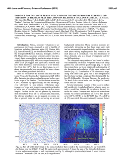
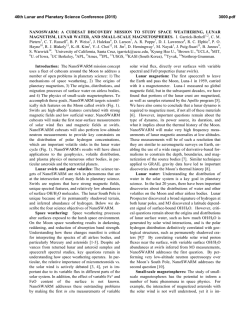
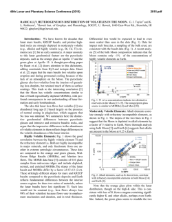
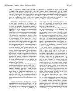
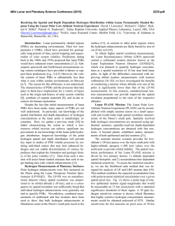
46th Lunar and Planetary Science Conference (2015)
1567.pdf
A NEW LOOK AT LUNAR REGOLITH PARTICLES WITH LIGHT, SCANNING ELECTRON AND XRAY ULTRA MICROSCOPY Carol Kiely*, (cak4@lehigh,edu), Gary Greenberg** ([email protected]) and
Christopher J. Kiely*, ([email protected]) *Department of Materials Science and Engineering, Lehigh University, 5
East Packer Avenue, Bethlehem, PA 18015, and **Institute for Astronomy, University of Hawaii.
Introduction: Forty five years of research has
provided a lot of information about the chemical and
physical properties of lunar soil [1]. Due to their size
(<400µm), the morphology of individual particles has
predominatly been studied using scanning electron
techniques. Two relatively new non-destructive
techniques were recently used to obtain images of
individual Apollo 11 lunar soil particles: namely deep
focus light microscopy and X-ray ultra microscopy [2].
The former yielded a collection of high resolution
micrographs in which the entire particle was in focus
and in color. The latter enabled the internal structure of
the whole particle be viewed without having to cut
slices or section it. Also, for the first time a light,
secondary electron, and x-ray micrographs of an
individual lunar agglutinate particle in the same
orientation were published. This provided a unique
insight into the morphology of the particle: the optical
image provided information on color and texture; the
scanning electron micrograph highlighted the surface
structure, and the high resolution x-ray image revealed
the internal structure showing the extent of the porosity
in the glassy matrix of the particle.
These three imaging techniques have now been
applied to particles isolated from a number of soils
collected from several different Apollo landing sites.
Results and Discussion: Glassy spherules have been
found in nearly all lunar soils. The most well known
and extensively analysed are the orange and black
spherules in the famous orange soil discovered on the
rim of Shorty Crater, and the green glass spherules that
were found in the clod-like boulders collected from the
flank of Mount Hadley Delta. Both of these are
believed to have been ejected from fire fountain
volcanoes that erupted 3.8 and 3.3 billion years ago
respectively. There are, however, plenty of other
spherules that are thought to be of impact origin.
The scanning electron and complementary x-ray
micrographs of three lunar spherules isolated from two
Apollo 15 soils (15101 and 15301) are shown in Figure
1. The first spherule (Fgiure 1a) is relatively smooth
with some mineral fragments attached to its surface. It
contains a large number of internal pores, the largest
having a diameter of ~45µm. It also contains an
inclusion of a more dense mineral. An x-ray rotational
movie of this particle revealed that these pores were
spherical and that the inclusion had a platelet
morphology. The second spherule (Figure 1b) contains
a myriad of tiny particles wrapped in impact glass. It
also contains a large number of internal pores which
are no longer spherical. Internal pore diameters range
from ~180µm to a few microns. The pores in both of
these spherules have almost certainly formed by the
nucleation of gas bubbles in the glass as the
temperature increased during micrometeorite impact.
The identity of the gas responsible for forming the
bubbles has not yet been determined although in the
case of impact spherules it is thought to be implanted
solar wind gases.
Nucleation and growth of gas bubbles in molten
glass is also the driving force behind fire fountain
volcanoes, and although pores have been found in
larger green glass volcanic spherules by other research
groups [3] the gas responsible was not identified. We
did not see any pores in the x-ray ultra micrographs of
smaller orange or green volcanic spherules isolated
from 74220 and 15427 soils.
The third spherule (Figure 1c) is the only one of its
type found so far in this study. It contains a large
number of tiny fragments that appear to be welded
Figure 1: Scanning electron and complementary x-ray
micrographs of three lunar spherules isolated from
Apollo 15 soils: 15101 (a) and (b), 15301 (c).
46th Lunar and Planetary Science Conference (2015)
together and wrapped in a skin impact melt glass.
We have analysed several other non-spherical
particles that have been formed by molten glass
splashing onto and subsequently encasing agglomerates
of smaller particles. In every case, the glass is highly
porous. A light, scanning electron and x-ray
micrograph of one of these particles is shown in Figure
2a. The light micrograph clearly shows that a splash of
green glass has partially encased an agglomerate of
much smaller beige colored fragments; the x-ray image
reveals the presence of a large number of internal
pores, and the SE micrograph shows that some of these
pores have broken through the surface of the glass. One
possible explanation for this morphology is that
implanted solar wind gases were released from the
underlying particles when they were coated in hot
molten glass.
The second particle, Figure 2b, is of a breccialike particle isolated from 74220 soil. The light
micrograph reveals the variation in color of the large
and small fragments; variations in structure of the
fracture surfaces of the individual fragments can be
seen in the SE micrographs, and the more absorbing
1567.pdf
fragments are highlighted in the X-ray image. Notice
also that there are no pores present in this particle.
Summary: The ability to collect complementary light,
SE and x-ray micrographs of the same particle in the
same orientation is valuable asset in the study of lunar
particles. Combining this information with stereo
optical and scanning electron anaglyphs, together with
rotational SE and X-ray movies leads to a much fuller
understanding of their morphology and how they were
formed. These are just a few examples of the many
high resolution micrographs taken during this study.
Acknowledgements: The lunar regolith samples were
kindly loaned to us through the NASA CAPTEM
program run by the Johnson Space Center.
References: [1] McKay D.S. et al. (1991). Chapter 7,
The Lunar Regolith, Lunar Sourcebook: A User’s
Guide to the Moon, 285-356, Cambridge University
Press. [2] Kiely C. et al, (2011) Microscopy and
Microanalysis, 17, 34-48. [3] Thomas-Keprta K.L. et
al. (2014) Proceedings of the 45th LPSC, 2507.
(a)
(b)
Figure 2. Light, scanning electron and X-ray micrographs of two individual lunar particles isolated from (a)15101
and (b)74220 soil.

© Copyright 2026