
Neural correlates of self-reflection
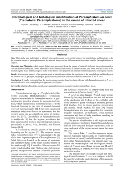
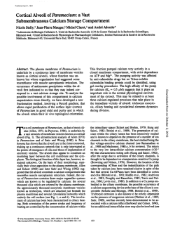
Brain (2002), 125, 1808±1814
Neural correlates of self-re¯ection
Sterling C. Johnson,1,2 Leslie C. Baxter,1 Lana S. Wilder,1 James G. Pipe,2 Joseph E. Heiserman2 and
George P. Prigatano1
Departments of 1Clinical Neuropsychology and 2Magnetic
Resonance Imaging, Barrow Neurological Institute, St
Joseph's Hospital and Medical Center, Phoenix, AZ, USA
Summary
The capacity to re¯ect on one's sense of self is an
important component of self-awareness. In this paper,
we investigate some of the neurocognitive processes
underlying re¯ection on the self using functional MRI.
Eleven healthy volunteers were scanned with echoplanar imaging using the blood oxygen level-dependent
contrast method. The task consisted of aurally delivered
statements requiring a yes±no decision. In the experimental condition, participants responded to a variety of
statements requiring knowledge of and re¯ection on
their own abilities, traits and attitudes (e.g. `I forget
important things', `I'm a good friend', `I have a quick
temper'). In the control condition, participants
responded to statements requiring a basic level of
semantic knowledge (e.g. `Ten seconds is more than a
Correspondence to: Sterling C. Johnson, PhD, Clinical
Neuropsychology, Barrow Neurological Institute, St
Joseph's Hospital and Medical Center, 222 W. Thomas
Road 315, Phoenix, AZ 85013, USA
E-mail: [email protected]
minute', `You need water to live'). The latter condition
was intended to control for auditory comprehension,
attentional demands, decision-making, the motoric
response, and any common retrieval processes.
Individual analyses revealed consistent anterior medial
prefrontal and posterior cingulate activation for all participants. The overall activity for the group, using a
random-effects model, occurred in anterior medial prefrontal cortex (t = 13.0, corrected P = 0.05; x, y, z, 0,
54, 8, respectively) and the posterior cingulate (t = 14.7,
P = 0.02; x, y, z, ±2, ±62, 32, respectively; 967 voxel
extent). These data are consistent with lesion studies of
impaired awareness, and suggest that the medial prefrontal and posterior cingulate cortex are part of a
neural system subserving self-re¯ective thought.
Keywords: fMRI; medial prefrontal cortex; posterior cingulate; self-awareness; self-re¯ection
Abbreviation: AMPFC = anterior medial prefrontal cortex
Introduction
The capacity to consciously re¯ect on one's sense of self is an
important aspect of self-awareness. A sense of self is a
collection of schemata regarding one's abilities, traits and
attitudes that guides our behaviours, choices and social
interactions. The accuracy of one's sense of self will impact
ability to function effectively in the world. A patient for
whom self-awareness is compromised may have a sense of
self regarding abilities and traits that is not congruent with
what others observe (Stuss, 1991; Prigatano, 1999). For
example, a brain-injured patient may feel he/she can competently return to the same level of employment when
observations by others indicate otherwise. When asked,
brain-injured patients often underestimate their own emotional dyscontrol, cognitive dif®culties and interpersonal
de®cits relative to a family member's rating of their abilities
(Prigatano, 1996). Inaccurate self-knowledge can signi®cantly impede efforts to rehabilitate brain-injured patients,
ã Guarantors of Brain 2002
since they may not appreciate the need for such treatment
(Sherer et al., 1998a, b).
Hughlings Jackson postulated that a sense of self is
dependent on the evolutionary development of the prefrontal
cortex (Meares, 1999). Lesion studies have generally supported this hypothesis. Damage to the anterior prefrontal
regions has been associated with impaired self-awareness for
the appropriateness of social interactions, judgement and
planning dif®culties (Prigatano and Schacter, 1991; Stuss,
1991), as well as impaired awareness of the mental states of
others (`theory of mind'; Stone et al., 1998; Stuss et al.,
2001). Impaired self-awareness appears to occur more
frequently following medial prefrontal damage (Damasio
et al., 1990), but may not be limited to this region.
Although much has been learned from lesion studies, to
date there is little functional imaging data on this topic. A
recent study found that patients with frontal dementia and a
fMRI of self re¯ection
change in personality functioning also exhibited greater right
prefrontal hypoperfusion (Miller et al., 2001) using SPECT
(single-photon emission computed tomography) scanning.
Studies using cognitive activation paradigms report activations to self-monitoring of current emotional or somatic states
(McGuire et al., 1996; Lane et al., 1997; Blakemore et al.,
2000; Gusnard et al., 2001). Activations in these studies were
within the anterior cingulate and paracingulate region,
Brodmann area (BA) 32 (Frith and Frith, 1999). To date, no
studies have addressed the process of conscious re¯ection on
one's own traits, abilities and attitudes that comprise a sense
of self.
For the paradigm reported in this paper, we used stimuli
that were similar to what one might ®nd on a self-report
personality or mood survey. We asked participants to answer
questions regarding stable traits, attitudes and abilities. The
control condition involved retrieval of general factual knowledge (semantic knowledge retrieval). In light of previous
lesion and imaging studies, we hypothesized that the medial
prefrontal cortex would be involved in this task.
Methods
Subjects
Eleven right-handed, healthy volunteer participants (four
females, seven males; mean age 35 years, SD = 12; mean
education 17 years, SD = 2.3) were recruited from employees
within the medical centre. The volunteers provided written
informed consent to participate in this institutional review
board-approved study.
Paradigm
In the functional MRI (fMRI) paradigm, the participants were
asked to make decisions about themselves on speci®c
statements requiring self-evaluation in the domains of
mood, social interactions, cognitive and physical abilities.
A standard set of statements was administered via headphones to each participant during scanning. The set included
items such as `I get angry easily', `I often forget things', `My
future is bright', `I'd rather be alone', `I catch on quickly', `I
can be trusted' and `I'm good at my job'. In the control
condition (used to control for auditory processing, attention,
language comprehension, decision making, the motor response and retrieval), participants made decisions about
statements of factual knowledge. Statements included items
such as `Ten seconds is more than a minute' and `You need
water to live'. Participants responded to each statement with a
`yes' (right hand) or `no' (left hand) button press. A constant
visual reminder of which button to press for each response
was displayed through the goggle projection system throughout the entire scan.
Statements were digitized and delivered aurally at 44.1
kHz. They were presented every 4 s in blocks of six for each
condition. The statements were, on average, 2 s in duration,
1809
leaving 2 s to respond. The experimental and control
conditions alternated over ®ve cycles. The task was presented
twice using equivalent forms (order of form administration
was counterbalanced across subjects).
Scanning technique
Participants were positioned in a GE 1.5 Tesla NVi scanner
with foam padding around the head to minimize participant
motion. Stimuli were delivered into the scanner via a
laptop computer connected to an MR-compatible goggle
and headphone system from Resonance Technology
(Northridge, CA, USA). The software Presentation (http://
www.neurobehaviouralsystems.com) was used for delivering
the auditory stimuli and recording responses. Input from the
scanner via a low amplitude electrical pulse allowed the
stimulus delivery software to monitor the scanner at every
slice acquisition and deliver the auditory stimuli with
accurate timing. A single-button MRI-compatible response
device was placed in each hand for making yes±no responses.
Imaging parameters
T2* weighted images were acquired with a gradient echo,
echo-planar pulse sequence to elicit blood oxygen leveldependent (BOLD) contrast. The scanning parameters were
as follows: TE (echo time) = 40 ms; TR (repetition time) =
3000 ms; ¯ip angle = 90°; acquisition matrix = 64 3 64
voxels; ®eld of view (FOV) = 240 mm. Thirty-two slices of
the brain were acquired axially within the TR at each time
point, with near isotropic voxel resolution of 3.75 3 3.75 3
4.2 mm. Eighty-two time points were collected over a 4 min
scanning run (images from the ®rst 6 s were discarded).
High-resolution structural images were acquired for overlay of the statistical results. The images were collected using
a SPGR (spoiled gradient) T1-weighted, 3D acquisition with
the following parameters: TR = 24 ms, TE = 6 ms, ¯ip angle =
40°, NEX = 1, slice thickness = 1.9 mm, 0 skip between
slices, FOV = 24 cm, in-plane resolution = 0.9375 mm2
voxels. The T1 and T2* weighted images were co-registered
using a least squares minimization routine.
Analysis
Images were analysed using Statistical Parametric Mapping
software (SPM99, University College London, UK; http://
www.®l.ion.ucl.ac.uk). Prior to statistical analysis, the timeseries of images were corrected for motion, normalized into a
standard atlas space (using the International Consortium for
Brain Mapping template as implemented in SPM99), and then
spatially smoothed using an 8-mm full-width at halfmaximum Gaussian kernel.
Individual time-series analysis was performed on each
participant. The boxcar model included bandpass ®ltering to
remove high and low frequency signal, and convolution with
a haemodynamic response function on a voxel by voxel basis
1810
S. C. Johnson et al.
Table 1 Coordinates (MNI space) and peak activation statistics for prefrontal and cingulate
cortex for each participant
Talairach coordinates
Participant
Region
x
y
z
t-value
P-value (corrected)
S1
AMPFC
PC
AMPFC
PC
AMPFC
PC
AMPFC
PC
AMPFC
PC
AMPFC
PC
AMPFC
PC
AMPFC
PC
AMPFC
PC
AMPFC
PC
AMPFC
PC
0
±4
4
±4
0
±4
±10
0
2
±4
6
±8
±6
±4
2
10
0
±4
8
14
±8
±4
66
±48
52
±74
54
±46
64
±58
60
±54
56
±55
60
±52
62
±58
76
±44
64
±54
60
±56
10
36
±4
38
18
±6
26
36
18
32
38
24
32
30
20
32
8
24
34
20
30
10
5.72
9.90
5.77
5.49
8.68
5.38
7.61
8.04
7.62
8.40
7.94
5.45
7.72
5.55
9.76
7.97
13.63
12.65
8.98
5.98
6.24
8.31
0.001
0.000
0.012
0.032
0.000
0.005
0.000
0.000
0.000
0.000
0.000
0.004
0.000
0.002
0.000
0.000
0.000
0.000
0.000
0.006
0.000
0.000
S2
S3
S4
S5
S6
S7
S8
S9
S10
S11
AMPFC = anterior medial prefrontal cortex; PC = posterior cingulate cortex; MNI = Montreal
Neurological Institute.
using the general linear model (Friston et al., 1995). The
individual results were of most interest to us. However, we
also characterized the average group response using a
random-effects approach (Holmes and Friston, 1998).
Results
Inspection of performance data indicated an equivalent
proportion of `yes' (right button) responses between conditions across participants (mean of 50% `yes' responses in the
self-re¯ection condition, mean of 51% `yes' responses in the
semantic condition). These proportions did not differ signi®cantly. The reaction time for the `self' condition was 659 ms,
and for the semantic condition it was 602 ms. The difference
between the two conditions was not signi®cant (P = 0.61).
All 11 participants individually activated the anterior
medial prefrontal cortex and posterior cingulate above a Pvalue threshold (corrected for multiple comparisons) of 0.05,
as shown in Table 1. Figure 1 depicts the individual
activations for the 11 subjects prior to any spatial standardization. Activation maps are overlayed on their own coregistered T1-weighted MRI scans. Peak activations in the
anterior medial prefrontal cortex (AMPFC) occurred just to
the right of midline in ®ve of the participants, three were just
left of midline, and the remaining three were at midline (see
Table 1 for spatially normalized activation coordinates).
A summary of the group activity is shown in Fig. 2. The
main effects of activation in the group analysis were highly
consistent with the individual analyses, as expected.
Activations included the AMPFC (t = 13.0, corrected P =
0.05; x, y, z, 0, 54, 8) and posterior cingulate (t = 14.7,
corrected P = 0.02; x, y, z, ±2, ±62, 32). No other regions
survived the correction for multiple comparisons.
Discussion
Consistent and robust anterior medial prefrontal and posterior
cingulate activation during self-re¯ection was observed in all
11 participants. While the peak of the activation varied
somewhat between individuals, the preponderance of activity
was always within AMPFC, BA 9 and 10, and posterior
cingulate, in the area of BA 23, 30 and 31. Activation of the
anterior medial prefrontal region was consistent with our
hypothesis and with lesion studies of patients with impaired
self-awareness (Stuss, 1991). The consistency and magnitude
of the activation was, however, somewhat greater than
expected.
Previous functional imaging studies involving self-evaluation have focused on appraising current internal states rather
than more stable traits (Frith and Frith, 1999). These prior
studies have collectively demonstrated anterior medial
prefrontal activation. Together, the current results and
previous studies suggest that mentalizing about the self,
fMRI of self re¯ection
Fig. 1 Midsagittal view of each participant showing prefrontal
activation during self-re¯ective thought. The activations are
superimposed on top of each participant's anatomical scan. Note
the consistent activation of the prefrontal cortex across subjects.
For the purposes of this display, the statistical threshold was set to
an uncorrected P-value of 0.0005. These results are prior to any
spatial standardization. The coordinates for the spatially
standardized results for each individual are shown in Table 1.
whether it be traits or current states, may activate the same or
similar medial frontal network.
Developmental theorists have written that a sense of self
begins in early childhood as a multidimensional set of
schemata, is de®ned further through accommodation of
experiences and perceptions in childhood and adolescence,
and gains permanence in early adulthood (Rychlak, 1981;
Kagan, 1982; Miller et al., 2001; Zeman, 2001). Piaget
de®ned schema as the potential to act or to be a certain way
(Rychlak, 1981). Over time, schemata may become unlinked
1811
to speci®c autobiographical events, and become part of a nonepisodic knowledge base, analogous to semantic knowledge.
Functional imaging studies of episodic autobiographical
memory retrieval have consistently reported posterior
cingulate activation (Maddock, 1999; Maddock et al., 2001;
Maguire et al., 2001). The posterior cingulate has reciprocal
connections with other memory areas, including the dorsolateral prefrontal cortex, the posterior parahippocampal
cortex, presubiculum and the entorhinal cortex, and with
several nuclei of the thalamus (Duvernoy, 1998; Morris et al.,
1999; Mesulam, 2000). The posterior cingulate has also been
shown to respond to a familiar face or voice (Shah et al.,
2001), retrieval of episodic information (Andreasen et al.,
1995; Wiggs et al., 1999) as well as retrieval of semantic
information (Pihlajamaki et al., 2000). Further, resting
hypometabolism in this region has been observed consistently
in very early-stage Alzheimer's disease, when dif®culty with
memory is the most prominent symptom (Minoshima et al.,
1997). Persons who are at genetic risk for Alzheimer's
disease also exhibit posterior cingulate hypometabolism
(Reiman et al., 1996).
Although this study reports activations in the posterior
cingulate, the paradigm differs from tasks of autobiographical
episodic memory in many respects. Autobiographical memory paradigms used in functional imaging experiments
generally entail recollection of vivid, personally relevant
episodes from the past. These experiments generally allow for
a relatively longer period of time, 4±20 s, for the participant
to retrieve the event as well as its context (Maguire et al.,
2001; Ryan et al., 2001) In the current study, participants
were allowed only 2 s to re¯ect and decide. There was little
time for participants to retrieve prior episodes and contexts on
which to base their subjective decisions. Furthermore, they
were speci®cally instructed to use their ®rst impressions
rather than ruminating on the question or justifying their
responses with facts.
The posterior cingulate appears not only to be important for
memory, but also for the perception and evaluation of
emotional stimuli. A recent review paper (Maddock, 1999)
observed that the posterior cingulate is the most frequently
activated region during evaluation of emotional salience of a
stimulus. Since this region is also activated in autobiographical retrieval, Maddock (1999) further argued that the
posterior cingulate may mediate an `interaction' between
memory retrieval and emotion. This notion is relevant to our
task, during which participants were required to retrieve and
re¯ect on self schemata that may have had an emotional
component. Measuring the emotional salience was beyond
the scope of this study, but would have been helpful in
interpreting the activations observed.
We note a consistent observation in research on emotion
and memory, that stimuli with greater emotional salience tend
to be better recalled (Cahill, 1997, 2000; Maddock and
Buonocore, 1997; Maddock et al., 2001). Lasting personal
memories typically have a salient affective tone, and it may
be dif®cult to separate the retrieval of content from the
1812
S. C. Johnson et al.
Fig. 2 Group activations for the self-re¯ection versus general knowledge conditions. This maximum intensity projection represents a
random-effects group analysis. Lower right: a midsagittal anatomical view is shown with the group activation superimposed. The results
are thresholded at an uncorrected P-value of 0.001, corresponding to a t-statistic of 4.14. The main effect of activation occurred in anterior
medial prefrontal cortex (maxima t = 13.0, corrected P = 0.05; x, y, z = 0, 54, 8, respectively; cluster size 2326 voxels) and the posterior
cingulate (maxima t = 14.7, corrected P = 0.02; x, y, z = ±2, ±62, 32; cluster size 976 voxels). Other activated regions at this threshold
included the thalamus (t = 7.53; x, y, z = 4, ±4, 12), bilateral posterolateral orbital cortex (right orbital: t = 7.67; x, y, z = 28, 18, ±18; left
orbital: t = 7.51; x, y, z = ±10, 6, 14), bilateral inferior temporal gyrus (right: t = 10.38; x, y, z = 52, ±6, ±24; left: t = 8.46; x, y, z = ±62,
±14, ±16) and bilateral cerebellum (right: t = 6.13; x, y, z = 24, ±74, ±38; left: t = 6.12; x, y, z = ±32, ±78, ±38).
accompanying emotional tone. Nevertheless, future studies
should attempt to control for the affective processes during
autobiographical memory retrieval in order to de®ne better
the role of the posterior cingulate.
There are other limitations to these data. Although we
instructed participants to respond with their ®rst impressions,
and not to rely on speci®c instances to answer each question,
we are unable to rule out episodic autobiographical retrieval
as a possible response strategy and source of activity in the
posterior cingulate. Also, although the reaction times for the
experimental and control conditions were equivalent, the self
condition may have been more psychologically uncomfortable and revealing than the control task, and therefore more
dif®cult.
Self-re¯ection, as we have described it here, can be
considered a metacognitive function (Stuss et al., 2001),
and our results may not be speci®c to self-re¯ection per se.
Two previous PET studies examining `theory of mind' tasks
reported anterior medial prefrontal and posterior cingulate
activity (Fletcher et al., 1995; Goel et al., 1995). The tasks
used in these studies required the participant to `mentalize'
about the beliefs and desires of others so as to predict their
behaviour. Fletcher et al. and Goel et al. found maximal
medial prefrontal activity at Talairach (x, y, z) locations ±12,
42, 40 and±6, 46, 28, respectively, slightly posterior and
superior to the activation reported in the present study (2, 54,
8). To address the issue of whether the activations seen here
represent generically metacognitive functions, or are in fact
speci®c to the self-re¯ective thought process, future research
should directly compare metacognitive tasks requiring
mentalizing about the self with tasks requiring mentalizing
about others.
We assumed that the healthy individuals in our study would
rate themselves accurately. While this assumption may be
reasonable for healthy, cognitively normal volunteers, it would
probably not hold for patients with altered self-awareness. In
fMRI of self re¯ection
such cases, other methods may be helpful to differentiate
between self-re¯ection and self-awareness, such as incorporating patient and caregiver ratings of the patient's abilities into
the statistical analysis. This work is underway in our laboratory.
Conclusion
The medial prefrontal cortex and posterior cingulate are
important brain regions for accessing a sense of self. The
frontal activation results are consistent with lesion studies in
patients with impaired self-awareness, as well as other
functional imaging tasks involving mentalizing about the
self or others. The current results suggest that studying
aspects of self-awareness is quite feasible, with functional
imaging using this type of paradigm.
Acknowledgements
We wish to thank Sarah Hahn, PhD, and Patty Puppe, RT, for
their efforts on this project. We also wish to thank Richard
Lane, MD, PhD, for review and critical discussion of an
earlier version of the manuscript. This study was supported in
part by the National Institute on Aging (AG18540), the
National Institute of Mental Health (MH65723), the Arizona
Alzheimer's Research Center, and the Barrow Neurological
Foundation.
References
Andreasen NC, O'Leary DS, Cizadlo T, Arndt S, Rezai K, Watkins
GL, et al. Remembering the past: two facets of episodic memory
explored with positron emission tomography. Am J Psychiatry
1995; 152: 1576±85.
Blakemore SJ, Wolpert D, Frith C. Why can't you tickle yourself?
[Review]. Neuroreport 2000; 11: R11±6.
Cahill L. The neurobiology of emotionally in¯uenced memory.
Implications for understanding traumatic memory. [Review]. Ann
NY Acad Sci 1997; 821: 238±46.
1813
Goel V, Grafman J, Sadato N, Hallett M. Modeling other minds.
Neuroreport 1995; 6: 1741±6.
Gusnard DA, Akbudak E, Shulman GL, Raichle ME. Medial
prefrontal cortex and self-referential mental activity: relation to a
default mode of brain function. Proc Natl Acad Sci USA 2001; 98:
4259±64.
Holmes AP, Friston KJ. Generalisability, random effects and
population inference. Neuroimage 1998; 7: S754.
Kagan J. The emergence of self. J Child Psychol Psychiatry 1982;
23: 363±81.
Lane RD, Fink GR, Chau PM, Dolan RJ. Neural activation during
selective attention to subjective emotional responses. Neuroreport
1997; 8: 3969±72.
Maddock RJ. The retrosplenial cortex and emotion: new insights
from functional neuroimaging of the human brain. [Review]. Trends
Neurosci 1999; 22: 310±6.
Maddock RJ, Buonocore MH. Activation of left posterior cingulate
gyrus by the auditory presentation of threat-related words: an fMRI
study. Psychiatry Res 1997; 75: 1±14.
Maddock RJ, Garrett AS, Buonocore MH. Remembering familiar
people: the posterior cingulate cortex and autobiographical memory
retrieval. Neuroscience 2001; 104: 667±76.
Maguire EA, Vargha-Khadem F, Mishkin M. The effects of
bilateral hippocampal damage on fMRI regional activations and
interactions during memory retrieval. Brain 2001; 124: 1156±70.
McGuire PK, Paulesu E, Frackowiak RS, Frith CD. Brain activity
during stimulus independent thought. Neuroreport 1996; 7:
2095±9.
Meares R. The contribution of Hughlings Jackson to an
understanding of dissociation. Am J Psychiatry 1999; 156: 1850±5.
Mesulam M-M. Behavioral neuroanatomy: large scale networks,
association cortex, frontal syndromes, the limbic system, and
hemispheric specializations. In: Mesulam MM, editor. Principles of
Behavioral and Cognitive Neurology. Oxford: Oxford University
Press; 2000. p. 1±173.
Cahill L. Neurobiological mechanisms of emotionally in¯uenced,
long-term memory. [Review]. Prog Brain Res 2000; 126: 29±37.
Miller BL, Seeley WW, Mychack P, Rosen HJ, Mena I, Boone K.
Neuroanatomy of the self: evidence from patients with
frontotemporal dementia. Neurology 2001; 57: 817±21.
Damasio AR, Tranel D, Damasio H. Individuals with sociopathic
behavior caused by frontal damage fail to respond autonomically to
social stimuli. Behav Brain Res 1990; 41: 81±94.
Minoshima S, Giordani B, Berent S, Frey KA, Foster NL, Kuhl DE.
Metabolic reduction in the posterior cingulate cortex in very early
Alzheimer's disease. Ann Neurol 1997; 42: 85±94.
Duvernoy HM. The Human Hippocampus. 2nd edn. Berlin:
Springer-Verlag; 1998.
Morris R, Petrides M, Pandya DN. Architecture and connections of
retrosplenial area 30 in the rhesus monkey (Macaca mulatta). Eur J
Neurosci 1999; 11: 2506±18.
Fletcher PC, Happe F, Frith U, Baker SC, Dolan RJ, Frackowiak RS,
et al. Other minds in the brain: a functional imaging study of `theory
of mind' in story comprehension. Cognition 1995; 57: 109±28.
Friston KJ, Holmes AP, Worsley KJ, Poline JP, Frith CD,
Frackowiak RSJ. Statistical parametric maps in functional
imaging: a general linear approach. Hum Brain Mapp 1995; 2:
189±210.
Frith CD, Frith U. Interacting mindsÐa biological basis. [Review].
Science 1999; 286: 1692±5.
Pihlajamaki M, Tanila H, Hanninen T, Kononen M, Laakso M,
Partanen K, et al. Verbal ¯uency activates the left medial temporal
lobe: a functional magnetic resonance imaging study. Ann Neurol
2000; 47: 470±6.
Prigatano GP. Behavioral limitations TBI patients tend to
underestimate: a replication and extension to patients with
lateralized dysfunction. Clin Neuropsychol 1996; 10: 191±201.
Prigatano GP. Disorders of self-awareness after brain injury. In:
1814
S. C. Johnson et al.
Prigatano GP. Principles of Neuropsychological Rehabilitation.
New York: Oxford University Press, 1999; p. 265±93.
Impaired awareness and employment outcome after traumatic brain
injury. J Head Trauma Rehabil 1998a; 13: 52±61.
Prigatano GP, Schacter DL, editors. Awareness of De®cit After
Brain Injury: Clinical and Theoretical Issues. New York: Oxford
University Press; 1991.
Sherer M, Boake C, Levin E, Silver BV, Ringholz G, High WM Jr.
Characteristics of impaired awareness after traumatic brain injury.
J Int Neuropsychol Soc 1998b; 4: 380±7.
Reiman EM, Caselli RJ, Yun LS, Chen K, Bandy D, Minoshima S,
et al. Preclinical evidence of Alzheimer's disease in persons
homozygous for the epsilon 4 allele for apolipoprotein E. New Engl
J Med 1996; 334: 752±8.
Stone VE, Baron-Cohen S, Knight RT. Frontal lobe contributions to
theory of mind. J Cogn Neurosci 1998; 10: 640±56.
Ryan L, Nadel L, Keil K, Putnam K, Schnyer D, Trouard T, et al.
Hippocampal complex and retrieval of recent and very remote
autobiographical memories: evidence from functional magnetic
resonance imaging in neurologically intact people. Hippocampus
2001; 11: 707±14.
Stuss DT. Disturbance of self-awareness after frontal system
damage. In: Prigatano GP, Schacter DL, editors. Awareness of
De®cit After Brain Injury: Clinical and Theoretical Issues. New
York: Oxford University Press; 1991. p. 63±83.
Stuss DT, Gallup GG Jr, Alexander MP. The frontal lobes are
necessary for `theory of mind'. Brain 2001; 124: 279±86.
Rychlak J. Two kinds of constructive theories: Jean Piaget and
George Kelly. In: Rychlak J, editor. Personality and psychotherapy.
Boston: Houghton Mif¯in; 1981; p. 664±753.
Wiggs CL, Weisberg J, Martin A. Neural correlates of semantic
and episodic memory retrieval. Neuropsychologia 1999; 37:
103±18.
Shah NJ, Marshall JC, Za®ris O, Schwab A, Zilles K, Markowitsch
HJ, et al. The neural correlates of person familiarity: a functional
magnetic resonance imaging study with clinical implications. Brain
2001; 124: 804±15.
Zeman A. Consciousness. [Review]. Brain 2001; 124: 1263±89.
Sherer M, Bergloff P, Levin E, High WM Jr, Oden KE, Nick TG.
Received May 14, 2001. Revised January 21, 2002.
Accepted February 28, 2002
© Copyright 2026