
Cytokine Activation During Attacks of the
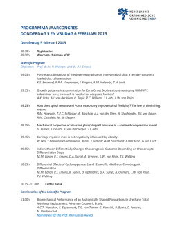
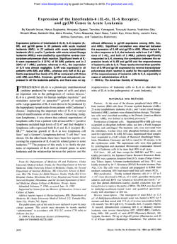
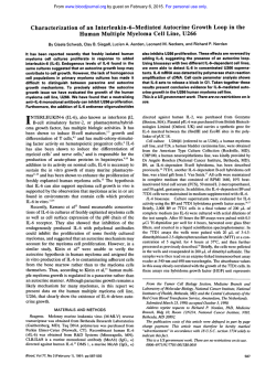
From www.bloodjournal.org by guest on February 6, 2015. For personal use only.
Cytokine Activation During Attacks of the Hyperimmunoglobulinemia D and
Periodic Fever Syndrome
BY Joost P.H. Drenth, Marcel van Deuren, Johanna van der Ven-Jongekrijg, Casper G. Schalkwijk,
and Jos W.M. van der Meer
The hyperimmunoglobulinemia D and periodic fever (hyperIgD) syndrome is typified by recurrent febrile attacks with
abdominal distress, joint involvement(arthralgias/arthritis),
headache, skin lesions, and anelevated serum IgDlevel
(>l00 U/mL). This familial disorder has been diagnosed in
59 patients, mainly from Europe. The pathogenesis of this
febrile disorder is unknown, but attacks are joined by an
acute-phase response. Because this response is considered
t o be mediated by
cytokines, we measured the acute-phase
proteins C-reactive protein (CRP) and soluble t y p e 4 phospholipase A2 (PLA2)together with circulating concentrations
and ex vivo production of the proinflammatory cytokines
interleukin-la (IL-la), IL-Ip, IL-6, and tumor necrosis factor
a (TNFa) and the inhibitorycompounds IL-l receptor antagonist flL-Ira). IL-10. and the
soluble TNF receptors p55(sTNFr
p551 and p75 (sTNFr p751 in 22 patients with the hyper-lgD
syndrome during attacks and remission. Serum CRP and
PLA2 concentrations were elevated during attacks (mean,
213 mg/L and1,452 ng/mL, respectively) and decreased between attacks. Plasma concentrations of IL-la, IL-lp, or IL10 were not increased during attacks. TNFa concentrations
were slightly, but significantly, higher with attacks (104 v
117 pg/mL). Circulating IL-6 values increased with attacks
(19.7 v 147.9 pg/mL) andcorrelated with CRP and PLA2values during the febriieattacks. The values of theantiinflammatory compounds IL-Ira, sTNFr p55, and sTNFr p75 were
significantly higher with attacks than between attacks, and
there was a significant positive correlation between each.
The ex-vivo production of TNFa, IL-Ip, and IL-Ira was significantly higher with attacks, suggesting that the monocytes/macrophages were already primed in vivo t o produce
increased amounts of thesecytokines. These findings point
t o an activation of the cytokine
network, and thissuggests
that these inflammatory mediators may contribute to the
symptoms of the hyper-lgD syndrome.
0 1995 by The American Societyof Hematology.
T
from a disturbed cytokine synthesis which may be responsible for some ofits clinical manifestationsand could also
contribute to the polyclonal serum IgD elevation.’” Therefore, we studied the pattern of circulating and ex vivo production of inflammatorymediators
1L-la,IL-ID.
IL-6,
TNFa, C-reactive protein (CRP), soluble type-I1 phospholipase A? (PLA,), and inhibitory compoundssuch as IL-I
receptor antagonist ( L l r a ) , IL-10, and soluble TNF receptors p55 (sTNFr p55) and p75 (sTNFr p7.5).
HE HYPERIMMUNOGLOBULINEMIA D and periodic fever syndrome (hyper-IgD) is a rare entity characterized by periodic occurrence of febrile attacks.’ All patients have a clearly elevated (po1y)clonal serum IgD (> 100
U/mL). So far,wehavediagnosed
59 patients (31 male
and 28 female) from nine, mainly European, countries. The
episodes last 3 to 7 days, occur every 4 to 6weeks,start
early, and persist throughout life, and, between attacks, patients are nonsymptomatic.’ Symptoms are not triggered by
known infectiousagentsandincludeabdominaldistress
(vomiting, diarrhea, and pain), joint involvement(arthralgia/
arthritis), skin lesions, headache, and lymphadenopathy. 2 ~ 4
The main feature of the disease is the abrupt onset of fever
that parallels with an acute-phase reaction as evidenced by
leukocytosis, neutrophilia, and a raised erythrocyte sedimentation rate.
The pathogenesis of the hyper-IgD syndrome is unclear,
but, because fever and the acute-phase reaction are considered to bemediated by cytokines such as interleukin- 1p (ILlo), IL-6, and tumor necrosis factor a (TNFa), it can be
speculated that symptoms in the hyper-IgD syndrome result
From the Department of Medicine, Division of General lnternal
Medicine, University Hospital St Radboud, Nijmegen, The Netherlands.
Submitted October 20, 1994; accepted February 4, 1995.
J.P.H.D.isarecipient
of U DutchOrganization f o r Scient$c
Research fellowshipfor Clinical Investigators (KWO 900-716-065).
Address reprint requests to Professor
Jos W.M. Van der Meer,
MD, Department of Medicine, Division of General Internal Medicine,UniversityHospital
St Radboud,P.O. Box 9101, 6500 HB
Nijmegen, The Netherlands.
The publicarion costsof this article were defrayedin part by puge
chargepayment. This article must thereforebeherebymarked
“advertisement” in accordance with 18 U.S.C. section 1734 solely to
indicate this fact.
0 1995 by The American Society of Hematology.
0006-4971/95/8512-0034$3.00/0
3586
MATERIALS AND METHODS
Patients. Thestudygroupconsisted
of 22 Dutch patients, 14
male and 8 female patients, with the hyper-IgD syndrome. Themean
age at the time of the study was 28.3 years (SD ? 14.4 years). The
diagnosis was made following standard clinical criteria, and a recent
review with a pertinent description of the clinical and biologic features of thehyper-IgDsyndromehasbeen
published elsewhere.’
recurrent febrileattacks
Briefly, all patientsweresufferingfrom
accompanied by a combination of the following features: lymphadenopathy,splenomegaly,arthralgiadarthritis,abdominalpainhorniting/diarrhea, and skin lesions. The attacks are self-limiting and last
3 to 7 days. The main clinical features of these patients are listed
in Table 1.
Bloodsamples. Samplingofplasma was performedduringattacks as well as during remission, using special endotoxin-free tubes
NJ). The
(Vacutainer
Systems;
Becton
Dickinson,
Rutherford,
plasma was immediately processed to avoid ex vivo cytokine excre(238°C not caused
tion.’ We defined attacks if the patient was febrile
by infection) and showed signs of active disease with symptoms as
mentioned before. Remission was defined as the absence of symptomsforat least 1 month. No medication,includingantipyretics,
were allowed during the study period.All patients underwent physical examination, and results are listed in Table 1. Cytokine production was measured using a whole blood culture system as described
48 pL EDTA-KI
elsewhere.” Briefly, two 4-mL tubes containing
and 250 pL aprotinin ( 1 0,000 kallikrein-inactivating units per mL;
Bayer, Leverkusen, Germany) were drawn.
One tube was incubated immediately; the other tube was incubated
after addition of SO p L lipopolysaccharide (LPS; Escherichia coli
serotype OSS:BS, final concentration 10 pglmL;Sigma,StLouis.
Blood, Vol 85, No 12 (June 15), 1995:pp 3586-3593
From www.bloodjournal.org by guest on February 6, 2015. For personal use only.
CVTOKINES IN HYPER-IGD SYNDROME
3587
Table 1. Main Clinical Features of 22 Patients With the Hyper-lgD and Periodic Fever Syndrome
Patient No.
l
2
3
4
5
6
7
8
9
10
11
12
13
14
15
16
17
18
19
20
21
22
Age
Sex
F
M
M
M
F
M
M
M
F
F
M
F
F
M
M
M
M
M
M
M
F
F
(yr)
32
30
34
69
42
49
35
36
24
43
20
16
22
13
21
8
12
31
27
7
24
21
Lymphadenopathy
Splenomegaly
Abdominal Distress
Skin Lesions
Arthritis
+
+
-
+
+
+
+
+
+
+
+
+
+
+
+
+
+
-
+
+
+
+
+
+
+
+
+
+
+
+
+
+
+
+
+
+
+
-
-
+
+
-
+
+
+
+
+
+
-
+
+
-
+
-
-
+
-
-
-
+
+
+
+
-
-
-
+
+
+
+
+
+
+
+
-
-
-
+
-
+
+
+
+
+
+
+
+
+
+
+
+
+
+
+
+
+
+
t
+
-
+
+
-
Abbreviations: +, present: -, absent; M, male; F, female.
MO). After 24 hours of incubation at 37°C both tubes are centrifuged
at 2,250g for 10 minutes and, second, at 15,OOOg for 5 minutes to
obtain platelet-poor plasma. Aliquots were stored at -70°C until
assay. Cytokine production with LPS stimulation was measured and
compared with production without LPS stimulation. Control values
were obtained from 10 healthy volunteers (20 to 45 years of age)
and were 6,847 2 2,175 pg/mL (IL-lP); 3,344 2 969 pg/mL
(TNFa), and 9,827 2 2,597 pg/mL (IL-ha). TNFa was determined
by radioimmunoassay as described by Van der Meer et all0 (lower
detection level, 20 pg/mL; upper detection limit, 10,OOO pg/mL).
IL- la and IL-Ip were measured by radioimmunoassay (without
chloroform extraction) according to Lisi et a l l ’ (&la lower detection limit, 5 pg/mL; IL-lp lower detection limit, 40 pg/mL). IL-lra
was determined by radioimmunoassay according to Poutsiaka et allz
(lower detection limit, 60 pg/mL). sTNFr were measured by an
enzyme-linked immunosorbent assay (ELISA [detection level, 80
pg/mL for sTNFr p55 and 300 pg/mL for sTNFr ~751;a kind gift
of H. Gallati, Hoffmann-La Roche, Basel, Switzerland). This assay
measures both free and total receptor-bound concentrations. Because
sTNFr is not produced ex vivo, its LPS-stimulated production was
not assessed in this study. IL-6 was measured by an ELISA as
described earlier (detection level, 14 ~ g / m L ) . IL-10
’~
was measured
by sandwich ELISA with the 4G6 and 5D11 mouse antihuman IL10 monoclonal antibodies for capture and the 5A10 antihuman L - l 0
monoclonal antibodies for detection (kindly provided by Medgenix,
Amersfoort, The Netherlands). The detection limit was 11 pg/mL.I4
CRP measurements were performed with an immunoturbidimetric
assay using an automatic analyzer HITACHI 747 (Hitachi CO,
Tokyo, Japan). The measuring range was from 3 to 440 mg/L.
Secreted nonpancreatic type-I1 PLAl antigen concentrations in
plasma were determined with an ELBA modified from Smith et
aL5 Two different monoclonal antibodies against human PLAz (a
kind gift from Dr F.B. Taylor, Jr. Oklahoma Medical Research Foundation, Oklahoma City, OK) were used as coating and catching
antibodies. Because previous experiments showed a very good correlation between results from ELISA measurements and those obtained
with cultured medium from Hepatoma G2 cells stimulated with hu-
man IL-6, only the former method was used (Schalkwijk CG, unpublished observations). The lower limit of detection was 1 ng/mL. To
minimize analytical errors, all samples from the same patient were
analyzed in the same run in duplicate.
Statistical analysis. The paired nonparametric Wilcoxon signed
rank test was used for statistical comparison of values obtained
during active disease versus remissions. The unpaired nonparametric
Mann-Whitney U test was used where appropriate. In patients with
multiple samples taken during separate episodes of active disease,
the mean value was used. Probability ( P ) values were calculated on
the basis of two-tailed tests, A correlation coefficient was calculated
with the Pearson’s correlation test. A P value of less than .05 was
considered to be the lowest level of significancy. Data are given in
mean 2 standard deviation (SD).
RESULTS
Results of circulating cytokines and other inflammatory
mediators are given in Figs 1 and 2. The CRF’ concentrations,
a reflection of the acute-phase response, clearly modulated
with the attacks in the patients with the hyper-IgD syndrome.
The CRP concentrations were uniformly high during attacks
(213.3 -C 98.2 mg/L) but decreased to low values (22.6 ?
26.8 mg/L) in between attacks ( P < ,0001; see Fig 1A).
Concentrations of PLAz increased to amean of 1,452 ?
1,665 ng/mL during the febrile attacks, as compared with
values during nonsymptomatic periods ( 1 13 2 240 ng/mL;
P < .0001; see Fig 1B). One patient even had a 253-fold
elevation (maximum, 6,066 ng/mL) of PLA2 during attack
as compared with convalescence. The concentrations of
PLAz and CRF’ obtained during febrile attacks showed a
very good correlation during attacks ( r = 0.74; P < .OOOl).
We could not detect measurable quantities of circulating
IL-la between and during attacks in patients with the hyperIgD syndrome (Table 2). The mean plasma concentrations
of IL-lP during active disease did not differ from the values
From www.bloodjournal.org by guest on February 6, 2015. For personal use only.
3588
DRENTH ET AL
400
?
A
B
I
aoo-
:l 000
\
m
S
100-
:l00
4
Q
100-
.l0
0 -
1
no
.&h
obtainedduring remissions. The TNFa concentrationsincreased from 104 ? 20 pg/mL to 1 17 + 15 pg/mL, a small
but significant increase ( P = ,0075). Nevertheless, the TNFa
concentrations during attack remained within normal values
25 pg/mL). IL-6 was also signifiof our laboratory (106 I
cantly elevated during active disease (147.9? 258.3 pg/mL;
P < .0001; see Fig 2A). Three patients
hadvalues above
150 pg/mL (471, 720, and 1,074 pg/mL, respectively). Exclusion of data from these 3 patients did result in a lower
mean IL-6 plasma concentration during attacks (54.9 + 27.2
pg/mL). However, the difference betweenvaluesobtained
during attacks and between attacks remained significant despite exclusion ( P < .0001). During febrile episodes, there
was a moderate, albeit significant, correlation between IL-6
and the acute-phase proteins CRP ( r = 0.47; P = .014) and
PLA2 ( r = 0.45; P = ,018).
No correlation could be detected between IL-6 and either
IL-10 or TNFa.
IL-IO, measured in plasma of8patients,
remained unchanged regardless of the phase of the disease (Table 2).
The anti-inflammatory compounds IL-Ira, sTNFr p55,and
sTNFr p75 were significantly increased during attacks compared with remissions (Fig 2B through D). The mean values
of IL-lra were 996 ? 329 pg/mL during attacks, whereas
167 pg/mL during remission ( P < .0001;
they were 300
see Fig 2B). Themean sTNFr p55 and sTNFr p75
concentrations during active disease increased to 5,043 ? 1,703 pg/
mL and 7,103 ? 2,378 pg/mL, respectively (both P < .0001;
see Fig 2C and D). The sTNFr p7YsTNFr p55 ratio did not
change withattacks (data not shown). We could not establish
a correlation between either the individual concentrations of
IL-Ira, sTNFr p55, or sTNFr p75 and the temperature
at the
time of blood drawing or the frequency of the attacks (data
not shown). During attacks, there was a significant correlation between circulating plasmaconcentrations of IL-Ira and
sTNFr p55 ( r = 0.48; P = .0231), IL-Ira and sTNFr p75 (Y
+
Fig 1. Individual serum CRP
(A) and PLAz (B) for 22 hyper-lgD
patients during and in between
attacks. For (A), the vertical lines
indicate mean ( 0 )2 SD. The vertical lines in (B) indicate median
and 95% confidence intervals. The increase was statistically significant for
CRP and PIA2
( P c .0001).
= 0.67; P = .0006), and sTNFr p55 and sTNFr p75
(r =
0.58; P = ,0041); however, no correlation could be detected
between individual CRP levels and IL-Ira, sTNFr p55, or
sTNFr p75 levels. In 3 patients, the course of the circulating
IL-Ira, sTNFr p55, and sTNFr p75 concentrations during
the first 5 days after onset of the attack was followed as
shown in Fig 3. The course of values for the various cytokines is as follows. sTNFr p55 and sTNFr p75 only decrease
3 days after onset of the febrile attack. IL-lra reaches the
highest values on the second dayof the attack and normalizes
within 5 days. IL-6 was rapidly cleared from thecirculation,
similar to the acute-phase proteins CRP and PLA2.
Ex vivo cytokine production. Figure4,upperpanel,
shows thenonstimulated production of the various cytokines
tested. The unstimulated production of TNFa between attackswas significantly higher as compared withthat for
healthy controls (564 5 304 v 225 ? 11 pg/mL). Only the
values for IL-lra were significantlyhigher during attacks
than during remissions, 3,642 2 1708 pg/mL as compared
with 2,048 ? 2,398 pg/mL ( P = .015). The productionof
IL- la in whole blood in the presence or absence of LPS
was negligible and did not differbetween attacks and remission. The LPS-stimulated production of I L - l p and TNFa in
patients with the hyper-IgD was significantly higher during
attacks as compared with that in remissions and for healthy
controls (Fig 4, lower panel; P < .0001).During remissions,
both the unstimulated and LPS-stimulatedproduction
of
TNFa, but not of IL-Ip, was significantly higher compared
with that of controls ( P < .OS). Fourteen patients had TNFa
concentrations beyond the upper detection limit of
10,000
pg/mL during attacks, compared with only 6 patients during
remission. LPS-stimulated productionof IL-6 during attacks
was similar to that measured during nonsymptomatic periods. The LPS-stimulated production of IL-lra was also significantly higher during attacks than in between attacks and
than that in healthy volunteers ( P < .0001).
From www.bloodjournal.org by guest on February 6, 2015. For personal use only.
3589
CYTOKINES IN HYPER-IGD SYNDROME
- 1600
-1000
a
2
!!
.-
A
l
I
104
a
m
-600
.((.drn.#k
1 - 1
10000
16000
C
D
11.600
~000
-10000
10
Fig 2. Circulating 11-6 (A), ILI r a (B), sTNFrp55(C),andsTNFr
p75 (D) in 22 hyper-lgD patients.
The individual values
are
represented by blackdotsand
were
obtained during andbetween attacks. In (A), the vertical lines indicate median ( + I and 95% confidence intervals. For (B through
D), the verticallinesare
the
was
mean (01 ? SD. The increase
statistically
significant
for
all
four cytokines ( P c .00011.
-7600
16
p
e5
4000
two
I
-6000
a
2
10
a
!i
a
-2600
0
During attacks, there were modest correlations between
IL-lra and IL-l@production ( r = 0.42; P = .027) and ILIra and L - 6 production ( r = 0.3; P = .027). The production
of IL-1@ correlated wellwith that of L - 6 during febrile
episodes (I = 0.53; P = .0057).
DISCUSSION
We investigated the pattern of circulating cytokines and
the ex vivo production of cytokines in 22 patients with the
hyper-IgD syndrome. We found evidence for a vigorous
acute-phase response in the hyper-IgD syndrome, as reflected by the significant increase of CRP and PLAl concentrations that subsided during remission. The significant ele-
vation of the proinflammatory cytokines IL-6 and, albeit very
modest, TNFa together with the elevation of the natural
cytokine inhibitors IL-lra, sTNFr p55, and sTNFr p75 point
to activation of the cytokine network during the attacks of
the hyper-IgD syndrome. Although it is not clear to what
extent these cytokines contribute to the clinical features of
the hyper-IgD syndrome, many of its symptoms are compatible with the effects of proinflammatory cytokines. However,
it is unlikely that only one factor can cause the clinical
picture; instead, activation of the cytokine network and
involvement of various cytokines probably contribute to the
pathogenesis of the attacks.
The elevation of these inflammatory mediators occurs re-
From www.bloodjournal.org by guest on February 6, 2015. For personal use only.
3590
DRENTH ET AL
10 and TNFa may be thedelay between onset of attack and
sampling. Although we sampled all our patients on their first
day with fever, thismayalready
have been toolate. The
Circulating Concentration (pg/mL) 2 SD
slight elevation of TNFa during attacks perhapsreflects the
Cytokine
Between Attacks
Attack
descendingslope of an earlierpeak. The increased CRP
IL-Iff
15
<5
levels at time of drawing of blood indicate an already ongoIL-IP
83.1 f 20.3
80.4 2 21.6
ing acute-phase response thought to be the consequence of
IL-IO*
20.3 2 21.1
14.8 2 3.7
the action of cytokines, and these may have already disap* Tested in 8 patients.
peared from the circulation. This hypothesis is in line with
the finding of increased cytokine inhibitors such as IL- Ira,
sTNFr p55, andsTNFrp75,
of which it isknown.from
experimental endotoxemia, that they can still be detected in
gardless of the severity of attacks, as reflected by body temthe circulation hours after the disappearance of IL-Io and
perature and CRP concentrations the
or frequency of attacks.
TNFmZ5-”
The sameholds for PLA2, which peaks l8 to 24
The absenceof any elevationof IL- 10 concentration,
a potent
hourslaterthan TNFa after endotoxin infusion in human
inhibitorof synthesis of cytokines, argues against an imvolunteers.2x
portant roleforthiscytokine
in the pathogenesis of the
In our study, elevation of IL-lra, sTNFr p55, and sTNFr
hyper-IgD syndrome.I6Lastly, there isincreased exvivo
p75
was present at the first day of an attack, and appreciable
production of IL-lp, TNFa,and L - l r a during attacks comconcentrations were found on the second and third day of
pared with nonsymptomatic periods,as measured in a whole
the attack. Endogenously produced IL-Ira, a major natural
blood culture system.
inhibitor of IL-1, may ameliorate the severity of attacks in
The acute-phase response in humans is considered to be
mediated by theproinflammatory cytokines, and we detected the hyper-IgD syndrome.29-” Because we did not find clear
elevations of IL-1 concentrations, it is impossible to judge
a modest but significant elevation of IL-6 together with a
whether the amount of IL-lra is adequate to alleviate the
significant correlation between IL-6 and
the acute-phase proacute inflammation. Our finding of predominant concentrateins CRP and PLA2 in the blood during the febrile
attacks.
tions of IL-1 antagonist recalls the results obtained in sysIn view of these findings,it can be speculatedthatthe
temic JRA, another periodic febrile disorder. In JRA, inhibiincreased plasma IL-6 concentration results from overprotion of IL-l bioactivity by serum andurine during febrile
duction rather than underutilization, because renal function
attacks was found.?’ Also, for TNFa, we merely find inhibiwas normal in allpatients. In addition, the elevated IL-6
tion,
as judged by the elevated concentrations of soluble
values are associated with increased CRP and PLA2values.
receptors for TNF.’3 sTNFrs bind TNF and forma biologic,
Underutilization would probably have resulted in low coninactive complex, blunting the deleterious effectsof TNFa.”
centrations of these acute-phase proteins. IL-6, as the major
How do ourresults of circulating cytokines comparewith
mediator for the regulation of acute-phase protein synthesis
measurements
in another periodic fever syndrome such as
in humans, also influences the glycosylation of these proteins.7.~7,~a
In recent studies in the hyper-IgD syndrome, we
familial Mediterranean fever (FMF)? In FMF patients, TNF
and IL-I activity has been investigated.
have shown elevated a1-3 fucosylation of the acute-phase
In a study with Sephardic Jews with FMF there was no
protein a,-acid glycoprotein (AGP), and this finding is comevidence
of TNFa bioactivityin plasma obtained during
patible with a role of IL-6 during the recurrent febrile atattack and remi~sion.’~Nevertheless, the investigators sugtacks.” In addition, IL-6 is a stimulus for PLA2 synthesis in
lackof
gesteda roleforTNFa in FMFandimputethe
the liver, and thestrongly elevated (1 3-fold) PLA2 concentraelevated
circulating
plasma
TNFa
to
the
short
half
life (6
tions found during the febrile attacks,
together with the correminutes)
and
the
possible
binding
to
soluble
receptors.
In a
lation between IL-6 and PLA2, corroborate with these findings.” By cleavage of arachidonic acid from cell membrane, similar study of Turkish FMFpatients, using ELISA, TNFa
was detected, albeit in modest concentrations (33.1 pg/mL
PLA2 in its turn is able to elicit an inflammatory response.
in betweenattacks compared with 62.2pg/mLduringatEnhanced PLA2 activity in serum propagates tissue damage
tacks).’s Here,again, one maywonderwhetherthevalues
through membrane digestion and liberation of cytotoxic fatty
measured during an attack
represent a tail of initially elevated
acids?’ For example, when administered intra-articulary in
TNF concentrations. Thehigherconcentrationswe
found
rats,it causes synovitis:* and serum PLA’ concentrations
(104
5
20
pg/mL)
and
attacks
(
1
17
-+ I5
during
remissions
correlate well with activity of juvenile rheumatoid arthritis
pg/mL) in the hyper-IgD syndrome are most likely due to
(JRA)in human^.'^ Furthermore,intradermal injection of
detection of both free TNF and TNF bound to sTNFr.
PLA2 causes local dermal inflammatory infiltrate^.'^ These
The role of IL-1 was tested in a study of 18 FMF patients,
biologic effects of PLA2 may be responsible for some
of the
9 symptomatic and 9 asymptomatic, and mean IL- 1 activity
clinical featuresof the hyper-IgD syndrome, because
arthritis
in patients (either during attack or remission) was comparaand cutaneousvasculitis are main symptoms of the disease.’,’
ble with the values obtained in controls.’*
Despite our efforts, we have
not been able to establish
We studiedthe effect ofbacterial LPS on the cytokine
which endogenous pyrogen is responsible for the fever in
productionasgeneratedin
whole blood. In contrast to Tthe hyper-IgD syndrome.
A possible reason for our failure to trace appreciable ele- cell mitogens such as concavalin A, in our experience, LPS
is a reliable stimulator of cytokine production. The effect of
vations of pyrogenic proinflammatory cytokines such as ILTable 2. Circulating Cytokinas in 22 Patients
With Hyper-lgD Syndrome
From www.bloodjournal.org by guest on February 6, 2015. For personal use only.
3591
CYTOKINES IN HYPER-IGDSYNDROME
1 a00
1090
..e
000
400
.a00
0
Do00
10.0
DO00
10.0
a000
*ooo
a000
1000
0
0
a00
a000
F
800
l000
0
B
Fig 3. Time courseof circulating concentrationsof 11-6 (Al. ILlra (B), sTNFr p55 (C),sTNFr p75
(D),
CRP (E),and PLA, (F) in 3 individual patientswith the hyperigD syndrome during the first 5
days of an attack.
1000
109
000
00
0
LPS is dose-dependent, and dosages at 10 &mL as used in
our study have yielded reproducible result^.^' It is of interest
that we detected an augmented LPS-stimulated ex vivo production of TNFa, E-lP, and IL-lra in our whole blood
culture system during attacks.
This enhanced production capacity in the hyper-IgD syndrome during attacks is in contrast with findings obtained in
various infectious diseases such as meningococcal sepsis and
typhoid fever, where the ex vivo production of TNF, IL-1,
and IL-6 is depressed during the acute stage and is gradually
restored during r e ~ o v e r y . ~ ' . ~ ~
Also, TNFa and IL-1 production, as stimulated by LPS,
0
has been reported to be markedly decreased during attacks
in ~ ~ . 3 4 , 3 6 . 4We
0 could confirm these results in three Fh4F
patients and detected decreased ex vivo production of TNFa,
IL-lP, and IL-lra during attacks (Drenth JPH, unpublished
observations). These findings clearly differ from the increased production during the febrile attacks of the hyperIgD syndrome. It is possible that the monocytes/macrophages in the hyper-IgD syndrome are already primed in
vivo to produce increased amounts of these cytokines. This
could be the result of increased concentrations of AGP and
CRP,because these acute-phase proteins have been shown
to potentiate the in vitro production of cytokines and of
From www.bloodjournal.org by guest on February 6, 2015. For personal use only.
3592
DRENTH ET AL
6000 t-
tional Hyper-IgD Study Group: Hyperimmunoglobulinemia D
and
periodicfever.Theclinicalspectrum
in aseries of SO patients.
5000
Medicine (Baltimore) 73:133. 1994
3 . Drenth JPH. Boom BW, Toonstra J, Van der Meer JWM. and
4000
the International Hyper-IgD Study Group: Cutaneous manifestations
M
a 3000
andhistologicfindings
in thehyperimmunoglobulinemiaDsyndrome. Arch Dermatol 1.3059. I994
2000
4. Loeliger AE, Kruize AA. Bijlsma JWJ, Derksen RHWM: Ar1000
thritis in hyperimmunoglobulinemia D. Ann Rheum Dis S2:81, 1993
S. Baumann H. Gauldie J: The acute phase response. lmmunol
0
IL-la
IL-1D
TNF
IL-lra
IL-6
Today IS:74, 1994
6 . Steel DM. Whitehead AS: Themajoracute phase reactants:
C-reactive protein. serum amyloid P component and serum amyloid
A protein. lmmunol Today 1S:XI. 1994
7. Van Dijk W, Turner CA. MackiewiczA: Changes in the glycosylation of acute-phase proteins in health and disease: Occurrence.
**
regulation and function. Glycosyl Dis 1:s. 1994
30000
T
X. Cannon JG. Nerad JL. Poutsiaka DD. Dinarello CC: Measuring
circulating cytokines. J Appl Physiol 75: 1897. 1993
20000
9. Van Deuren M, van der Ven-Jongekrijg. Keuter M. Demacker
3
".
PNM, Van derMeerJWM:Cytokineproduction
in whole blood
M
a
cultures. J Int Fed Clin Chem S:216, 1993
10000
10. Van der Meer JWM. Endres S. Lonnemann G. Cannon JG.
lkejima T, Okusawa S. Gelfand JA, Dinarello CA: Concentrations
of immunoreactive human necrosis factor alpha produced by human
n
I L - l a IL-1D
TNF
IL-6
IL-lra
mononuclear cells in vitro. J Leukoc Biol 43216. 1988
I I . Lisi PJ. Chu CW. Koch CA, Endres S. Lonneman G. DinaFig 4. Unstimulated fupperpanel) and net LPS-stimulatedproducrello CA: Development and use of radio immunoassay for human
tion flower panel) of IL-la, IL-lp, TNFa, IL-6, and IL-lra in a whole
interleukin-113. Lymphokine Res 6:229. 1987
blood culture system incubated for 24 hours. Data are shown as the
12. Poutsiaka DD. Clark BD, Vannier E. Dinarello CA: Producmean -C SD and are derived from experiments with 22 hyper-lgD
tion of interleukin-l receptor antagonist and interleukin-lo
by pepatients during (0)and in between attacks (a)and in 10 healthy
ripheral blood mononuclear cells is differentially regulated. Blood
volunteers (0).'P < .05 compared with healthy controls; "P < .05
compared with values obtained between attacks.
78: 127s. 199 I
13. Barrera P, Boerbooms AMT. JanssenEM,SauerweinRW,
Gallati H, Mulder J. De BooTh,Demacker PNM. Van de Putte
LBA. Van der Meer JWM: Circulatingsoluble tumor necrosis factor
TNFa in parti~ular.".'~In addition. CRP and, to lesser exreceptors, interleukin-2 receptors, tumor necrosis factor a. and intent, AGP are able to induce the synthesis of IL-I ra in vitro.'?
terleukin-6levels in rheumatoidarthritis.Longitudinalevaluation
We have shown earlier that serum AGP concentrations are
duringmethotrexateandazathioprine
therapy. Arthritis Rheum
markedly elevated during attacks of the hyper-IgD syndrome
36: 1072, I993
and are somewhat elevated during remission." In contrast.
14. MarchantA,Devitre J, By1B.De
Groote D. Vincent JL.
lower concentrations of the acute-phase protein AGP are
sepsis.
Lancet
Goldman M: Interleukin- 10 production during
found in FMF, and this perhaps contributes to the diminished
343:707. 1994
15. Smith GM. Ward RL, McGuigan L, Rajkovic IA. Scott K F
LPS-stimulated cytokine production.w These dataunderMeasurement of human phospholipase A2 in arthritis plasma using
score differences in pathogenesis of the disorders. Elsea newly developed sandwich ELISA. Br J Rheumatol 31:17S. 1992
where, we have already alluded to the geneticdifferences
16. De Waal Malefyt R, Abrams J. Bennett B, Figdor CC. De
between FMF and the hyper-IgD syndrome."' Additional
Vries
JE: Interleukin-IO(IL-IO) inhibits cytokine synthesis
by human
studies are needed to further elucidate the role of the acutemonocytes: An autoregulatory role of IL-I 0 produced by monocytes.
phase proteins in cytokine regulation inthe periodic fever
J Exp Med 174:1209. 1991
syndromes and to delineate the molecular basis of the fever.
17. CastellJV,Andus T. Kunz D. Heinrich PC: Interleukin-6.
The major regulator of acute-phaseprotein synthesis in man and rat.
ACKNOWLEDGMENT
Ann N Y Acad Sci SS7:87. 1989
We are indebted to GerardPesmanandMarielleSpruytenburg
18. Pos 0. Moshage HJ, Yap SH, Snieders JPM. AardenLA. Van
(Laboratory of Endocrinology and Reproduction, University HospiCool J. Boers W, Brugman AM, Van Dijk W: Effect of monocytic
tal St Radboud. Nijmegen, The Netherlands) for the analysis of ILproducts. recombinant interleukin- I , and recombinant interleukinla, IL-Io. TNFa,and IL-Ira. We are grateful toMarie-CCcile Dek6 onglycosylation of a,-acid glycoprotein:Studies with primary
ker for her help with collecting the sera.
hepatocyte cultures and rats. Inflammation 13:41S. 1989
19. Havenaar EC, Drenth JPH. Van Ommen ECR. Van der Meer
REFERENCES
JWM, Van Dijk J: Glycosylation of a,-Acid glycoprotein in hyperI . Van der Meer JWM. Vossen JM, Rad1 J, Van Nieuwkoop JA,
immunoglobulinemia D and periodic fever syndrome. Evidence for
Meyer CJLM, Lobatto S. Van Furth R: Hyperimmunoglobulinemia
persistentinflammation.
Clin lmmunollmmunopathol
1995 (in
D and periodic fever: A new syndrome. Lancet I : 1087. 1984
press)
2. Drenth JPH, Haagsma CJ,Van der Meer JWM, and the Interna20. Crow1 RM. Stoller TJ. Conroy RR, StonerCR:Induction
.
2
1
** I
From www.bloodjournal.org by guest on February 6, 2015. For personal use only.
CYTOKINES IN HYPER-IGD SYNDROME
of phospholipase A2 gene expression in human hepatoma cells by
mediators of the acute phase response. J Biol Chem 266:2647, 1991
21. Pruzanski W, Vadas P: Phospholipase A2.A mediator between proximal and distal effectors of inflammation. Immunol Today
12:143, 1991
22. Vadas P, Pruzanski W, Kim J, Fornasier V: The proinflammatory effect of intra-articular injection of soluble human and venom
phospholipase Al. Am J Path01 134:807, 1989
23. Pruzanski W, Albin-Cook K, Laxer R, MacMillan J, Stefanski
E, Vadas P, Silverman ED: Phospholipase A2 in juvenile rheumatoid
arthritis: Correlation to disease type and activity. J Rheumatol
21:1951, 1994
24. Pruzanski W, Vadas P, Fornasier V: Inflammatory effect of
intradermal administration of soluble phospholipase A2 in rabbits. J
Invest Dermatol 86380, 1986
25. Spinas GA, Bloesch D, Kaufmann M T , Keller U, Dayer JM:
Induction of plasma inhibitors of interleukin 1 and TNF-cr activity
by endotoxin administration to normal humans. Am J Physiol
259:R993, 1990
26. Van Zee W, Kohno T, Fischer E, Rock CS, Rock GS, Moldawer LL, Lowry S F Tumor necrosis factor soluble receptors circulate during experimental and clinical inflammation and can protect
against excessive tumor necrosis factor (Y in vitro and in vivo. Proc
Natl Acad Sci USA 89:4845, 1992
27. Granowitz EV, Santos AA, Poutsiaka DD, Cannon JG, Wilmore DW, Wolff SM, Dinarello CA: Production of interleukin-lreceptor antagonist during experimental endotoxaemia. Lancet
338:1423, 1991
28. Pruzanski W, Wilmore DW, Suffredini A, Martich GD, Hoffman AG, Browning JL, Stefanski E, Sterny B, Vadas P Hyperphospholipasemia A2 in human volunteers challenged with intravenous
endotoxin. Inflammation 16561, 1992
29. Dinarello CA: Interleukin-l and interleukin-l antagonism.
Blood 77:1627, 1991
30. Dinarello CA, Thompson RC: Blocking IL-1: Interleukin 1
receptor antagonist in vivo andin vitro. Immunol Today 12:404,
1991
3 1. Arend W P : Interleukin 1 receptor antagonist. A new member
of the interleukin 1 family. J Clin Invest 881445, 1991
32. F’rieur AM, Kaufmann MT, Griscelli C, Dayer JM: Specific
interleukin-l inhibitor in serum and urine of children with systemic
juvenile chronic arthritis. Lancet 2:1240, 1987
33. Van Deuren M, Dofferhof ASM, Van der Meer JWM:Cytokines and the response to infection. J Pathol 168:349, 1992
3593
34. Schattner A, Lachmi M, Livneh A, Pras M: Tumor necrosis
factor in familial Mediterranean fever. Am J Med 90:434, 1991
35. Ozylikan E, Simsek H, Telatar H: Tumor necrosis factor in
familial Mediterranean fever. Am J Med 92579, 1992 (letter)
36. Rozenbaum M, Katz R, Rozner I, Pollack S: Decreased interleukin 1 activity released from circulating monocytes of patients
with familial Mediterranean fever during in vitro stimulation by
lipopolysaccharide. J Rheumatol 19:416, 1992
37. Nerad J, Griffiths JK, Van der Meer J W M , Endres S, Poutsiaka DD, Keusch GT, Bennish M, Salam MA, Dinarello CA, Cannon
CA: Interleukin-1/3(IL-lP), IL-I receptor antagonist and TNFcr production in whole blood. J Leukoc Biol 52:687, 1992
38. Van Deuren M, Van der Ven-Jongekrijg J, Demacker PNM,
Bartelink AKM, Van Dalen R, Sauerwein RW, Gallati H, Vannice
JL, Van der Meer JWM: Differential expression of proinflammatory
cytokines and their inhibitors during the course of meningococcal
infections. J Infect Dis 169:157, 1994
39. Keuter M, Dharmana E, Hussein Gasem M, Van der VenJongekrijg J, Djokomoeljanto R, Dolmans WMV, Demacker P,
Sauenvein R, Gallati H,Van der Meer JWM: Patterns of proinflammatory cytokines and inhibitors during typhoid fever. J Infect
Dis 169:1306, 1994
40. Schattner A The reply. Am J Med 92580, 1992 (letter)
41. Boutten A, Dehoux M, Deschenes M, Rouzeau JD, Bories
PN, Durand G: @,-Acidglycoprotein potentiates lipopolysaccharideinduced secretion of interleukin-1 8, interleukin-6 and tumor necrosis factor-cr by humanmonocytes and alveolar and peritoneal macrophages. Eur J Immunol 22:2687, 1992
42. Ballou SP, Lozanski G: Induction of inflammatory cytokine
release from cultured human monocytes by C-reactive protein. Cytokine 4361, 1992
43. Tilg H, Vannier E, Vachino G, Dinarello CA, Mier W: Antiinflammatory properties of hepatic acute phase proteins: Preferential
induction of interleukin 1 (IL-l) receptor antagonist over IL-lP
synthesis by human peripheral blood mononuclear cells. J Exp Med
178:1629, 1993
44. Mellinkhof SM, Snodgrass RW, Schwabe AD, Mead JF,
Weimer HE, Frankland M: Familial Mediterranean fever. Plasma
protein abnormalities, low fat diet, and possible implications in
pathogenesis. Ann Intern Med 56: 171, 1962
45. Drenth JPH, Mariman ECM, Van der Velde-Visser SD, Ropers HH, Van der Meer JWM: Location of the gene causing hyperimmunoglobulinemia D and periodic fever syndrome differs from that
for familial Mediterranean fever. Hum Genet 94:616, 1994
From www.bloodjournal.org by guest on February 6, 2015. For personal use only.
1995 85: 3586-3593
Cytokine activation during attacks of the hyperimmunoglobulinemia
D and periodic fever syndrome
JP Drenth, M van Deuren, J van der Ven-Jongekrijg, CG Schalkwijk and JW van der Meer
Updated information and services can be found at:
http://www.bloodjournal.org/content/85/12/3586.full.html
Articles on similar topics can be found in the following Blood collections
Information about reproducing this article in parts or in its entirety may be found online at:
http://www.bloodjournal.org/site/misc/rights.xhtml#repub_requests
Information about ordering reprints may be found online at:
http://www.bloodjournal.org/site/misc/rights.xhtml#reprints
Information about subscriptions and ASH membership may be found online at:
http://www.bloodjournal.org/site/subscriptions/index.xhtml
Blood (print ISSN 0006-4971, online ISSN 1528-0020), is published weekly by the American
Society of Hematology, 2021 L St, NW, Suite 900, Washington DC 20036.
Copyright 2011 by The American Society of Hematology; all rights reserved.


© Copyright 2026