
Subacute Radiation Dermatitis Secondary to Cardiac
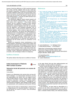
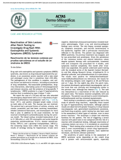
IMAGES IN
CARDIOLOGY
Subacute Radiation Dermatitis Secondary to Cardiac
Resynchronization Device Implantation
Radiodermitis Subaguda Secundaria a Implante de Resincronizador Cardíaco
EDGAR ANTEZANA CHÁVEZ, DARÍO DI TOROMTSAC, CARLOS LABADETMTSAC
The images correspond to a 53-year-old male patient, referred to our center to have his cardiac resynchronization
therapy defibrillator (CRT-D) checked. He had a history of
idiopathic dilated cardiomyopathy, permanent atrial fibrillation with complete left bundle branch block, heart failure in FC III, and severe left ventricular dysfunction (EF
= 23%).
In 2012, the patient underwent the first CRT-D implantation in his home province. Surgery was prolonged, lasting
for about 6 hours. The coronary sinus catheter could not be
placed, so the device was programmed as defibrillator.
Physical examination revealed an ulcerated tumor lesion with raised erythematous edges and fibrinous base of
15 × 12 cm in the right dorsal region, which, as referred by
the patient, had appeared about 6 months after the procedure.
The location of the lesion coincided with the left anterior oblique position of the fluoroscopic tube, commonly used
for coronary sinus catheter placement.
Given the nature of the lesion, it seems to be subacute
radiation dermatitis. Radiation dermatitis usually occurs
within 2 to 12 months after the procedure.
Subacute lesions include from erythema and local scaling, similar to that in acute radiation dermatitis, to necrotic
ulceration of the deep skin layers. Lesions also involve irrigation, and cause long-term pigmentary changes and loss of
skin annexes. About 20% of the cases can progress to ulcers
with irregular edges and neoplastic lesions. (1, 2)
Its occurrence depends on the radiation dose in the area,
requiring > 8 Gy for lesions similar to those observed in this
case. (3) Several factors can increase sensitivity to radiation, in addition to individual susceptibility, certain drugs
(amiodarone, fibrates, actinomycin, and methotrexate), collagen diseases, immunosuppressive diseases, diabetes, and
malfunctioning of radioscopy equipment. (4, 5) Of all these
factors, only amiodarone intake was present in our patient.
On the other hand, some skin areas are more vulnerable
than others, mainly the axillary, pectoral, and dorsal areas,
as was the case in our patient. (6)
Conflicts of interest
None declared.
REFERENCES
1. Stone M, Robson K, LeBoit P. Subacute radiation dermatitis from
fluoroscopy during coronary artery stenting: Evidence for cytotoxic
lymphocyte mediated apoptosis. J Am Acad Dermatol 1998;38:333-6.
http://doi.org/dvc734
2. Herrera E, Moreno A, Requena L, Rodríguez JL. Dermatopatología: Correlación clínico-patológica. 1.ª ed. Grupo Menarini; 2007.
p. 550-3.
3. Mettler FA Jr, Koenig TR, Wagner LK, Kelsey CA. Radiation
injuries after fluoroscopic procedures. Semin Ultrasound CT MR
2002;23:428-42. http://doi.org/fsv2vx
4. Rodríguez I, Fernández D, Rovira I, Fuentes ME. Radiodermitis crónica secundaria a cateterismo cardíaco. Actas Dermosifiliogr
2001;92:291-5. http://doi.org/f2h88s
5. Villanueva Ramos TI, Alcalá Pérez D, Vega González MT, Pedralta
Pedrero ML, Medina Bojórquez A, Barrera Cruz A y cols. Guía de
práctica clínica para prevención y tratamiento de la radiodermitis
aguda. Dermatol Rev Mex 2012;56:3-13.
6. Nahass GT. Acute radiodermatitis after radiofrequency catheter
ablation. J Am Acad Dermatol 1997;36:881-4. http://doi.org/fj6d5z
Rev Argent Cardiol 2014;82:508 http://dx.doi.org/10.7775/rac.v82.i6.4874
Address for reprints: Dr. Edgar Antezana - Corbeta Pi y Margal 750 - (1155) CABA, Argentina - Tel. 011 4121-0821 - e-mail: [email protected]
Hospital de Agudos “Dr. Cosme Argerich” - Department of Electrophysiology
MTSAC
Full Member of the Argentine Society of Cardiology


© Copyright 2026