
CASE AND RESEARCH LETTERS Reactivation of Skin Lesions
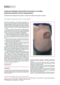
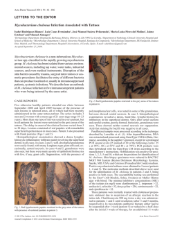
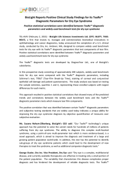
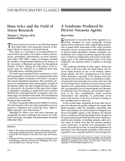
Documento descargado de http://www.actasdermo.org el 08/11/2016. Copia para uso personal, se prohíbe la transmisión de este documento por cualquier medio o formato. Actas Dermosifiliogr. 2016;107(9):781---798 CASE AND RESEARCH LETTERS Reactivation of Skin Lesions After Patch Testing to Investigate Drug Rash With Eosinophilia and Systemic Symptoms (DRESS) Syndrome夽 Reactivación de las lesiones cutáneas por pruebas epicutáneas en el estudio de un síndrome de DRESS To the Editor: Drug rash with eosinophilia and systemic symptoms (DRESS) syndrome, also known as drug-induced hypersensitivity syndrome, is an uncommon severe reaction with a late onset and prolonged course that is sometimes fatal (10%-40%). The pathogenesis of this condition is complex, and various factors are believed to interact, including accumulation of drug metabolites, HLA-associated predisposition, drugvirus interactions, alternating cycles of immunosuppression and immune recovery, and the presence of drug-specific T lymphocytes.1 DRESS syndrome is a type IV hypersensitivity reaction that is mediated by type 2 helper T cells.2 We report the case of a 24-year-old man who attended the emergency department with a 2 to 3---day history of generalized erythematous maculopapular rash (Figs. 1 and 2), fever >39 ◦ C, and painful enlarged lymph nodes (>2 cm) on both sides of his neck. The mucosa was not involved. The patient had a personal history of asthma, anxiety, and chronic prostatitis and had been receiving montelukast, inhaled salmeterol/fluticasone, and bromazepam for more than a year. In addition to his usual treatment, he had received trimethoprim-sulfamethoxazole during the previous month. The blood workup revealed leukocytosis of up to 21 300 cells/L with 25% eosinophils and acute hepatitis (aspartate aminotransferase, 91 U/L; alanine aminotransferase, 599 U/L; and ␥-glutamyl transpeptidase, 248 U/L); the remaining values were within the normal range. The results of serology testing for hepatotropic viruses were 夽 Please cite this article as: Córdoba S, Navarro-Vidal B, MartínezMorán C, Borbujo J. Reactivación de las lesiones cutáneas por pruebas epicutáneas en el estudio de un síndrome de DRESS. Actas Dermosifiliogr. 2016;107:781---783. negative. Abdominal ultrasound examination revealed moderate splenomegaly. Chest x-ray and electrocardiogram findings were normal. The skin biopsy revealed spongiosis, lymphatic exocytosis, and necrotic keratinocytes in the epidermis, together with a perivascular mononuclear infiltrate in the dermis. The patient was diagnosed with DRESS syndrome, his previous treatment was stopped, and he was prescribed oral prednisone. Given the persistence of the cutaneous lesions and altered laboratory values despite systemic therapy with corticosteroids, treatment with oral ciclosporin was prescribed for 4 months, until symptoms resolved completely. One month after treatment was suspended, the patient underwent patch testing with the standard series of the Spanish Contact Dermatitis and Skin Allergy Research Group, trimethoprim 2% in dimethyl sulfoxide, and sulfamethoxazole 2% in petrolatum. The results were positive for methylchloroisothiazolinone/methylisothiazolinone (no relevance); the results for the remaining allergens were negative at 48 and 96 hours and at 7 days. On the eighth day after the patch tests, the patient developed a generalized maculopapular rash on the trunk that was clinically and histologically similar to the previous rash, although less intense (Fig. 3). The rash was interpreted as a reactivation of the rash induced by the patch tests. The lesions resolved completely with oral prednisone. The patient was advised to avoid trimethoprimsulfamethoxazole and has remained asymptomatic. Patch testing is considered a useful diagnostic tool in the study of adverse drug reactions,1 especially those caused by type IV hypersensitivity mechanisms. Although positive results are observed with greater frequency in drug-induced maculopapular rash, generalized exanthematic pustulosis, and fixed drug eruptions, patch testing has also been used in the assessment of other types of reaction, even in more severe reactions.2 The technique is simple and relatively harmless for the patient. The risk of triggering a severe reaction with patch tests is low compared with the risk of a reaction with intradermal testing. Although reactivation of cutaneous lesions can occur, it is considered an uncommon finding.3---5 Patch testing after evaluation of the risk-benefit ratio is even recommended in severe drug hypersensitivity reactions, including DRESS syndrome, toxic epidermal necrolysis, and Steven-Johnson syndrome.6 The sensitivity of patch testing in the assessment of DRESS syndrome varies depending on the culprit drug and is greater with anticonvulsant agents, especially 1578-2190/© 2016 Elsevier España, S.L.U. and AEDV. All rights reserved. Documento descargado de http://www.actasdermo.org el 08/11/2016. Copia para uso personal, se prohíbe la transmisión de este documento por cualquier medio o formato. 782 Figure 1 CASE AND RESEARCH LETTERS Erythematous maculopapular rash on the trunk. carbamazepine. In the case of other drugs involved in the syndrome, the sensitivity of patch testing is considered low.2,6 Despite its limited sensitivity, patch testing is an appropriate way of assessing drug-induced reactions before performing prick tests, intradermal tests, and even drug challenge tests, which carry greater risks for the patient. Figure 3 Lesions of lesser intensity similar to those of the previous rash. Reactivation after patch tests. We present a case of DRESS syndrome caused by trimethoprim-sulfamethoxazole. The patient developed a reactivation of the skin lesions induced by patch tests. The low concentration of drug in the patches, the poor availability of tests, or the fact that the reaction was triggered by a metabolite and not by the drug itself could explain the negative result in the patch tests. Nevertheless, percutaneous absorption from the patches was sufficient to cause a generalized reaction. Conflicts of Interest The authors declare that they have no conflicts of interest. References Figure 2 Purpuric erythematous maculopapular lesions on the lower limbs. 1. De la Torre C, Suh Oh HJ. Novedades en el diagnóstico de las toxicodermias. Actas Dermosifiliograf. 2013;104:782---8. 2. De Santiago F, Gonçalo M, Vieira R, Coelho S, Figueiredo A. Epicutaneous patch testing in drug hypersensitivity syndrome (DRESS). Contact Dermatitis. 2010;62:47---53. 3. Vaillant L, Camenen I, Lorette G. Patch testing with carbamazepine: Reinduction of an exfoliative dermatitis. Arch Dermatol. 1989;152:299. 4. Wolkenstein P, Chosidow O, Flechet ML, Robbiola O, Paul M, Dume L, et al. Patch testing in severe cutaneous adverse drug reactions including Steven-Johnson syndrome and toxic epidermal necrolysis. Contact Dermatitis. 1996;35:234---6. 5. Mashiah T, Brenner S. A systemic reaction to patch testing for the evaluation of acute generalized exanthematous pustulosis. Arch Dermatol. 2003;139:1181---3. 6. Buyuktiryaki AB, Bezirganoglu H, Sahiner UM, Yavuz ST, Tuncer A, Kara A, et al. Patch testing is an effective method for the diagnosis of carbamazepine-induced drug reaction, eosinophilia and systemic symptoms (DRESS) syndrome in an 8-year-old girl. Australas J Dermatol. 2012;53:274---7. Documento descargado de http://www.actasdermo.org el 08/11/2016. Copia para uso personal, se prohíbe la transmisión de este documento por cualquier medio o formato. CASE AND RESEARCH LETTERS S. Córdoba,∗ B. Navarro-Vidal, C. Martínez-Morán, J. Borbujo Servicio de Dermatología, Hospital Universitario de Fuenlabrada, Fuenlabrada, Madrid, Spain Secondary Syphilis Presenting as Oral Lesions and Posterior Placoid Chorioretinitis in an Immunocompetent Patient夽 Lesiones orales y coriorretinitis placoide posterior como forma de presentación de sífilis secundaria en un paciente inmunocompetente To the Editor: Within the broad range of manifestations that syphilis can cause, lesions on the oral mucosa and eye involvement are very rare, particularly as isolated lesions. We describe a patient with oral lesions and eye involvement, with no associated skin lesions. The patient was a 59-year-old man, with no past history of interest. He was referred from another center to investigate a 1-year history of oral mucosal symptoms presenting as odynophagia, a sensation of excess mucus secretion, and painful erosive lesions in the oral cavity and pharynx. The patient had also been seen in ophthalmology for a decrease in visual acuity over the previous 3 months. Physical examination revealed erosive lesions on the hard palate and on the right tonsillar pillar (Fig. 1) and difficulty swallowing. On ophthalmologic examination, a large, circular, yellowish placoid lesion was observed on the pigmented epithelium of the retina, at the macula. The lesion extended to the upper temporal vascular arches of the right eye (Fig. 2A). Visual acuity was 20/50. This was initially considered as a chorioretinitis of uncertain origin. Biopsy of an oral lesion showed epidermal hyperplasia with a dense mixed inflammatory infiltrate with a predominance of plasma cells that extended into the deep dermis and was localized mainly around the blood vessels. Lymphocyte exocytosis was also observed. A provisional diagnosis of secondary syphilis was made, and staining with antitreponemal antibody showed the presence of numerous spirochetes in the mucosa and in smaller numbers in the submucosa. Syphilis serology in blood was positive (rapid plasma reagin, 1:128; treponema pallidum hemagglutination test, +). On further questioning, the patient denied previous skin lesions but did state that about 18 months earlier he had had an erosion on the glans that had been diagnosed as genital herpes at other 夽 Please cite this article as: Plana-Pla A, Pelegrín-Colás L, BielsaMarsol I, Ferrandiz-Foraster C. Lesiones orales y coriorretinitis placoide posterior como forma de presentación de sífilis secundaria en un paciente inmunocompetente. Actas Dermosifiliogr. 2016;107:783---784. 783 Corresponding author. E-mail address: [email protected] (S. Córdoba). ∗ center. Based on these findings, we made a diagnosis of posterior placoid chorioretinitis of probable syphilitic origin. HIV serology was negative and lumbar puncture showed a slight increase in the protein content of the cerebrospinal fluid (64 mg/dL), with normal cellularity and glucose levels and positive reagin antibodies titers (Venereal Disease Research Laboratory). A diagnosis was therefore made of secondary syphilis with central nervous system involvement (neurosyphilis) and the patient was treated with penicillin G sodium at a dose of 4 million IU intravenously every 4 hours for 14 days. The clinical course was favorable, with resolution of the oral lesions and a clear improvement in the ocular lesion (Fig. 2B). Syphilis can cause oral lesions in all of its stages, but they are most common in the secondary stage (30% of patients). However, the absence of concomitant skin manifestations, as in our case, is very rare.1 The oral lesions of secondary syphilis are clinically very heterogeneous and typically nonspecific; they include macules, papules, plaques and ulcers, which may be single or multiple, and they are usually associated with nonspecific pharyngitis, tonsillitis, or laryngitis, sometimes with palpable lymph nodes.2 These lesions most frequently occur together with the typical cutaneous manifestations of the disease, and usually resolve within 3 to 12 weeks.3 In the present case, however, the lesions were isolated and persisted for at least a year. In a review of the literature, we found isolated case reports of oral lesions without associated skin manifestations,3,4 but none had a clinical course of more than 5 months in patients who, like ours, were immunocompetent. Isolated lesions of syphilis on the oral mucosa are a considerable diagnostic challenge, and the histology of these lesions must therefore be carefully evaluated for any detail that will help to reach the diagnosis. In addition, the patient must be questioned closely about noncutaneous features of Figure 1 pillar. Erosions on the hard palate and the right tonsillar


© Copyright 2026